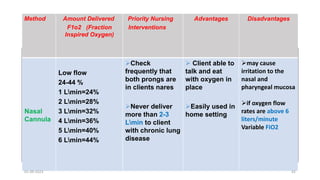
Oxygenation Needs
Mr. Ramprasad S M.Sc (N), SCEM
Associate Professor
Medical Surgical Nursing, Neurosciences Nursing
Trainer In Skills Lab, FRT, ENLS, BLS, TCLS, GFATM, Research Guide,
Reviewer (IJMR/IJNR/IJOGN), (Member TNAI/NTAI/IPA/NSS/IRS)
01-09-2023 1
Factors affecting Respiration
• The following factors affects the normal respiration of
the individual. They are,
a. Internal factors
b. External factors
01-09-2023 2
Conditions that affecting the
AIRWAY
01-09-2023 3
1. Allergies
A condition that occur when immune system
respond to foreign particles, such as pollens, dust,
certain food, chemicals, etc.
Allergies are characterized by rashes, cough, sneeze,
running nose, chest congestion, wheezing etc.
01-09-2023 4
2. Bronchial Asthma
A condition characterized by inflammation,
bronchospasm, and edema airway.
Characterized by, cyanosis, rapid pulse, cough,
shortness of breath.
01-09-2023 5
3. Common Cold
Infection of the upper respiratory tract results in
narrowing of the airway.
Common characteristics are,
Cough, running nose, fever, congestion, nose block
01-09-2023 6
4. Bronchitis
It is an inflammatory condition of the bronchial tube.
Common signs are
Production of the mucus, chest tightness, low grade
fever,
01-09-2023 7
Conditions that affecting the
Movement of the Airway
01-09-2023 8
1. Dyspnea/Shortness of breath
It is defined as the sensation of breathlessness or
decrease breathing.
Tight feeling in chest, caused by asthma, heart failure,
and lung disease.
01-09-2023 9
Grading
Grade 1 :occurs when strenuous activity
Grade 2 : person cant climb the stairs
Grade 3 : occurs in usual activities
Grade 4 : requires assistance during ADL
Grade 5 : occurs at rest
01-09-2023 10
Causes
Anemia/
Asthma
Heart and lung disease
Obesity/rib injury/ poisoning
01-09-2023 11
2. Tachypnea
Condition that occurs when respiratory rate more than
20 per minute
01-09-2023 12
Causes
Exercise
Increased physical activity
Allergy
Pneumonia / TB
01-09-2023 13
Symptoms
Shortness of breath
LOC
Confusion
Sweating
01-09-2023 14
3. Bradypnea
A condition where heart rate less than 16 per minute
Caused by,
Opiods , smoking, alcoholism, sleep apnea, neuro
muscular disorder
01-09-2023 15
Symptoms,
Itching , dry skin, hair loss, dizziness, cyanosis, coma
01-09-2023 16
Conditions Affecting Diffusion
01-09-2023 17
1. Pulmonary Edema
Abnormal accumulation and collection of fluid in the
lung tissue or in the alveolar space.
01-09-2023 18
Causes are,
Severe HTN, MI, cardiomyopathy, kidney failure
01-09-2023 19
Clinical Features are
Central cyanosis (lips /nails)
Blood mixed secretions
Dyspnea
Respiratory distress
01-09-2023 20
Grunting and wheezing sound
Pale skin
Rapid heart rate
01-09-2023 21
Causes
Cardiogenic causes
artherosclerosis, HTN, MI, cardiomyopathy
Non cardiogenic causes
pneumonectomy , renal failure, toxic inhalation
01-09-2023 22
2. COPD
Condition where airflow is obstructed due to
emphysema chronic bronchitis, or both
01-09-2023 23
Causes are,
Chronic smoking, infections , obesity , asthma, air
pollution , occupation exposure
01-09-2023 24
Features are,
Chronic cough, sputum, chest pain, dyspnea,
hemoptysis, weakness,fatigue
01-09-2023 25
A condition characterized by collapse of the lungs, may be acute or
chronic.
Atelectasis
01-09-2023 26
Caused due to blockage in the air passages or pressure on the lungs by
outside. The other risk factors are,
Anesthesia, foreign object in the airway, lung disease, tumor
01-09-2023 27
Factors affecting Oxygen Transport
01-09-2023 28
It is an inability of the heart to pump adequate blood to the parts of the
body.
The primary causes are CHD, HTN, VHD,Cardiomyopathies,
Arrythmias
1. Heart Failure
01-09-2023 29
Other causes,
Pulmonary embolism, anemia, anesthesia, increase sodium intake.
01-09-2023 30
Nursing Interventions To Promote
Oxygenation
01-09-2023 31
Oxygen Administration
01-09-2023 32
it is to treat the deficiency in oxygen among the individual but not to
treat the underlying causes. This is important to keep the healthy level
of tissue oxygen.
Introduction
01-09-2023 33
To reduce the effect of anoxemia
To maintain healthy tissue level oxygen
Purposes
01-09-2023 34
Basics of Anantomy
01-09-2023 35
Low Flow System High Flow System
Nasal cannula Venturi mask
Simple mask T-Piece
NRVM/NRBM
Partial Rebreather mask
Methods of oxygen administration
01-09-2023 36
Basic Oxygen Delivery Methods
Nasal cannula and face mask are noninvasive interventions.
Useful to deliver O2 if the patient is breathing on their own
Nasal Cannula
(nasal prongs)
Simple face mask Face Mask with
Reservoir bag
Bag-Valve-Mask
(BVM / AMBU bag)
Page 7
01-09-2023 37
1. Nasal cannula
2. Nasal prongs
3. Oxygen mask
4. Oxygen tent
5. Venturi mask
6. Oxygen hood
Methods of oxygen administration
01-09-2023 38
A plastic tube that used to administer oxygen which connected to the
oxygen cylinder. The maximum flow of the oxygen will be upto 4L/Min
Nasal cannula
01-09-2023 39
01-09-2023 40
It is a disposable.
plastic devise with two protruding prongs for
insertion into the nostrils, connected to an
oxygen source.
Used for low-medium concentrations of
Oxygen (24-44%).
01-09-2023 41
Disadvantages
Advantages
Priority Nursing
Interventions
Amount Delivered
F1o2 (Fraction
Inspired Oxygen)
Method
may cause
irritation to the
nasal and
pharyngeal mucosa
if oxygen flow
rates are above 6
liters/minute
Variable FIO2
 Client able to
talk and eat
with oxygen in
place
Easily used in
home setting
Check
frequently that
both prongs are
in clients nares
Never deliver
more than 2-3
Lmin to client
with chronic lung
disease
Low flow
24-44 %
1 Lmin=24%
2 Lmin=28%
3 Lmin=32%
4 Lmin=36%
5 Lmin=40%
6 Lmin=44%
Nasal
Cannula
01-09-2023 42
The types of face mask are
The Simple Oxygen Mask
The Partial Rebreather Mask:
The Non Rebreather Mask:
The Venturi Mask:
Face mask
01-09-2023 43
01-09-2023 44
The simple Oxygen mask
Simple mask is made of clear,
flexible , plastic or rubber that
can be molded to fit the face.
01-09-2023 45
The simple Oxygen mask
It delivers 35% to 60% oxygen .
A flow rate of 6 to 10 liters per minute.
Often it is used when an increased delivery of oxygen is
needed for short periods
(i.e., less than 12 hours).
01-09-2023 46
Disadvantages
Advantages
Priority Nursing
Interventions
Amount
Delivered
F1o2 (Fraction
Inspired Oxygen)
Method
Tight seal required to
deliver higher concentration
Difficult to keep mask in
position over nose and
mouth
Potential for skin
breakdown (pressure,
moisture)
Wasting
Uncomfortable for pt while
eating or talking
Expensive with nasal tube
Can provide
increased
delivery of
oxygen for
short period of
time
Monitor client
frequently to check
placement of the
mask.
Support client if
claustrophobia is
concern
Secure physician's
order to replace
mask with nasal
cannula during meal
time
Low Flow
6-10 Lmin
35%-60%
Simple
mask
01-09-2023 47
The partial rebreather mask
The mask is have with a reservoir bag must
inflated during both inspiration & expiration
 It collection of the first parts of the patients'
exhaled air.
It is used to deliver oxygen concentrations up
to 80%.
01-09-2023 48
The partial rebreather mask
The oxygen flow rate must be
maintained at a minimum of 6
L/min to ensure that the patient
does not rebreathe large amounts
of exhaled air.
The remaining exhaled air exits
through vents.
01-09-2023 49
The non rebreather mask
 This mask provides the highest
concentration of
oxygen (95-100%) at a flow rate6-15
L/min.
It is similar to the partial rebreather
mask
except two one-way valves prevent
conservation of exhaled air.
The bag is an oxygen reservoir
01-09-2023 50
Disadvantages
Advantages
Priority Nursing
Interventions
Amount Delivered
F1o2 (Fraction
Inspired Oxygen)
Method
Requires
tight seal
(eating and
talking difficult,
uncomfortable
Not as drying
to mucous
membranes
PT can inhale
room air
through
openings in
mask if
oxygens
supply is
briefly
interrupted
Set flow rate
so mask
remains tow-
thirds full
during
inspiration
Keep
reservoir bag
free of twists
or kinks
Low Flow
6 Lmin
75%-80%
oxygen
Partial
Rebreather
Mask
01-09-2023 51
Disadvantages
Advantages
Priority Nursing
Interventions
Amount
Delivered
F1o2
Method
Impractical for long
term Therapy
Malfunction can cause
CO2 buildup
suffocation
Expensive
Feeling of suffocation
Uncomfortable
Costly
Delivers the
highest possible
oxygen
concentration
Suitable for pt
breathing
spontaneous with
sever hypoxemia
Maintain flow rate so
reservoir bag collapses
only slightly during
inspiration
Check that valves and
rubber flaps are
function properly (open
during expiration )
Monitor SaO2 with
pulse oximeter
Low
Flow
6-15 L
min
80%-
100%
Non
rebreather
MASK
01-09-2023 52
Venturi mask
 It is high flow concentration of
oxygen.
Oxygen from 40 - 50%
At liters flow of 4 to 15 L/min.
01-09-2023 53
Disadvantages
Advantages
Priority
Nursing
Interventions
Amount
Delivered
F1o2
Method
uncomfortable
Risk for skin
irritation
produce respiratory
depression in COPD
patient with high oxygen
concentration 50%
Delivers most
precise oxygen
concentration
Doesn’t dry
mucous
membranes
(humidity
Requires
careful
monitoring to
verify F1O2 at
flow rate
ordered
Check that
air intake
valves are not
blocked
Oxygen from
40-- 50%
of 4 to 15
L/min.
Venturi
Mask
01-09-2023 54
T-piece
Used on end of ET
tube when weaning
from ventilator
Provides accurate
FIO2
Provides good
humidity
01-09-2023 55
Side effect & complication of oxygen therapy
Oxygen toxicity
Retrolental fibroplasia
Absorption atelectasis
01-09-2023 56
oxygen toxicity
It is a condition in which ventilator failure
occurs due to inspiration of a high
concentration of oxygen for aprolonged
period of time.
Oxygen concentration greater than 50% over 24 to 48 hours can
cause pathological changes in the lungs.
01-09-2023 57
Signs and symptoms of oxygen toxicity
Non-productive cough.
• Nausea and vomiting.
• Sub sternal chest pain.
• Fatigue.
• Nasal stuffiness.
01-09-2023 58
Sore throat.
• Hypoventilation.
. Nasal congestion.
. Dyspnea.
. Inspiration pain
01-09-2023 59
Evaluation
Breathing pattern regular and at normal rate.
 pink color in nail beds, lips, conjunctiva of eyes.
 No confusion, disorientation, difficulty with cognition.
Arterial oxygen concentration or hemoglobin
 Oxygen saturation within normal limits.
01-09-2023 60
Documentation
Date and time oxygen started.
Method of delivery.
Oxygen concentration and flow rate.
Patient observation.
Add oro nasal care to the nursing care plan
01-09-2023 61
Oxygen Delivery and Maintaining the Airway
Page 9
Delivery Method O2 lpm O2 % Skill level Airway Patency
BASIC
Nasal Cannula 1-4 24 - 36 Minimal
Conscious patient with patent airway,
If the patient is breathing spontaneously
Simple Mask 5-10 40 - 60 Minimal
Non-Rebreather Mask 10-15 60 - 90 Minimal
Bag Valve Mask (BVM) /
AMBU bag
>15 20 - 95 Basic
Reduced consciousness (or unconscious)
Consider insertion of nasal or oral airway
Manual positive pressure ventilation (PPV)
ADVANCED
LMA, ETT and Crico-
thyroidotomy >15 21 - 100 Advanced
Unconscious (if not, needs sedation) and
airway not protected
Start with manual PPV with bag valve.
Connect to ventilator when available
01-09-2023 62
Summary
Oxygen Administration can be done under the following ways.
Invasive and Non Invasive
Supraglotic and Infra Glottis
Slow flow rate and High flow rate
01-09-2023 63
Maintaining The Airway
Methods of Maintaining Airway Patency
01-09-2023 64
The following methods are used to maintain the airway,
1. Positioning
2. Manoeuvres
a. Head Tilt and Chin lift
b. Jaw Thrust
3. Suction
4. Use of airway adjuncts
a. Nasopharyngeal Airway
b. Oropharyngeal Airway
01-09-2023 65
5. Advanced Airway Methods
a. Laryngeal Mask Airway (LMA)
b. Endotracheal Intubation (ETT)
c. Cricothyroidotomy
01-09-2023 66
• Patient positioning:
• Manual airway maneuvers
• Towel support under shoulders
• Suctioning
• Supplemental oxygen
• Airway adjuncts
• Advanced airway placement
67
01-09-2023 67
Airway Management Techniques
• Airway Maneuvers: Chin lift / jaw thrust (most basic)
• Airway adjuncts: Oral, nasal
• Bag-Valve-Mask (BVM)
• Supraglottic Airways
• Endotracheal intubation
• Cricothyrotomy (most advanced)
68
01-09-2023 68
Airway Management – The Basics
The tongue is the commonest cause of airway obstruction.
69
01-09-2023 69
Jaw thrust
head tilt
chin lift
Head Tilt
+
Chin Lift
Or
Jaw Thrust
Head tilt and chin lift contraindicated in:
- Trauma
- History of neck surgery or cervical spine pathology
Only jaw thrust
Airway Management – Open the Airway!
01-09-2023 70
Airway Adjuncts
Airway adjuncts should be used as early as possible when needed
Nasopharyngeal Airway (NPA) Oropharyngeal Airway (OPA)
• In conscious and
unconscious patients
• Only in unconscious
patients
• DON’T use if gag reflex is
intact
01-09-2023 71
Nasopharyngeal Airway
1. Can be used both conscious and unconscious patients
2. Used easily and effectively
3. Check the appropriate size of the NPA
4. Position the airway accurately
5. Contraindicated in nasal fractures
01-09-2023 72
01-09-2023 73
Adult 6 to 9 cm.
small adult 6 to 7
medium size adult 7 to 8 cm
large adult 8 to 9 cm
NPA airway sizes
01-09-2023 74
Oropharyngeal Airway
1. Can be used only in unconscious patients
2. Avoid using when the Gag reflex in intact
3. Avoid in oral trauma/spasm of mouth muscles
4. Check the appropriate size of the OPA
5. Position the airway accurately
6. Contraindicated in conscious patient
01-09-2023 75
OPA airway sizes
01-09-2023 76
Neonatal 00
Infant 0
Child 1
Small adult 2
Adult 3
Large adult
4
5
6
01-09-2023 77
Endotracheal Intubation
https://www.youtube.com/watch?v=70fhNvE6aGQ
01-09-2023 78
ENDOTRACHEAL INTUBATION
Endotracheal Intubation is placement of special
tube in trachea. Endotracheal intubation is employed to
relieve upper airway obstruction, maintain a patent
airway or treat impending or actual respiratory failure
from any cause.
01-09-2023 79
Indications/Purposes
To secure airway
To supply oxygen
For general anesthesia
For cardiopulmonary resuscitation
For ventilator therapy in ICU
01-09-2023 80
Articles
Endotracheal tubes
Laryngoscope blades
Oral airway
Suction devices: catheters and tonsil tip
Intravascular catheters, spinal needle, or bone marrow
needle.
Oro gastric tube
01-09-2023 81
Laryngoscope handles and blades
Scissors ,Gloves, Syringes, Needles,
01-09-2023 82
Pre-Procedure
I. Assess current respiratory status and history including
Reason for intubation.
Response to intubation.
II. Auscultate breath sound bilaterally
III. Evaluate respiratory effort
Respiratory rate
Respiratory pattern
01-09-2023 83
V. Auscultate breath sound bilaterally.
VI. Evaluate respiratory effort.
Respiratory rate, Respiratory pattern,
Chest expansion: equal and bilateral
01-09-2023 84
VIII. Observe colour and tissue perfusion.
IX. Assess heart rate.
Assess activity and alertness, irritability, confusion, fatigue,
lethargy, inappropriate behavior and coma.
Assess child and family's understanding of need, purpose and
functioning of intubation.
01-09-2023 85
Procedure
1. Confirm availability of all equipment
2. Wear PPE
3. Perform hand washing
4. Don gloves
5. Take laryngoscope and place inside the throat to visualize the glottis
6. Take appropriate sized ET tube and put bougie into it
7. Inflate the ET tube and check its efficacy
8. Bend the ET tube and insert into the mouth upto trachea
01-09-2023 86
Procedure (Contd…)
9. Once the ET tube is placed remove the styllet or bougie and inflate the tube
with 5-7 ml of air.
10. Secure the ET Tube with the adhesive plaster and confirm its placemat by
auscultation.
11. Check the inflation by using AMBU (chest should expand)
12. Do 5 point auscultation including epigastrium (sound will be absent at
epigastrium)
01-09-2023 87
CHEST PHYSIOTHERAPY
01-09-2023 88
Definition
Chest physiotherapy (CPT) is a therapeutic
intervention applied to help natural airway clearance
mechanisms (cough and ciliary cleansing mechanisms)
reduce or prevent blockage of airways by thick,
tenacious mucus
01-09-2023 89
Purposes/Indication
• Assist in coughing
• Reeducate breathing muscles
• Improve ventilation of the lungs
01-09-2023 90
Contraindications
• Increased ICP
• Unstable head or neck injury
• Active haemorrhage or heamoptysis
• Recent spinal injury
• Rib fracture
• Uncontrolled hypertension
• Thoracic surgeries
01-09-2023 91
Articles Needed
Tilt bed and/or pillows
Towels or thick pad
Sputum cup/tissue
Stethoscope
01-09-2023 92
Types of CPT
The procedure of chest physiotherapy includes the
following types,
1. Percussion
2. Chest Vibration
3. Deep breathing Exercises
4. Postural drainage
5. Coughing
01-09-2023 93
Preprocedure care
• Identify physical indications for CPT.
• Identify possible contraindications for CPT.
• Determine child's age, developmental level, ability to
understand procedure, and ability to cooperate.
• Assess diversional activities child might enjoy during CPT.
• Determine parent's under-standing of and ability to perform
CPT at home.
• Assess ability of older child to do self-physiotherapy
01-09-2023 94
Percussion/tapotment/clapping/cupping
• Therapist and parents should wash hands before
treatment.
• Administer bronchodilators as ordered before CPT.
• Select areas of lungs to be percussed.
• Treatments may be split up into sections and
performed at various times throughout the day
01-09-2023 95
• Assist person in assuming proper positioning, placing
pillows for comfort, support and to maintain position.
• Perform percussion using cupped hands. Clap
rhythmically over specific area to be drained. Mold hands
side by side.
01-09-2023 96
Alternate hands while clapping. Clapping should be
vigorous, not painful. Percuss 1 minute over lung segment to
be drained.
01-09-2023 97
Procedure
1. Check the right patient
2. Explain the procedure to the patient
3. Check the last meal that he has consume
4. Wash hands and dry
5. Inform the patient to perform diaphragmatic
breathing
6. Position the patient
01-09-2023 98
Percussion
1. clap with cupped hands over the chest wall for over 1-2
min in each lung area
2. the percussion should be from lower ribs to shoulder on
the back and from lower ribs to top of the chest in the front
3. Avoid percussion on spine, liver , kidney, spleen, breast
and clavicle.
01-09-2023 99
01-09-2023 100
01-09-2023 101
Vibration
1. place hand /palm down on chest area to be drained with one
hand over the other or fingers together.
2. Patient should inhale and exhale slowly through pursed lips
3. Vibrate for 5 exhalations in each lungs after 3-4 vibrations
encourage patient to cough and clear the secretion
01-09-2023 102
Post Procedure
1. Allow patient to rest
2. Auscultate and check the breath sound
3. Repeat cycle for 10-15 minutes based on patient condition
4. Wash hands
5. Comfort the patient
01-09-2023 103
Special Considerations
1. CPT should be perform 1 hour before meals and 1-3 hours after meal
2. Administer bronchodilator and MDI 15 minutes earlier
3. Check breathing pattern and cyanosis in him
4. Administer pain killers 15-20 minutes before
5. Stop procedure if there is tachycardia/fall in BP/chest pain
01-09-2023 104
Postural Drainage
Also called as bronchial drainage, an intervention
for airway clearance by mobilizing secretions in
one or more lung segments to the central airway
by placing the patient in various positions.
01-09-2023 105
Indications
1. Cystic fibrosis
2. Bronchiectasis
3. Pulmonary diseases
4. Patient with artificial airway
01-09-2023 106
Contraindications
1. Severe hemoptysis
2. Cardiac arrhythmias
3. Neurosurgery patients
4. Unstable angina
01-09-2023 107
5. Plural effusion
6. Pneumothorax
7. Pulmonary embolism
01-09-2023 108
Preparations
1. Check the clinical findings and indications for
PD
2. Tilt the patient to drain the secretions
3. Lower lobes requires the PD than upper lobe
01-09-2023 109
4. Provide bronchodilators or Nebulizer
5. The PD can be scheduled 3-4 times a day based
on degree of lung congesion
6. Time is before breakfast/lunch and bed time
01-09-2023 110
Care of Chest Drainage
01-09-2023 111
01-09-2023 112
01-09-2023 113
• Parietal pleura: Inside membrane of the rib cage
• Pulmonary or visceral pleura: Outside membrane of the lungs
• The two membranes are separated by a lubricating fluid (pleural
fluid) which enables them to slide against each other. Lung can
expand and contract
• Vacuum or “negative pressure” in the pleural space keeps the two
pleurae together. If fluid or air enters the pleural space, lungs cannot
fully expand during each respiratory cycle
Chest TUBE Placement
Normal Breathing Mechanics
Diaphragm
contracts down &
draws air into the
trachea, bronchi,
bronchioles, and
alveoli. Negative
pressure between
the pleurae is
approximately
-8cmH2O
Diaphragm relaxes,
rises, and pushes air
& CO2 back out the
same route. Negative
pressure between
the pleurae is
approximately -
4cmH2O
INHALATION EXHALATION
01-09-2023 116
• Pneumothorax: Air in the pleural space (trauma, lung
disease, procedure complications)
• Hemothorax: blood in the pleural space (blunt or penetrating
trauma)
• Hemopneumothorax: air & Blood in the pleural space
• Tension pneumothorax: occurs when injured tissue forms a
one way valve or flap allowing air to enter but preventing it
from escaping naturally, (emergent situation)
Indications For Chest Tube Insertion
•Pleural effusion: excessive fluid in pleural space
•Empyema: collection of pus d/t infection
•Chylothorax: lymphatic fluid accumulation in the
pleural space (trauma, expanding tumor, surg in the
mediastinum) – milky white pleural fluid
01-09-2023 117
Insertion Site
Superior Tube Placement:
Removal of Air (ie.
Pneumothorax)
Inferior Tube Placement:
Removal of Fluids (ie.
Hemothorax, Empyema)
Posterior Tube Placement:
Loculated (confined pocket)
fluid or air
Insertion Site
“Safe Triangle”
Tube placed in the midaxillary line
btw 4th and 5th Ribs should
successfully drain fluid or vent air
CHEST TUBE SYSTEM
3 Bottle System
Patient
Suction
Collection
Bottle
Water
Seal
Suction
Control
Collection Bottle and Water Seal
Collection Chamber: Collects drainage
Water Seal: Maintained at 2cm, One way valve; Prevents air or
fluid from returning to the chest
Patient
SUCTION
01-09-2023 123
Nursing Assessments
and Interventions
Assessment
USE THE ACRONYM
S = Site
T = Tubing
O = Output
P = Patency
STOP
S = Site
Check Site:
• Ensure tube is in place,
• No S&S of infection
• Dressing CDI, change q48h & PRN wet
or soiled. Sterile technique, occlusive
dressing
 Subcutaneous emphysema
Subcutaneous Emphysema (Air
Underneath the Skin)
Subcutaneous emphysema
(air trapped under skin)
a. Palpate – Crepitis (Rice Krispies)
b. Notify MD
c. Continuous Monitoring
Check the Dressing & Site
sTOP
T = Tubing
 Connections are secured
 No dependent loops, kinks
 Straighten periodically
 Keep the drainage system
below patient’s level
STOP
O = Output
 Document amount, type and color
 Check the level regularly
 Document in Epic – LDA
 Use the white write-on column on
the drainage chamber to mark
drainage include date & time
(Check with your unit to see how
often)
STOP
P = Patency (A: water seal)
 Assess the water seal with the suction off
 If water seal level is too high, it will be more difficult
for air to leave the chest (aspirate water via grommet)
 If the water seal level is too low, it leaves the water
seal chamber at risk for exposure to air and can cause
a pneumothorax (add sterile water via grommet)
STOP
P = Patency (B: Bubbling)
Bubbling means there is a leak in the system unless the
patient has a pneumothorax.
• Intermittent bubbling may indicate air leak from
pleural space, will resolve when lung re-expands
• Continuous bubbling suspect a leak in the system
STOP
P = Patency (C: fluctuations)
 Normal:
 Inspiration the water seal level will reach -10cm
 Expiration the water seal level will reach -4cm of
negative pressure normally
 If fluctuations stop, the tubing may be obstructed (kinks,
pt lying on tubing, stuck in railing, etc.)
 If more than 2cm of water the drainage will decrease
 If less than 2cm of water there is risk of pneumothorax
Assess pulmonary status
• Respiration Rate
• Work of Breathing
• Breath Sounds
• Continuous SpO2
Monitoring
At the bedside
• Always keep drain BELOW the
chest for gravity drainage
• Creates a pressure gradient with
relatively higher pressure in the
chest
• Fluid moves from an area of
higher pressure to an area of
lower pressure
At the bedside –
Emergency actions
Dislodged tube from pt.
• Cover open insertion site with
Vaseline gauze at peak
inspiration
• Cover with 4x4’s gauze & tape
three sides only
• Notify M.D. STAT
• chart event
Dislodged from collection
device
• place the end of the tube into
bottle of sterile water to create a
one way valve.
• Prepare new drainage device
and connect to pt tubing
Items to be kept at bedside
• 2 clamps
• Vaseline gauze
• 4x4 gauze dressing
• Non-porous tape
• Sterile water bottle or NS
Clamping
Clamp Only When
 Changing the device (the drainage system)
 Checking for an air leak
 Tubing is disconnected and sterile water is out of reach
 Checking lung re-expansion prior to chest tube removal
DO NOT Clamp
 Ambulating Patient (will increase risk of tension
pneumothorax)
 Transport – Disconnect suction tubing at Suction Control Stop
cock, leave open
Chest Tube removal
INDICATIONS:
• Improved respiratory status
• Symmetrical rise and fall of chest
• Bilateral Breath Sounds
• Decreased chest tube drainage
• Absence of bubbling in the water seal chamber during
inspiration
• Improved CXR findings
Chest Tube removal
Process:
• Premedicate to relieve pain
• Teach valsalva maneuver (preformed before tube removal
to prevent air from reentering pleural space)
• Supplies: sterile gloves, goggles, gown, mask, dressing
supplies, sterile suture removal kit, wide occlusive tape.
• Semi-Fowlers position
• Practitioner clamps chest tube, pt peforms
Valsalva maneuver, chest tube removed
• Occlusive dressing applied
• Post CT removal CXR – ensure lung
remains fully inflated
01-09-2023 141
Pulse Oximetry
Pulse Oximeter is used to measure the level of
oxygen saturation in the body that shows the
amount of oxygen that hemoglobin is carrying in
the blood
01-09-2023 142
Normal oxygen saturation in adult is 95%-100%
01-09-2023 143
01-09-2023 144
Working Principles
01-09-2023 145
01-09-2023 146
01-09-2023 147
01-09-2023 148

oxygenation needs.pptx

  • 1.
    Oxygenation Needs Mr. RamprasadS M.Sc (N), SCEM Associate Professor Medical Surgical Nursing, Neurosciences Nursing Trainer In Skills Lab, FRT, ENLS, BLS, TCLS, GFATM, Research Guide, Reviewer (IJMR/IJNR/IJOGN), (Member TNAI/NTAI/IPA/NSS/IRS) 01-09-2023 1
  • 2.
    Factors affecting Respiration •The following factors affects the normal respiration of the individual. They are, a. Internal factors b. External factors 01-09-2023 2
  • 3.
    Conditions that affectingthe AIRWAY 01-09-2023 3
  • 4.
    1. Allergies A conditionthat occur when immune system respond to foreign particles, such as pollens, dust, certain food, chemicals, etc. Allergies are characterized by rashes, cough, sneeze, running nose, chest congestion, wheezing etc. 01-09-2023 4
  • 5.
    2. Bronchial Asthma Acondition characterized by inflammation, bronchospasm, and edema airway. Characterized by, cyanosis, rapid pulse, cough, shortness of breath. 01-09-2023 5
  • 6.
    3. Common Cold Infectionof the upper respiratory tract results in narrowing of the airway. Common characteristics are, Cough, running nose, fever, congestion, nose block 01-09-2023 6
  • 7.
    4. Bronchitis It isan inflammatory condition of the bronchial tube. Common signs are Production of the mucus, chest tightness, low grade fever, 01-09-2023 7
  • 8.
    Conditions that affectingthe Movement of the Airway 01-09-2023 8
  • 9.
    1. Dyspnea/Shortness ofbreath It is defined as the sensation of breathlessness or decrease breathing. Tight feeling in chest, caused by asthma, heart failure, and lung disease. 01-09-2023 9
  • 10.
    Grading Grade 1 :occurswhen strenuous activity Grade 2 : person cant climb the stairs Grade 3 : occurs in usual activities Grade 4 : requires assistance during ADL Grade 5 : occurs at rest 01-09-2023 10
  • 11.
    Causes Anemia/ Asthma Heart and lungdisease Obesity/rib injury/ poisoning 01-09-2023 11
  • 12.
    2. Tachypnea Condition thatoccurs when respiratory rate more than 20 per minute 01-09-2023 12
  • 13.
  • 14.
  • 15.
    3. Bradypnea A conditionwhere heart rate less than 16 per minute Caused by, Opiods , smoking, alcoholism, sleep apnea, neuro muscular disorder 01-09-2023 15
  • 16.
    Symptoms, Itching , dryskin, hair loss, dizziness, cyanosis, coma 01-09-2023 16
  • 17.
  • 18.
    1. Pulmonary Edema Abnormalaccumulation and collection of fluid in the lung tissue or in the alveolar space. 01-09-2023 18
  • 19.
    Causes are, Severe HTN,MI, cardiomyopathy, kidney failure 01-09-2023 19
  • 20.
    Clinical Features are Centralcyanosis (lips /nails) Blood mixed secretions Dyspnea Respiratory distress 01-09-2023 20
  • 21.
    Grunting and wheezingsound Pale skin Rapid heart rate 01-09-2023 21
  • 22.
    Causes Cardiogenic causes artherosclerosis, HTN,MI, cardiomyopathy Non cardiogenic causes pneumonectomy , renal failure, toxic inhalation 01-09-2023 22
  • 23.
    2. COPD Condition whereairflow is obstructed due to emphysema chronic bronchitis, or both 01-09-2023 23
  • 24.
    Causes are, Chronic smoking,infections , obesity , asthma, air pollution , occupation exposure 01-09-2023 24
  • 25.
    Features are, Chronic cough,sputum, chest pain, dyspnea, hemoptysis, weakness,fatigue 01-09-2023 25
  • 26.
    A condition characterizedby collapse of the lungs, may be acute or chronic. Atelectasis 01-09-2023 26
  • 27.
    Caused due toblockage in the air passages or pressure on the lungs by outside. The other risk factors are, Anesthesia, foreign object in the airway, lung disease, tumor 01-09-2023 27
  • 28.
    Factors affecting OxygenTransport 01-09-2023 28
  • 29.
    It is aninability of the heart to pump adequate blood to the parts of the body. The primary causes are CHD, HTN, VHD,Cardiomyopathies, Arrythmias 1. Heart Failure 01-09-2023 29
  • 30.
    Other causes, Pulmonary embolism,anemia, anesthesia, increase sodium intake. 01-09-2023 30
  • 31.
    Nursing Interventions ToPromote Oxygenation 01-09-2023 31
  • 32.
  • 33.
    it is totreat the deficiency in oxygen among the individual but not to treat the underlying causes. This is important to keep the healthy level of tissue oxygen. Introduction 01-09-2023 33
  • 34.
    To reduce theeffect of anoxemia To maintain healthy tissue level oxygen Purposes 01-09-2023 34
  • 35.
  • 36.
    Low Flow SystemHigh Flow System Nasal cannula Venturi mask Simple mask T-Piece NRVM/NRBM Partial Rebreather mask Methods of oxygen administration 01-09-2023 36
  • 37.
    Basic Oxygen DeliveryMethods Nasal cannula and face mask are noninvasive interventions. Useful to deliver O2 if the patient is breathing on their own Nasal Cannula (nasal prongs) Simple face mask Face Mask with Reservoir bag Bag-Valve-Mask (BVM / AMBU bag) Page 7 01-09-2023 37
  • 38.
    1. Nasal cannula 2.Nasal prongs 3. Oxygen mask 4. Oxygen tent 5. Venturi mask 6. Oxygen hood Methods of oxygen administration 01-09-2023 38
  • 39.
    A plastic tubethat used to administer oxygen which connected to the oxygen cylinder. The maximum flow of the oxygen will be upto 4L/Min Nasal cannula 01-09-2023 39
  • 40.
  • 41.
    It is adisposable. plastic devise with two protruding prongs for insertion into the nostrils, connected to an oxygen source. Used for low-medium concentrations of Oxygen (24-44%). 01-09-2023 41
  • 42.
    Disadvantages Advantages Priority Nursing Interventions Amount Delivered F1o2(Fraction Inspired Oxygen) Method may cause irritation to the nasal and pharyngeal mucosa if oxygen flow rates are above 6 liters/minute Variable FIO2  Client able to talk and eat with oxygen in place Easily used in home setting Check frequently that both prongs are in clients nares Never deliver more than 2-3 Lmin to client with chronic lung disease Low flow 24-44 % 1 Lmin=24% 2 Lmin=28% 3 Lmin=32% 4 Lmin=36% 5 Lmin=40% 6 Lmin=44% Nasal Cannula 01-09-2023 42
  • 43.
    The types offace mask are The Simple Oxygen Mask The Partial Rebreather Mask: The Non Rebreather Mask: The Venturi Mask: Face mask 01-09-2023 43
  • 44.
  • 45.
    The simple Oxygenmask Simple mask is made of clear, flexible , plastic or rubber that can be molded to fit the face. 01-09-2023 45
  • 46.
    The simple Oxygenmask It delivers 35% to 60% oxygen . A flow rate of 6 to 10 liters per minute. Often it is used when an increased delivery of oxygen is needed for short periods (i.e., less than 12 hours). 01-09-2023 46
  • 47.
    Disadvantages Advantages Priority Nursing Interventions Amount Delivered F1o2 (Fraction InspiredOxygen) Method Tight seal required to deliver higher concentration Difficult to keep mask in position over nose and mouth Potential for skin breakdown (pressure, moisture) Wasting Uncomfortable for pt while eating or talking Expensive with nasal tube Can provide increased delivery of oxygen for short period of time Monitor client frequently to check placement of the mask. Support client if claustrophobia is concern Secure physician's order to replace mask with nasal cannula during meal time Low Flow 6-10 Lmin 35%-60% Simple mask 01-09-2023 47
  • 48.
    The partial rebreathermask The mask is have with a reservoir bag must inflated during both inspiration & expiration  It collection of the first parts of the patients' exhaled air. It is used to deliver oxygen concentrations up to 80%. 01-09-2023 48
  • 49.
    The partial rebreathermask The oxygen flow rate must be maintained at a minimum of 6 L/min to ensure that the patient does not rebreathe large amounts of exhaled air. The remaining exhaled air exits through vents. 01-09-2023 49
  • 50.
    The non rebreathermask  This mask provides the highest concentration of oxygen (95-100%) at a flow rate6-15 L/min. It is similar to the partial rebreather mask except two one-way valves prevent conservation of exhaled air. The bag is an oxygen reservoir 01-09-2023 50
  • 51.
    Disadvantages Advantages Priority Nursing Interventions Amount Delivered F1o2(Fraction Inspired Oxygen) Method Requires tight seal (eating and talking difficult, uncomfortable Not as drying to mucous membranes PT can inhale room air through openings in mask if oxygens supply is briefly interrupted Set flow rate so mask remains tow- thirds full during inspiration Keep reservoir bag free of twists or kinks Low Flow 6 Lmin 75%-80% oxygen Partial Rebreather Mask 01-09-2023 51
  • 52.
    Disadvantages Advantages Priority Nursing Interventions Amount Delivered F1o2 Method Impractical forlong term Therapy Malfunction can cause CO2 buildup suffocation Expensive Feeling of suffocation Uncomfortable Costly Delivers the highest possible oxygen concentration Suitable for pt breathing spontaneous with sever hypoxemia Maintain flow rate so reservoir bag collapses only slightly during inspiration Check that valves and rubber flaps are function properly (open during expiration ) Monitor SaO2 with pulse oximeter Low Flow 6-15 L min 80%- 100% Non rebreather MASK 01-09-2023 52
  • 53.
    Venturi mask  Itis high flow concentration of oxygen. Oxygen from 40 - 50% At liters flow of 4 to 15 L/min. 01-09-2023 53
  • 54.
    Disadvantages Advantages Priority Nursing Interventions Amount Delivered F1o2 Method uncomfortable Risk for skin irritation producerespiratory depression in COPD patient with high oxygen concentration 50% Delivers most precise oxygen concentration Doesn’t dry mucous membranes (humidity Requires careful monitoring to verify F1O2 at flow rate ordered Check that air intake valves are not blocked Oxygen from 40-- 50% of 4 to 15 L/min. Venturi Mask 01-09-2023 54
  • 55.
    T-piece Used on endof ET tube when weaning from ventilator Provides accurate FIO2 Provides good humidity 01-09-2023 55
  • 56.
    Side effect &complication of oxygen therapy Oxygen toxicity Retrolental fibroplasia Absorption atelectasis 01-09-2023 56
  • 57.
    oxygen toxicity It isa condition in which ventilator failure occurs due to inspiration of a high concentration of oxygen for aprolonged period of time. Oxygen concentration greater than 50% over 24 to 48 hours can cause pathological changes in the lungs. 01-09-2023 57
  • 58.
    Signs and symptomsof oxygen toxicity Non-productive cough. • Nausea and vomiting. • Sub sternal chest pain. • Fatigue. • Nasal stuffiness. 01-09-2023 58
  • 59.
    Sore throat. • Hypoventilation. .Nasal congestion. . Dyspnea. . Inspiration pain 01-09-2023 59
  • 60.
    Evaluation Breathing pattern regularand at normal rate.  pink color in nail beds, lips, conjunctiva of eyes.  No confusion, disorientation, difficulty with cognition. Arterial oxygen concentration or hemoglobin  Oxygen saturation within normal limits. 01-09-2023 60
  • 61.
    Documentation Date and timeoxygen started. Method of delivery. Oxygen concentration and flow rate. Patient observation. Add oro nasal care to the nursing care plan 01-09-2023 61
  • 62.
    Oxygen Delivery andMaintaining the Airway Page 9 Delivery Method O2 lpm O2 % Skill level Airway Patency BASIC Nasal Cannula 1-4 24 - 36 Minimal Conscious patient with patent airway, If the patient is breathing spontaneously Simple Mask 5-10 40 - 60 Minimal Non-Rebreather Mask 10-15 60 - 90 Minimal Bag Valve Mask (BVM) / AMBU bag >15 20 - 95 Basic Reduced consciousness (or unconscious) Consider insertion of nasal or oral airway Manual positive pressure ventilation (PPV) ADVANCED LMA, ETT and Crico- thyroidotomy >15 21 - 100 Advanced Unconscious (if not, needs sedation) and airway not protected Start with manual PPV with bag valve. Connect to ventilator when available 01-09-2023 62
  • 63.
    Summary Oxygen Administration canbe done under the following ways. Invasive and Non Invasive Supraglotic and Infra Glottis Slow flow rate and High flow rate 01-09-2023 63
  • 64.
    Maintaining The Airway Methodsof Maintaining Airway Patency 01-09-2023 64
  • 65.
    The following methodsare used to maintain the airway, 1. Positioning 2. Manoeuvres a. Head Tilt and Chin lift b. Jaw Thrust 3. Suction 4. Use of airway adjuncts a. Nasopharyngeal Airway b. Oropharyngeal Airway 01-09-2023 65
  • 66.
    5. Advanced AirwayMethods a. Laryngeal Mask Airway (LMA) b. Endotracheal Intubation (ETT) c. Cricothyroidotomy 01-09-2023 66
  • 67.
    • Patient positioning: •Manual airway maneuvers • Towel support under shoulders • Suctioning • Supplemental oxygen • Airway adjuncts • Advanced airway placement 67 01-09-2023 67
  • 68.
    Airway Management Techniques •Airway Maneuvers: Chin lift / jaw thrust (most basic) • Airway adjuncts: Oral, nasal • Bag-Valve-Mask (BVM) • Supraglottic Airways • Endotracheal intubation • Cricothyrotomy (most advanced) 68 01-09-2023 68
  • 69.
    Airway Management –The Basics The tongue is the commonest cause of airway obstruction. 69 01-09-2023 69
  • 70.
    Jaw thrust head tilt chinlift Head Tilt + Chin Lift Or Jaw Thrust Head tilt and chin lift contraindicated in: - Trauma - History of neck surgery or cervical spine pathology Only jaw thrust Airway Management – Open the Airway! 01-09-2023 70
  • 71.
    Airway Adjuncts Airway adjunctsshould be used as early as possible when needed Nasopharyngeal Airway (NPA) Oropharyngeal Airway (OPA) • In conscious and unconscious patients • Only in unconscious patients • DON’T use if gag reflex is intact 01-09-2023 71
  • 72.
    Nasopharyngeal Airway 1. Canbe used both conscious and unconscious patients 2. Used easily and effectively 3. Check the appropriate size of the NPA 4. Position the airway accurately 5. Contraindicated in nasal fractures 01-09-2023 72
  • 73.
  • 74.
    Adult 6 to9 cm. small adult 6 to 7 medium size adult 7 to 8 cm large adult 8 to 9 cm NPA airway sizes 01-09-2023 74
  • 75.
    Oropharyngeal Airway 1. Canbe used only in unconscious patients 2. Avoid using when the Gag reflex in intact 3. Avoid in oral trauma/spasm of mouth muscles 4. Check the appropriate size of the OPA 5. Position the airway accurately 6. Contraindicated in conscious patient 01-09-2023 75
  • 76.
  • 77.
    Neonatal 00 Infant 0 Child1 Small adult 2 Adult 3 Large adult 4 5 6 01-09-2023 77
  • 78.
  • 79.
    ENDOTRACHEAL INTUBATION Endotracheal Intubationis placement of special tube in trachea. Endotracheal intubation is employed to relieve upper airway obstruction, maintain a patent airway or treat impending or actual respiratory failure from any cause. 01-09-2023 79
  • 80.
    Indications/Purposes To secure airway Tosupply oxygen For general anesthesia For cardiopulmonary resuscitation For ventilator therapy in ICU 01-09-2023 80
  • 81.
    Articles Endotracheal tubes Laryngoscope blades Oralairway Suction devices: catheters and tonsil tip Intravascular catheters, spinal needle, or bone marrow needle. Oro gastric tube 01-09-2023 81
  • 82.
    Laryngoscope handles andblades Scissors ,Gloves, Syringes, Needles, 01-09-2023 82
  • 83.
    Pre-Procedure I. Assess currentrespiratory status and history including Reason for intubation. Response to intubation. II. Auscultate breath sound bilaterally III. Evaluate respiratory effort Respiratory rate Respiratory pattern 01-09-2023 83
  • 84.
    V. Auscultate breathsound bilaterally. VI. Evaluate respiratory effort. Respiratory rate, Respiratory pattern, Chest expansion: equal and bilateral 01-09-2023 84
  • 85.
    VIII. Observe colourand tissue perfusion. IX. Assess heart rate. Assess activity and alertness, irritability, confusion, fatigue, lethargy, inappropriate behavior and coma. Assess child and family's understanding of need, purpose and functioning of intubation. 01-09-2023 85
  • 86.
    Procedure 1. Confirm availabilityof all equipment 2. Wear PPE 3. Perform hand washing 4. Don gloves 5. Take laryngoscope and place inside the throat to visualize the glottis 6. Take appropriate sized ET tube and put bougie into it 7. Inflate the ET tube and check its efficacy 8. Bend the ET tube and insert into the mouth upto trachea 01-09-2023 86
  • 87.
    Procedure (Contd…) 9. Oncethe ET tube is placed remove the styllet or bougie and inflate the tube with 5-7 ml of air. 10. Secure the ET Tube with the adhesive plaster and confirm its placemat by auscultation. 11. Check the inflation by using AMBU (chest should expand) 12. Do 5 point auscultation including epigastrium (sound will be absent at epigastrium) 01-09-2023 87
  • 88.
  • 89.
    Definition Chest physiotherapy (CPT)is a therapeutic intervention applied to help natural airway clearance mechanisms (cough and ciliary cleansing mechanisms) reduce or prevent blockage of airways by thick, tenacious mucus 01-09-2023 89
  • 90.
    Purposes/Indication • Assist incoughing • Reeducate breathing muscles • Improve ventilation of the lungs 01-09-2023 90
  • 91.
    Contraindications • Increased ICP •Unstable head or neck injury • Active haemorrhage or heamoptysis • Recent spinal injury • Rib fracture • Uncontrolled hypertension • Thoracic surgeries 01-09-2023 91
  • 92.
    Articles Needed Tilt bedand/or pillows Towels or thick pad Sputum cup/tissue Stethoscope 01-09-2023 92
  • 93.
    Types of CPT Theprocedure of chest physiotherapy includes the following types, 1. Percussion 2. Chest Vibration 3. Deep breathing Exercises 4. Postural drainage 5. Coughing 01-09-2023 93
  • 94.
    Preprocedure care • Identifyphysical indications for CPT. • Identify possible contraindications for CPT. • Determine child's age, developmental level, ability to understand procedure, and ability to cooperate. • Assess diversional activities child might enjoy during CPT. • Determine parent's under-standing of and ability to perform CPT at home. • Assess ability of older child to do self-physiotherapy 01-09-2023 94
  • 95.
    Percussion/tapotment/clapping/cupping • Therapist andparents should wash hands before treatment. • Administer bronchodilators as ordered before CPT. • Select areas of lungs to be percussed. • Treatments may be split up into sections and performed at various times throughout the day 01-09-2023 95
  • 96.
    • Assist personin assuming proper positioning, placing pillows for comfort, support and to maintain position. • Perform percussion using cupped hands. Clap rhythmically over specific area to be drained. Mold hands side by side. 01-09-2023 96
  • 97.
    Alternate hands whileclapping. Clapping should be vigorous, not painful. Percuss 1 minute over lung segment to be drained. 01-09-2023 97
  • 98.
    Procedure 1. Check theright patient 2. Explain the procedure to the patient 3. Check the last meal that he has consume 4. Wash hands and dry 5. Inform the patient to perform diaphragmatic breathing 6. Position the patient 01-09-2023 98
  • 99.
    Percussion 1. clap withcupped hands over the chest wall for over 1-2 min in each lung area 2. the percussion should be from lower ribs to shoulder on the back and from lower ribs to top of the chest in the front 3. Avoid percussion on spine, liver , kidney, spleen, breast and clavicle. 01-09-2023 99
  • 100.
  • 101.
  • 102.
    Vibration 1. place hand/palm down on chest area to be drained with one hand over the other or fingers together. 2. Patient should inhale and exhale slowly through pursed lips 3. Vibrate for 5 exhalations in each lungs after 3-4 vibrations encourage patient to cough and clear the secretion 01-09-2023 102
  • 103.
    Post Procedure 1. Allowpatient to rest 2. Auscultate and check the breath sound 3. Repeat cycle for 10-15 minutes based on patient condition 4. Wash hands 5. Comfort the patient 01-09-2023 103
  • 104.
    Special Considerations 1. CPTshould be perform 1 hour before meals and 1-3 hours after meal 2. Administer bronchodilator and MDI 15 minutes earlier 3. Check breathing pattern and cyanosis in him 4. Administer pain killers 15-20 minutes before 5. Stop procedure if there is tachycardia/fall in BP/chest pain 01-09-2023 104
  • 105.
    Postural Drainage Also calledas bronchial drainage, an intervention for airway clearance by mobilizing secretions in one or more lung segments to the central airway by placing the patient in various positions. 01-09-2023 105
  • 106.
    Indications 1. Cystic fibrosis 2.Bronchiectasis 3. Pulmonary diseases 4. Patient with artificial airway 01-09-2023 106
  • 107.
    Contraindications 1. Severe hemoptysis 2.Cardiac arrhythmias 3. Neurosurgery patients 4. Unstable angina 01-09-2023 107
  • 108.
    5. Plural effusion 6.Pneumothorax 7. Pulmonary embolism 01-09-2023 108
  • 109.
    Preparations 1. Check theclinical findings and indications for PD 2. Tilt the patient to drain the secretions 3. Lower lobes requires the PD than upper lobe 01-09-2023 109
  • 110.
    4. Provide bronchodilatorsor Nebulizer 5. The PD can be scheduled 3-4 times a day based on degree of lung congesion 6. Time is before breakfast/lunch and bed time 01-09-2023 110
  • 111.
    Care of ChestDrainage 01-09-2023 111
  • 112.
  • 113.
    01-09-2023 113 • Parietalpleura: Inside membrane of the rib cage • Pulmonary or visceral pleura: Outside membrane of the lungs • The two membranes are separated by a lubricating fluid (pleural fluid) which enables them to slide against each other. Lung can expand and contract • Vacuum or “negative pressure” in the pleural space keeps the two pleurae together. If fluid or air enters the pleural space, lungs cannot fully expand during each respiratory cycle
  • 114.
  • 115.
    Normal Breathing Mechanics Diaphragm contractsdown & draws air into the trachea, bronchi, bronchioles, and alveoli. Negative pressure between the pleurae is approximately -8cmH2O Diaphragm relaxes, rises, and pushes air & CO2 back out the same route. Negative pressure between the pleurae is approximately - 4cmH2O INHALATION EXHALATION
  • 116.
    01-09-2023 116 • Pneumothorax:Air in the pleural space (trauma, lung disease, procedure complications) • Hemothorax: blood in the pleural space (blunt or penetrating trauma) • Hemopneumothorax: air & Blood in the pleural space • Tension pneumothorax: occurs when injured tissue forms a one way valve or flap allowing air to enter but preventing it from escaping naturally, (emergent situation)
  • 117.
    Indications For ChestTube Insertion •Pleural effusion: excessive fluid in pleural space •Empyema: collection of pus d/t infection •Chylothorax: lymphatic fluid accumulation in the pleural space (trauma, expanding tumor, surg in the mediastinum) – milky white pleural fluid 01-09-2023 117
  • 118.
    Insertion Site Superior TubePlacement: Removal of Air (ie. Pneumothorax) Inferior Tube Placement: Removal of Fluids (ie. Hemothorax, Empyema) Posterior Tube Placement: Loculated (confined pocket) fluid or air
  • 119.
    Insertion Site “Safe Triangle” Tubeplaced in the midaxillary line btw 4th and 5th Ribs should successfully drain fluid or vent air
  • 120.
  • 121.
  • 122.
    Collection Bottle andWater Seal Collection Chamber: Collects drainage Water Seal: Maintained at 2cm, One way valve; Prevents air or fluid from returning to the chest Patient SUCTION
  • 123.
  • 124.
  • 125.
    Assessment USE THE ACRONYM S= Site T = Tubing O = Output P = Patency
  • 126.
    STOP S = Site CheckSite: • Ensure tube is in place, • No S&S of infection • Dressing CDI, change q48h & PRN wet or soiled. Sterile technique, occlusive dressing  Subcutaneous emphysema
  • 127.
    Subcutaneous Emphysema (Air Underneaththe Skin) Subcutaneous emphysema (air trapped under skin) a. Palpate – Crepitis (Rice Krispies) b. Notify MD c. Continuous Monitoring
  • 128.
  • 129.
    sTOP T = Tubing Connections are secured  No dependent loops, kinks  Straighten periodically  Keep the drainage system below patient’s level
  • 130.
    STOP O = Output Document amount, type and color  Check the level regularly  Document in Epic – LDA  Use the white write-on column on the drainage chamber to mark drainage include date & time (Check with your unit to see how often)
  • 131.
    STOP P = Patency(A: water seal)  Assess the water seal with the suction off  If water seal level is too high, it will be more difficult for air to leave the chest (aspirate water via grommet)  If the water seal level is too low, it leaves the water seal chamber at risk for exposure to air and can cause a pneumothorax (add sterile water via grommet)
  • 132.
    STOP P = Patency(B: Bubbling) Bubbling means there is a leak in the system unless the patient has a pneumothorax. • Intermittent bubbling may indicate air leak from pleural space, will resolve when lung re-expands • Continuous bubbling suspect a leak in the system
  • 133.
    STOP P = Patency(C: fluctuations)  Normal:  Inspiration the water seal level will reach -10cm  Expiration the water seal level will reach -4cm of negative pressure normally  If fluctuations stop, the tubing may be obstructed (kinks, pt lying on tubing, stuck in railing, etc.)  If more than 2cm of water the drainage will decrease  If less than 2cm of water there is risk of pneumothorax
  • 134.
    Assess pulmonary status •Respiration Rate • Work of Breathing • Breath Sounds • Continuous SpO2 Monitoring
  • 135.
    At the bedside •Always keep drain BELOW the chest for gravity drainage • Creates a pressure gradient with relatively higher pressure in the chest • Fluid moves from an area of higher pressure to an area of lower pressure
  • 136.
    At the bedside– Emergency actions Dislodged tube from pt. • Cover open insertion site with Vaseline gauze at peak inspiration • Cover with 4x4’s gauze & tape three sides only • Notify M.D. STAT • chart event Dislodged from collection device • place the end of the tube into bottle of sterile water to create a one way valve. • Prepare new drainage device and connect to pt tubing
  • 137.
    Items to bekept at bedside • 2 clamps • Vaseline gauze • 4x4 gauze dressing • Non-porous tape • Sterile water bottle or NS
  • 138.
    Clamping Clamp Only When Changing the device (the drainage system)  Checking for an air leak  Tubing is disconnected and sterile water is out of reach  Checking lung re-expansion prior to chest tube removal DO NOT Clamp  Ambulating Patient (will increase risk of tension pneumothorax)  Transport – Disconnect suction tubing at Suction Control Stop cock, leave open
  • 139.
    Chest Tube removal INDICATIONS: •Improved respiratory status • Symmetrical rise and fall of chest • Bilateral Breath Sounds • Decreased chest tube drainage • Absence of bubbling in the water seal chamber during inspiration • Improved CXR findings
  • 140.
    Chest Tube removal Process: •Premedicate to relieve pain • Teach valsalva maneuver (preformed before tube removal to prevent air from reentering pleural space) • Supplies: sterile gloves, goggles, gown, mask, dressing supplies, sterile suture removal kit, wide occlusive tape.
  • 141.
    • Semi-Fowlers position •Practitioner clamps chest tube, pt peforms Valsalva maneuver, chest tube removed • Occlusive dressing applied • Post CT removal CXR – ensure lung remains fully inflated 01-09-2023 141
  • 142.
    Pulse Oximetry Pulse Oximeteris used to measure the level of oxygen saturation in the body that shows the amount of oxygen that hemoglobin is carrying in the blood 01-09-2023 142
  • 143.
    Normal oxygen saturationin adult is 95%-100% 01-09-2023 143
  • 144.
  • 145.
  • 146.
  • 147.
  • 148.

Editor's Notes

  • #115 In short, The chest tube is placed in the Pleural space between lungs and chest wall.
  • #116 Under Normal Conditions the pleural space has Negative pressure to allow the lungs to expand and contract. When a condition arises in which there is positive pressure in the peural space, a chest drain may be required.
  • #119 So, when you think gravity, the heavier things are going to be found on the bottom where as air will have a tendency to float upwards.
  • #120 Safe Triangle: an area lateral to the nipple line bordered by the latissimus dorsi, the lateral border of the pectoralis major, and the apex of the triangle below the axilla
  • #122 The concept of the 3 bottle system is what brought about the water seal chest drain system that we currently use today.
  • #128 Subcutaneous Emphysema: arises as pleural space air leaks into subcutaneous tissue, when this happens tissues of the neck, face, and chest wall swell. Notify MD because tube placement or suction level may be evaluated.
  • #129 Check the dressing, Change every 48 hours and when needed if soiled or coming loose. At the site, check for s/s of infection (redness, drainage from the site)
  • #141 Post CT removal: Monitor- Resp status, vital signs, Site for drainage, pt’s comfort level