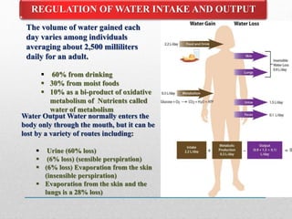
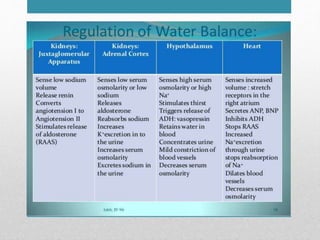
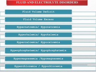
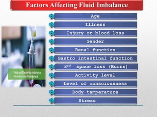
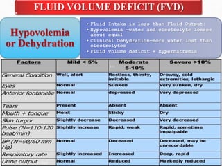
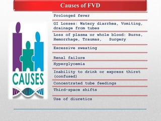
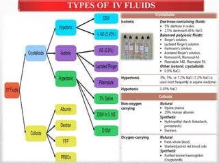
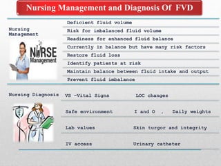
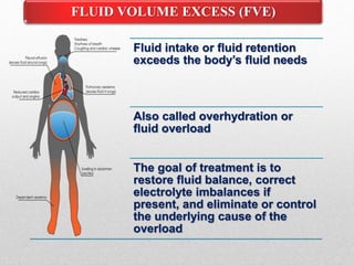
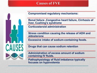
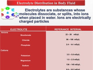
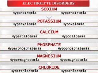
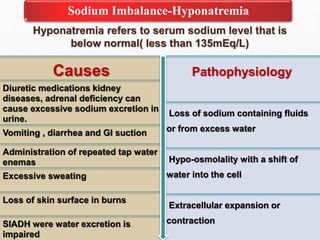
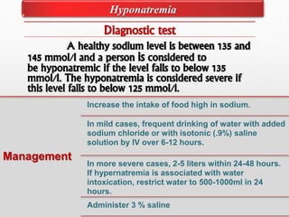
FLUID AND ELECTROLYTE
IMBALANCE
MANEESH
Fluid and electrolyte with in the body are
necessary to maintain health and function
in all body system.
INTRODUCTION
• Water is the primary content of our body. It accounts for
about 50-60% of body weight in the adult. Water content
varies with body mass, gender and age. In people with
over 65 years of age body water may decrease up to 45-50
% of total body weight.
Water Contents Of The Body
Body fluids Distribution
• Intracellular fluid(ICF) 40% body weight
• Extracellular fluid(ECF) 20% body weight
• Interstitial fluid - 15% body weight
• Plasma -5% body weight
MOVEMENT OF BODY FLUIDS
• Fluids and electrolyte constantly shift from
compartment to compartment to facilitate
body processes such as tissue
oxygenation, acid base balance and urine
formation
Mechanisms controlling fluid
& electrolyte movement
• Diffusion
• Facilitateddiffusion
• Active transport (SodiumPotassiumPump)
• Osmosis
DIFFUSION
 FACILITATED DIFFUSION
 ACTIVE TRANSPORT
Sodium potassium pump
It is the movement of fluids or
substances across the membrane
OSMOSIS
Osmotic Movement of Fluids
Movement of water from low
solute to high through a semipermeable
membrane until concentration gradient
is equal
Fluid Movement in Capillaries
As plasma flows through the capillary bed,
four factors determine if fluid moves out of the
capillary and into the interstitial space or if fluid
moves back into the capillary from the interstitial
space.
 Water balance is the result of
interaction of thirst & ADH to
maintain a stable plasma tonicity
(OP)
 The sensation of thirst promotes
water intake
 ADH regulates urinary water
excretion.
REGULATION OF WATER BALANCE
REGULATION OF WATER INTAKE AND OUTPUT
The volume of water gained each
day varies among individuals
averaging about 2,500 milliliters
daily for an adult.
 60% from drinking
 30% from moist foods
 10% as a bi-product of oxidative
metabolism of Nutrients called
water of metabolism
Water Output Water normally enters the
body only through the mouth, but it can be
lost by a variety of routes including:
 Urine (60% loss)
 (6% loss) (sensible perspiration)
 (6% loss) Evaporation from the skin
(insensible perspiration)
 Evaporation from the skin and the
lungs is a 28% loss)
Hypothalamic -
Pituitary regulation
Renal regulation
Adrenal cortical
regulation
Cardiac regulation
Gastro-intestinal
regulation
Fluid Volume Deficit
Fluid Volume Excess
Hypernatremia/ Hyponatremia
Hyperkalemia/ Hypokalemia
Hypercalcemia/ Hypocalcemia
Hyperphosphatemia/ Hypophosphatemia
Hypermagnesemia /Hypomagnesemia
Hyperchloremia / Hypochloremia
FLUID AND ELECTROLYTE DISORDERS
Age
Illness
Injury or blood loss
Gender
Renal function
Gastro intestinal function
3rd space loss (Burns)
Activity level
Level of consciousness
Body temperature
Stress
Factors Affecting Fluid Imbalance
• Fluid Intake is less than Fluid Output:
• Hypovolemia —water and electrolyte losses
about equal
• Clinical Dehydration—more water lost than
electrolytes
• Fluid volume deficit + hypernatremia
Hypovolemia
or Dehydration
FLUID VOLUME DEFICIT (FVD)
Causes of FVD
Prolonged fever
GI Losses: Watery diarrhea, Vomiting,
drainage from tubes
Loss of plasma or whole blood: Burns,
Hemorrhage, Traumas, Surgery
Excessive sweating
Renal failure
Hyperglycemia
Inability to drink or express thirst
(confused)
Concentrated tube feedings
Third-space shifts
Use of diuretics
Acute weight loss , Decreased skin turgor
Oliguria , Concentrated urine
Postural hypotension , Weak,rapid heart rate
Flattened neck veins , Increased temperature
Decreased central venous pressure , Cramps
Cool, clammy skin related to peripheral vasoconstriction
Thirst Anorexia , Nausea
Lassitude , Muscle weakness
Clinical Manifestations of FVD
Assessment
Health history
Physical Assessment
Diagnosis Test
Serum osmolality
Hemoglobin and Hematocrit
Urine specific gravity
CVP readings
Assessment and Diagnosis Test of FVD
Treatment
Fluid management
Correct with oral fluid
replacement
Oral rehydration therapy
IV therapy
Antiemetic drugs
Antidiarrheal drugs
Medical Management Of FVD
TYPES OF IV FLUIDS
Nursing
Management
Deficient fluid volume
Risk for imbalanced fluid volume
Readiness for enhanced fluid balance
Currently in balance but have many risk factors
Restore fluid loss
Identify patients at risk
Maintain balance between fluid intake and output
Prevent fluid imbalance
Nursing Diagnosis VS –Vital Signs LOC changes
Safe environment I and O , Daily weights
Lab values Skin turgor and integrity
IV access Urinary catheter
Nursing Management and Diagnosis Of FVD
Fluid intake or fluid retention
exceeds the body’s fluid needs
Also called overhydration or
fluid overload
The goal of treatment is to
restore fluid balance, correct
electrolyte imbalances if
present, and eliminate or control
the underlying cause of the
overload
FLUID VOLUME EXCESS (FVE)
Compromised regulatory mechanisms:
Renal failure ,Congestive heart failure, Cirrhosis of
liver, Cushing’s syndrome
Corticosteroid administration
Stress condition causing the release of ADH and
aldosterone
Excessive intake of sodium-containing foods.
Drugs that can cause sodium retention
Administration of excess amount of sodium
containing IV fluids.
Pathophysiology of fluid imbalance typically
focuses on hypovolemia
Causes of FVE
Increase in total body weight causes weight gain
over a short period of time.
Peripheral edema
Distended neck veins and peripheral lines.
CVP over 11 cm H2O
Crackles and wheezes in lungs
Polyuria
Ascites, pleural effusion
Bounding pulse
Pulmonary edema
Clinical Manifestations of FVE
Assessment and Diagnosis Test of FVE
Assessment Health history
Risk factors such as medication, heart failure,
renal diseases etc.
Physical Assessment
Weight, vital signs, peripheral pulses, jugular
venous distention, edema, lung sounds, urine
output and mental status
Diagnosis
Test
Serum osmolality
Urine specific gravity
CVP readings
.
Medical Management Of FVE
 Fluid Management
 Fluid intake should be restricted in clients who have
fluid volume excess.
 Dietary Management
 Sodium restricted diet is prescribed. e.g. Fruits and
vegetables, low salt breads, unsalted popcorns, fresh
meat .
 Nursing Management
 Assess the presence or worsening of FVE.
 Encourage sodium restrictions
 Monitor the clients response to diuretics.
 Teach self monitoring of weight
 Maintain strict intake output chart
 Skin care.
 Pharmacological Management
 Pharmacological measures to manage fluid Counter
regulatory hormones (e.g. angiotensin II, sympathetic
hormones, vasopressin)
Nursing Diagnosis Of FVE
Fluid volume overload related to
decreased cardiac output as evidence
by ejection fraction of 35%,
Edema in lower extremities,
Jugular distention,
Bilateral crackles,
Weight gain, and pleural effusions
noted in lungs bilaterally.
Water intoxication:
Water intoxication also known as water poisoning or
dilutional hyponatremia, is a potentially fatal disturbance
in brain functions that results when the normal balance of
electrolytes in the body is pushed outside safe limits by
over-hydration.
Third Spacing:
Occurs when fluid accumulates in areas that normally
have no fluid or minimal amount of fluid, such as with
ascites, and edema associated with burns.
Electrolytes are substances whose
molecules dissociate, or splits, into ions
when placed in water. Ions are electrically
charged particles
ELECTROLYTE REFERANCE INTERVAL
Anions
Bicarbonate
Chloride
Phosphate
Cations
Potassium
Magnesium
Sodium
22 – 26 mEq/L
96 – 106 mEq/L
2.4 - 4.4 mEq/L
3.5 – 5.0 mEq/L
1.5 – 2.5 mEq/L
136 – 145 mEq/L
Electrolyte Distribution in Body Fluid
CHLORIDE
Hyperchloremia Hypochloremia
MAGNESIUM
Hypermagnesemia Hypomagnesemia
PHOSPHATE
Hyperphosphatemia Hypophosphatemia
CALCIUM
Hypercalcemia Hypocalcemia
POTASSIUM
Hyperkalemia Hypokalemia
SODIUM
Hyponatremia Hypernatremia
ELECTROLYTE DISORDERS
Hyponatremia refers to serum sodium level that is
below normal( less than 135mEq/L)
Sodium Imbalance-Hyponatremia
Causes
Diuretic medications kidney
diseases, adrenal deficiency can
cause excessive sodium excretion in
urine.
Vomiting , diarrhea and GI suction
Administration of repeated tap water
enemas
Excessive sweating
Loss of skin surface in burns
SIADH were water excretion is
impaired
Pathophysiology
Loss of sodium containing fluids
or from excess water
Hypo-osmolality with a shift of
water into the cell
Extracellular expansion or
contraction
TYPES
Hypovolemic hypernatremia: sodium
loss is higher than water loss
Euvolemic hypernatremia: sodium
levels are almost normal
Hypervolemic hypernatremia: total
weight gain increases
Redistributive hypernatremia:
Water shifts from the intracellular to
the extracellular compartment, with a
resultant dilution of sodium.
Clinical Manifestations
Muscle cramps
Weakness
Fatigue
Anorexia, nausea, vomiting
Abdominal cramps
Diarrhea
If sodium falls below 120mEq/L
Headache, altered sensorium,
personality changes
Irritability, tremors, depression
Muscle twitching
Hyponatremia
Diagnostic test
A healthy sodium level is between 135 and
145 mmol/l and a person is considered to
be hyponatremic if the level falls to below 135
mmol/l. The hyponatremia is considered severe if
this level falls to below 125 mmol/l.
Management
Increase the intake of food high in sodium.
In mild cases, frequent drinking of water with added
sodium chloride or with isotonic (.9%) saline
solution by IV over 6-12 hours.
In more severe cases, 2-5 liters within 24-48 hours.
If hypernatremia is associated with water
intoxication, restrict water to 500-1000ml in 24
hours.
Administer 3 % saline
Hyponatremia
Nursing Diagnosis
Risk for acute confusion related
to electrolyte imbalance
Risk for injury related to altered
sensorium and decreased level
of consciousness
Risk for electrolyte
imbalance related to excessive
loss of sodium and/or excessive
intake or retention of water
Potential complication: severe
neurologic changes
Nursing management
Identify clients with risk for
hyponatremia
Monitor fluid losses and gains
Monitor presence of anorexia,
nausea, vomiting and abdominal
cramping
Check specific gravity of urine
Monitor client with cardiovascular
diseases
Avoid giving large water
supplements to clients receiving
isotonic tube feeding
Use extreme caution when
administering hypertonic saline
solution
Hyponatremia
Condition in which the serum sodium level
rises more than 145mEqlL
Sodium Imbalance-Hypernatremia
Causes
Water deprivation in clients who are
unable to respond to thirst due to
altered mental status
Hypertonic tube feeding with inadequate
water supplement
Increased insensible water loss
Excessive ingestion of salt
Excessive parenteral administration of
sodium containing solution.
Profuse sweating
Diabetes insipidus
Excessive water loss with diarrhea
Heat stroke
Pathophysiology
Due to etiological factors
An elevated serum sodium may
occur with water or sodium gain
Hypernatremia causes
hyperosmolarity
Hyperosmolarity causes a shift
of water out of cells
Cellular dehydration
CLINICALFEATURES
• Thirst. If it is not relieved
it can alter the neurologic
function
• Restlesness
• Agitation
• Irritability
• Dry mouth
• Flushed skin
• Disorientation
• Progress into seizures
,coma and death
TYPES OF
HYPERNATREMIA
Hypovolemic hypernatremia(low
volume)
• Inadequate intake of water
,excessive loss of water from urinary
tract, severe watery diarrhea are the
major causes.
Hypervolemic hypernatremia(high
volume)
• Salt poisoning is the major
cause in children
Euvolemic hypernatremia:
• Total body weight decreased
Hypernatremia
Hypernatremia
MANAGEMENT
Monitor serum electrolytes
Restrict fluids
Strict intake and output chart
Give plenty of oral fluids especially plain
water.
Monitor vitals signs
•Risk for injury related
to altered sensorium and
seizures
•Risk for fluid volume
deficit related to excessive
intake of sodium and/or loss
of water
•Risk for electrolyte
imbalance related to excessive
intake of sodium and/or loss
of water
•Potential complication:
seizures and coma leading to
irreversible brain damage
Nursing
Diagnosis
Hypernatremia
• Measure serum sodium levels
every 6 hrs. or daily
• Identify clients risk for
hypernatremia
• Monitor fluid losses and gains
• If tube feeding is used , give
sufficient water
• Record fluid intake and
output accurately, checking
body fluid loss to prevent
dehydration
Nursing
Management
Potassium is the major ICF cations, with 98% of the body potassium being
intracellular. Potassium concentration within muscle cells is approximately
140 mEq/L; potassium concentration in ECF is 3.5 to 5.0 mEq/L.
Potassium
Functions of Potassium
Disruptions in the dynamic equilibrium between ICF and ECF potassium often
cause clinical problems. Potassium regulates intracellular osmolality and
promotes cellular growth. Potassium is required for glycogen to be deposited in
muscle and liver cells. Potassium also plays a role in acid-base balance .
Sources of Potassium
Diet is the source of potassium. The typical Western diet contains approximately
50 to 100 mEq of potassium daily, mainly from fruits, dried fruits, and vegetables.
Many salt substitutes used in low-sodium diets contain substantial potassium.
Hyperkalemia can be classified according to serum potassium level are
 Mild (5.5–6.5 mmol/l)
 Moderate (6.5–7.5 mmol/l)
 Severe (>7.5 mmol/l)
Potassium Imbalance-Hyperkalemia
CAUSES
Impaired renal excretion, untreated
renal failure
Traumatic injuries
Massive intake of potassium
Potassium sparing diuretic therapy
Acid base imbalances
Inadequate urine output
Rapid IV administration of potassium
Pathophysiology
Reduced renal excretion.
Excessive intake or
leakage of potassium
from the intracellular
space.
Acute and chronic renal
failure.
Massive tissue breakdown
as in habdomyolysis.
CLINICAL
FEATURES
• ECG changes: Tall
peaked T wave,
depressed ST segment
• Arrhythmias, cardiac
arrest
• Muscle fatigue
• Weakness
• Nausea
• Skeletal muscle
weakness and paralysis
• Hyper activity of smooth
muscles, leading to GI
disturbances.
ASSESMENT
• History collection includes
current manifestations,
duration of symptom and
precipitating factors of salt
substitutes and potassium
supplements.
• Assess the apical and
peripheral pulses, muscle
strength, ECG pattern
• Tests serum electrolytes,
RFT, ABG
Hyperkalemia
MANAGEMENT
Management of
hyperkalemia mainly focus on returning the
serum potassium and to stabilize the
conducting system of heart.
IV Calcium gluconate, 1amp slow IV over 10
minutes
Insulin: Bolus injection of short acting
insulin; if blood glucose is<250mg/dl, 25 gm
of glucose should also be given
Sodium bicarbonate
Diuretics
Dialysis
Oral potassium binders
Hyperkalemia
• Activity intolerance related
to skeletal muscle weakness
• Risk for decreased cardiac
output related to
hyperkalemia
• Risk for ineffective health
maintenance related to
inadequate knowledge of
recommended diet
• Excess fluid volume related to
renal failure
Nursing
Diagnosis
• Check vital signs
• Continuous cardiac
monitoring to identify any
arrhythmias
• Instruct the patient to
avoid food items rich in
potassium like banana,
tomatoes, green leafy
vegetables, apricots,
oranges etc.
• Repeat electrolytes
• Avoid administration of
potassium conserving
diuretics, potassium
supplements
Nursing
Management
Hyperkalemia
Hypokalemia
Hypokalemia is when blood's potassium levels are too low.
Hypokalemia is generally defined as a serum potassium
level of less than 3.5 mEq/L (3.5 mmol/L).
CAUSES
Decreased intake of potassium rich food
Vomiting
Renal excreation
Nasogastric suctioning
Adrenal gland secrets excess
aldosterone.
Pathophysiology
Inadequate potassium
intake
Increased potassium
excretion
Shift of potassium from
the extracellular to the
intracellular space.
Hypokalemia
Multisystem Effects of Hypokalemia
Musculoskeletal: Fatigue, leg cramps, muscle weakness
Gastrointestinal: Nausea, vomiting anorexia
Cardiovascular: Dysrrythmias, irregular pulse and ECG
changes
Urinary : Dilute urine, polyuria , polydypsia
Neurologic: Confusion ,depression lethargy
Respiratory: Respiratory arrest(severe)
•Administration of inj.
KCl 40mEq/l in iv fluids
•Oral potassium
supplements
•Use potassium sparing
diuretics if diuretic
therapy is going on
•Treat diarrhea or
vomiting
•Discontinue diuretics or
laxatives
Management
•Advice to report any
chest discomfort
•Advice to take more
potassium rich food
like banana, oranges
•Advice to take oral
potassium
supplements properly
•Be aware of clients at
risk of hypokalemia
Nursing
Management
Hypokalemia
CALCIUM IMBALANCES
Calcium is necessary for the metabolic process
of bones and teeth . Normal serum level is 8.5-
10mg/dl.
Responsible for contraction, formation of bones etc
For oocyte activation
For myocardial contraction
Vital role in nerve impulse transmission
Major component in blood clotting systems.
ROLES OF CALCIUM
Serum calcium levels exceeds 11mg/dl
HYPERCALCEMIA
CAUSES
Over activity of parathyroid
gland
Excessive intake of calcium
supplements
Cancer: lung and breast
cancer increase risk of
hypercalcemia
Hereditary factors
Increased reabsorption of
calcium from bones
Renal failure decreases the
amount of calcium loss
CLINICAL MANIFESTATION
Fatigue
Weakness
Anorexia ,nausea , vomiting
Irritability
Headache
Abdominal pain and
constipation
Decreased neuromuscular
excitability
HYPERCALCEMIA
Nursing Intervention
Monitor clients at risk of
hypercalcemia
Increase client mobilization
Encourage fluid intake
Discourage excessive conception of
milk and products
MANAGEMENT
Restore calcium levels
Treat the underlying cause.
Administer calcitonin
Decreased calcium level <8mg/dl
HYPOCALCEMIA
CAUSES
Other electrolyte imbalance
Massive administration of banked blood
Medications like diuretics anticonvulsants
CLINICAL FEATURES
Tetany
Trousseaurs sign: carpel spasm induced by inflating a bp
cuff above systolic pressure for 5 minit's
Chvostek’s sign: contraction of facial muscle in response to
a light tap over the facial nerve in front of the ear.
HYPOCALCEMIA
MANAGEMENT
Oral calcium supplements
IV Calcium gluconate
Diet rich in calcium
Nursing Management
Be aware of clients risk for hypocalcemia
Be prepared to take seizure precautions
Safe administration of calcium
supplements
Educate people about osteoporosis
Phosphate Imbalance
Phosphate imbalances are seen in
patients with acute kidney injury which alters
kidneys ability to excrete phosphate. Normal
serum level is 2.5-4.5mg/dl.
HYPOPHOSPHATEMIA
Serum phosphate level is <1.2mg/dl
CAUSES
Alcoholism,
Burns
Starvation
Diuretic use
CLINICAL FEATURES
Muscle weakness
Fatigue
Confusion
Bone pain
HYPOPHOSPHATEMIA
MANAGEMENT
Increase intake of diary products
Phosphate supplements in diet
Treat underline cause
IV phosphate is administered when serum phosphate
is <1mgldL
HYPERPHOSPHATEMIA
Plasma phosphate level > 4.5mE/L
CLINICAL
FEATURES
Anorexia, Nausea
Vomiting, Muscle cramps
Pain, Parasthesia
TREATMENT
Limiting high phosphate containing foods like milk and milk
products.
Administer calcium or aluminum products for the execration
of phosphate
MAGNESIUM IMBALANCES
Magnesium is an intracellular cation. it’s
required for the ATP of Sodium- Potassium pump.
Serum level is 1.5-2mE/L
HYPERMAGNESEMIA
Serum level above
2.1mEq/l
CAUSES
Women receiving milk of
magnesia
Renal failure
HYPERMAGNESEMIA
MANAGEMENT Avoid magnesium containing
food like banana, orange,
peanuts etc.
Avoid administering magnesium.
CLINICAL
FEATURES
Nausea
Vomiting
Lethargy
HYPOMAGNESEMIA
Serum level drops
below 1.3mEq/L
HYPOMAGNESEMIA
Chronic alcoholism
Prolonged starvation or
fasting.
Critically ill patients
Malabsorption syndrome
HYPOMAGNESEMIA
CLINICALFEATURES
lethargy and
weakness
tremor
carpopedal
spasm
muscle
cramps
tetany
MANAGEMENT
IV Magnesium
correction
Oral correction of
magnesium
CLORIDE IMBALANCES
Hypochloremia Chloride level less
than 95 mEq/L.
The normal adult value for chloride is 97-107
mEq/L.
CAUSES
Loss of body fluids from prolonged
vomiting, diarrhea, sweating or high fevers.
Drugs such as: bicarbonate,
corticosteroids, diuretics, and laxatives.
.
CLINICAL
FEATURES
dehydration.
weakness or fatigue.
difficulty breathing.
diarrhea or vomiting, caused by fluid loss
HYPOCLOREMIA
MANAGEMENT
• Nonsteroidal anti-inflammatory drugs
• Hydrochloric acid (HCl) and carbonic anhydrase
inhibitors
• Administer normal saline or half strength saline to
replace chloride.
• Stop diuretics
HYPERCLOREMIA
HYPERCLOREMIA
Chloride levels at or
above 110 mEq/L
CAUSES
• Loss of body fluids from prolonged
vomiting, diarrhea, sweating or high
fever (dehydration).
• High levels of blood sodium.
• Kidney failure
• Diabetes insipidus
HYPERCLOREMIA
• Dehydration - due to diarrhea, vomiting, sweating.
• Hypertension.
• Cardiovascular dysfunction Edema
• Weakness.
• Thirst
CLINICAL FEATURES
MANAGEMENT
Drinking 2–3 quarts of fluid every day
Receiving intravenous fluids
Eating a better, more balanced diet
Avoiding alcohol, caffeine, and aspirin.
Fluid and electrolyte imbalance

Fluid and electrolyte imbalance

  • 1.
  • 2.
    Fluid and electrolytewith in the body are necessary to maintain health and function in all body system. INTRODUCTION
  • 3.
    • Water isthe primary content of our body. It accounts for about 50-60% of body weight in the adult. Water content varies with body mass, gender and age. In people with over 65 years of age body water may decrease up to 45-50 % of total body weight. Water Contents Of The Body
  • 4.
    Body fluids Distribution •Intracellular fluid(ICF) 40% body weight • Extracellular fluid(ECF) 20% body weight • Interstitial fluid - 15% body weight • Plasma -5% body weight
  • 5.
    MOVEMENT OF BODYFLUIDS • Fluids and electrolyte constantly shift from compartment to compartment to facilitate body processes such as tissue oxygenation, acid base balance and urine formation Mechanisms controlling fluid & electrolyte movement • Diffusion • Facilitateddiffusion • Active transport (SodiumPotassiumPump) • Osmosis
  • 6.
    DIFFUSION  FACILITATED DIFFUSION ACTIVE TRANSPORT Sodium potassium pump It is the movement of fluids or substances across the membrane
  • 7.
    OSMOSIS Osmotic Movement ofFluids Movement of water from low solute to high through a semipermeable membrane until concentration gradient is equal
  • 8.
    Fluid Movement inCapillaries As plasma flows through the capillary bed, four factors determine if fluid moves out of the capillary and into the interstitial space or if fluid moves back into the capillary from the interstitial space.
  • 9.
     Water balanceis the result of interaction of thirst & ADH to maintain a stable plasma tonicity (OP)  The sensation of thirst promotes water intake  ADH regulates urinary water excretion. REGULATION OF WATER BALANCE
  • 10.
    REGULATION OF WATERINTAKE AND OUTPUT The volume of water gained each day varies among individuals averaging about 2,500 milliliters daily for an adult.  60% from drinking  30% from moist foods  10% as a bi-product of oxidative metabolism of Nutrients called water of metabolism Water Output Water normally enters the body only through the mouth, but it can be lost by a variety of routes including:  Urine (60% loss)  (6% loss) (sensible perspiration)  (6% loss) Evaporation from the skin (insensible perspiration)  Evaporation from the skin and the lungs is a 28% loss)
  • 11.
    Hypothalamic - Pituitary regulation Renalregulation Adrenal cortical regulation Cardiac regulation Gastro-intestinal regulation
  • 15.
    Fluid Volume Deficit FluidVolume Excess Hypernatremia/ Hyponatremia Hyperkalemia/ Hypokalemia Hypercalcemia/ Hypocalcemia Hyperphosphatemia/ Hypophosphatemia Hypermagnesemia /Hypomagnesemia Hyperchloremia / Hypochloremia FLUID AND ELECTROLYTE DISORDERS
  • 16.
    Age Illness Injury or bloodloss Gender Renal function Gastro intestinal function 3rd space loss (Burns) Activity level Level of consciousness Body temperature Stress Factors Affecting Fluid Imbalance
  • 17.
    • Fluid Intakeis less than Fluid Output: • Hypovolemia —water and electrolyte losses about equal • Clinical Dehydration—more water lost than electrolytes • Fluid volume deficit + hypernatremia Hypovolemia or Dehydration FLUID VOLUME DEFICIT (FVD)
  • 18.
    Causes of FVD Prolongedfever GI Losses: Watery diarrhea, Vomiting, drainage from tubes Loss of plasma or whole blood: Burns, Hemorrhage, Traumas, Surgery Excessive sweating Renal failure Hyperglycemia Inability to drink or express thirst (confused) Concentrated tube feedings Third-space shifts Use of diuretics
  • 19.
    Acute weight loss, Decreased skin turgor Oliguria , Concentrated urine Postural hypotension , Weak,rapid heart rate Flattened neck veins , Increased temperature Decreased central venous pressure , Cramps Cool, clammy skin related to peripheral vasoconstriction Thirst Anorexia , Nausea Lassitude , Muscle weakness Clinical Manifestations of FVD
  • 20.
    Assessment Health history Physical Assessment DiagnosisTest Serum osmolality Hemoglobin and Hematocrit Urine specific gravity CVP readings Assessment and Diagnosis Test of FVD
  • 21.
    Treatment Fluid management Correct withoral fluid replacement Oral rehydration therapy IV therapy Antiemetic drugs Antidiarrheal drugs Medical Management Of FVD
  • 22.
  • 23.
    Nursing Management Deficient fluid volume Riskfor imbalanced fluid volume Readiness for enhanced fluid balance Currently in balance but have many risk factors Restore fluid loss Identify patients at risk Maintain balance between fluid intake and output Prevent fluid imbalance Nursing Diagnosis VS –Vital Signs LOC changes Safe environment I and O , Daily weights Lab values Skin turgor and integrity IV access Urinary catheter Nursing Management and Diagnosis Of FVD
  • 24.
    Fluid intake orfluid retention exceeds the body’s fluid needs Also called overhydration or fluid overload The goal of treatment is to restore fluid balance, correct electrolyte imbalances if present, and eliminate or control the underlying cause of the overload FLUID VOLUME EXCESS (FVE)
  • 25.
    Compromised regulatory mechanisms: Renalfailure ,Congestive heart failure, Cirrhosis of liver, Cushing’s syndrome Corticosteroid administration Stress condition causing the release of ADH and aldosterone Excessive intake of sodium-containing foods. Drugs that can cause sodium retention Administration of excess amount of sodium containing IV fluids. Pathophysiology of fluid imbalance typically focuses on hypovolemia Causes of FVE
  • 26.
    Increase in totalbody weight causes weight gain over a short period of time. Peripheral edema Distended neck veins and peripheral lines. CVP over 11 cm H2O Crackles and wheezes in lungs Polyuria Ascites, pleural effusion Bounding pulse Pulmonary edema Clinical Manifestations of FVE
  • 27.
    Assessment and DiagnosisTest of FVE Assessment Health history Risk factors such as medication, heart failure, renal diseases etc. Physical Assessment Weight, vital signs, peripheral pulses, jugular venous distention, edema, lung sounds, urine output and mental status Diagnosis Test Serum osmolality Urine specific gravity CVP readings .
  • 28.
    Medical Management OfFVE  Fluid Management  Fluid intake should be restricted in clients who have fluid volume excess.  Dietary Management  Sodium restricted diet is prescribed. e.g. Fruits and vegetables, low salt breads, unsalted popcorns, fresh meat .  Nursing Management  Assess the presence or worsening of FVE.  Encourage sodium restrictions  Monitor the clients response to diuretics.  Teach self monitoring of weight  Maintain strict intake output chart  Skin care.  Pharmacological Management  Pharmacological measures to manage fluid Counter regulatory hormones (e.g. angiotensin II, sympathetic hormones, vasopressin)
  • 29.
    Nursing Diagnosis OfFVE Fluid volume overload related to decreased cardiac output as evidence by ejection fraction of 35%, Edema in lower extremities, Jugular distention, Bilateral crackles, Weight gain, and pleural effusions noted in lungs bilaterally.
  • 30.
    Water intoxication: Water intoxicationalso known as water poisoning or dilutional hyponatremia, is a potentially fatal disturbance in brain functions that results when the normal balance of electrolytes in the body is pushed outside safe limits by over-hydration. Third Spacing: Occurs when fluid accumulates in areas that normally have no fluid or minimal amount of fluid, such as with ascites, and edema associated with burns.
  • 31.
    Electrolytes are substanceswhose molecules dissociate, or splits, into ions when placed in water. Ions are electrically charged particles ELECTROLYTE REFERANCE INTERVAL Anions Bicarbonate Chloride Phosphate Cations Potassium Magnesium Sodium 22 – 26 mEq/L 96 – 106 mEq/L 2.4 - 4.4 mEq/L 3.5 – 5.0 mEq/L 1.5 – 2.5 mEq/L 136 – 145 mEq/L Electrolyte Distribution in Body Fluid
  • 32.
    CHLORIDE Hyperchloremia Hypochloremia MAGNESIUM Hypermagnesemia Hypomagnesemia PHOSPHATE HyperphosphatemiaHypophosphatemia CALCIUM Hypercalcemia Hypocalcemia POTASSIUM Hyperkalemia Hypokalemia SODIUM Hyponatremia Hypernatremia ELECTROLYTE DISORDERS
  • 33.
    Hyponatremia refers toserum sodium level that is below normal( less than 135mEq/L) Sodium Imbalance-Hyponatremia Causes Diuretic medications kidney diseases, adrenal deficiency can cause excessive sodium excretion in urine. Vomiting , diarrhea and GI suction Administration of repeated tap water enemas Excessive sweating Loss of skin surface in burns SIADH were water excretion is impaired Pathophysiology Loss of sodium containing fluids or from excess water Hypo-osmolality with a shift of water into the cell Extracellular expansion or contraction
  • 34.
    TYPES Hypovolemic hypernatremia: sodium lossis higher than water loss Euvolemic hypernatremia: sodium levels are almost normal Hypervolemic hypernatremia: total weight gain increases Redistributive hypernatremia: Water shifts from the intracellular to the extracellular compartment, with a resultant dilution of sodium. Clinical Manifestations Muscle cramps Weakness Fatigue Anorexia, nausea, vomiting Abdominal cramps Diarrhea If sodium falls below 120mEq/L Headache, altered sensorium, personality changes Irritability, tremors, depression Muscle twitching Hyponatremia
  • 35.
    Diagnostic test A healthysodium level is between 135 and 145 mmol/l and a person is considered to be hyponatremic if the level falls to below 135 mmol/l. The hyponatremia is considered severe if this level falls to below 125 mmol/l. Management Increase the intake of food high in sodium. In mild cases, frequent drinking of water with added sodium chloride or with isotonic (.9%) saline solution by IV over 6-12 hours. In more severe cases, 2-5 liters within 24-48 hours. If hypernatremia is associated with water intoxication, restrict water to 500-1000ml in 24 hours. Administer 3 % saline Hyponatremia
  • 36.
    Nursing Diagnosis Risk foracute confusion related to electrolyte imbalance Risk for injury related to altered sensorium and decreased level of consciousness Risk for electrolyte imbalance related to excessive loss of sodium and/or excessive intake or retention of water Potential complication: severe neurologic changes Nursing management Identify clients with risk for hyponatremia Monitor fluid losses and gains Monitor presence of anorexia, nausea, vomiting and abdominal cramping Check specific gravity of urine Monitor client with cardiovascular diseases Avoid giving large water supplements to clients receiving isotonic tube feeding Use extreme caution when administering hypertonic saline solution Hyponatremia
  • 37.
    Condition in whichthe serum sodium level rises more than 145mEqlL Sodium Imbalance-Hypernatremia Causes Water deprivation in clients who are unable to respond to thirst due to altered mental status Hypertonic tube feeding with inadequate water supplement Increased insensible water loss Excessive ingestion of salt Excessive parenteral administration of sodium containing solution. Profuse sweating Diabetes insipidus Excessive water loss with diarrhea Heat stroke Pathophysiology Due to etiological factors An elevated serum sodium may occur with water or sodium gain Hypernatremia causes hyperosmolarity Hyperosmolarity causes a shift of water out of cells Cellular dehydration
  • 38.
    CLINICALFEATURES • Thirst. Ifit is not relieved it can alter the neurologic function • Restlesness • Agitation • Irritability • Dry mouth • Flushed skin • Disorientation • Progress into seizures ,coma and death TYPES OF HYPERNATREMIA Hypovolemic hypernatremia(low volume) • Inadequate intake of water ,excessive loss of water from urinary tract, severe watery diarrhea are the major causes. Hypervolemic hypernatremia(high volume) • Salt poisoning is the major cause in children Euvolemic hypernatremia: • Total body weight decreased Hypernatremia
  • 39.
    Hypernatremia MANAGEMENT Monitor serum electrolytes Restrictfluids Strict intake and output chart Give plenty of oral fluids especially plain water. Monitor vitals signs
  • 40.
    •Risk for injuryrelated to altered sensorium and seizures •Risk for fluid volume deficit related to excessive intake of sodium and/or loss of water •Risk for electrolyte imbalance related to excessive intake of sodium and/or loss of water •Potential complication: seizures and coma leading to irreversible brain damage Nursing Diagnosis Hypernatremia • Measure serum sodium levels every 6 hrs. or daily • Identify clients risk for hypernatremia • Monitor fluid losses and gains • If tube feeding is used , give sufficient water • Record fluid intake and output accurately, checking body fluid loss to prevent dehydration Nursing Management
  • 41.
    Potassium is themajor ICF cations, with 98% of the body potassium being intracellular. Potassium concentration within muscle cells is approximately 140 mEq/L; potassium concentration in ECF is 3.5 to 5.0 mEq/L. Potassium Functions of Potassium Disruptions in the dynamic equilibrium between ICF and ECF potassium often cause clinical problems. Potassium regulates intracellular osmolality and promotes cellular growth. Potassium is required for glycogen to be deposited in muscle and liver cells. Potassium also plays a role in acid-base balance . Sources of Potassium Diet is the source of potassium. The typical Western diet contains approximately 50 to 100 mEq of potassium daily, mainly from fruits, dried fruits, and vegetables. Many salt substitutes used in low-sodium diets contain substantial potassium.
  • 42.
    Hyperkalemia can beclassified according to serum potassium level are  Mild (5.5–6.5 mmol/l)  Moderate (6.5–7.5 mmol/l)  Severe (>7.5 mmol/l) Potassium Imbalance-Hyperkalemia CAUSES Impaired renal excretion, untreated renal failure Traumatic injuries Massive intake of potassium Potassium sparing diuretic therapy Acid base imbalances Inadequate urine output Rapid IV administration of potassium Pathophysiology Reduced renal excretion. Excessive intake or leakage of potassium from the intracellular space. Acute and chronic renal failure. Massive tissue breakdown as in habdomyolysis.
  • 43.
    CLINICAL FEATURES • ECG changes:Tall peaked T wave, depressed ST segment • Arrhythmias, cardiac arrest • Muscle fatigue • Weakness • Nausea • Skeletal muscle weakness and paralysis • Hyper activity of smooth muscles, leading to GI disturbances. ASSESMENT • History collection includes current manifestations, duration of symptom and precipitating factors of salt substitutes and potassium supplements. • Assess the apical and peripheral pulses, muscle strength, ECG pattern • Tests serum electrolytes, RFT, ABG Hyperkalemia
  • 44.
    MANAGEMENT Management of hyperkalemia mainlyfocus on returning the serum potassium and to stabilize the conducting system of heart. IV Calcium gluconate, 1amp slow IV over 10 minutes Insulin: Bolus injection of short acting insulin; if blood glucose is<250mg/dl, 25 gm of glucose should also be given Sodium bicarbonate Diuretics Dialysis Oral potassium binders Hyperkalemia
  • 45.
    • Activity intolerancerelated to skeletal muscle weakness • Risk for decreased cardiac output related to hyperkalemia • Risk for ineffective health maintenance related to inadequate knowledge of recommended diet • Excess fluid volume related to renal failure Nursing Diagnosis • Check vital signs • Continuous cardiac monitoring to identify any arrhythmias • Instruct the patient to avoid food items rich in potassium like banana, tomatoes, green leafy vegetables, apricots, oranges etc. • Repeat electrolytes • Avoid administration of potassium conserving diuretics, potassium supplements Nursing Management Hyperkalemia
  • 46.
    Hypokalemia Hypokalemia is whenblood's potassium levels are too low. Hypokalemia is generally defined as a serum potassium level of less than 3.5 mEq/L (3.5 mmol/L). CAUSES Decreased intake of potassium rich food Vomiting Renal excreation Nasogastric suctioning Adrenal gland secrets excess aldosterone. Pathophysiology Inadequate potassium intake Increased potassium excretion Shift of potassium from the extracellular to the intracellular space.
  • 47.
    Hypokalemia Multisystem Effects ofHypokalemia Musculoskeletal: Fatigue, leg cramps, muscle weakness Gastrointestinal: Nausea, vomiting anorexia Cardiovascular: Dysrrythmias, irregular pulse and ECG changes Urinary : Dilute urine, polyuria , polydypsia Neurologic: Confusion ,depression lethargy Respiratory: Respiratory arrest(severe)
  • 48.
    •Administration of inj. KCl40mEq/l in iv fluids •Oral potassium supplements •Use potassium sparing diuretics if diuretic therapy is going on •Treat diarrhea or vomiting •Discontinue diuretics or laxatives Management •Advice to report any chest discomfort •Advice to take more potassium rich food like banana, oranges •Advice to take oral potassium supplements properly •Be aware of clients at risk of hypokalemia Nursing Management Hypokalemia
  • 49.
    CALCIUM IMBALANCES Calcium isnecessary for the metabolic process of bones and teeth . Normal serum level is 8.5- 10mg/dl. Responsible for contraction, formation of bones etc For oocyte activation For myocardial contraction Vital role in nerve impulse transmission Major component in blood clotting systems. ROLES OF CALCIUM
  • 50.
    Serum calcium levelsexceeds 11mg/dl HYPERCALCEMIA CAUSES Over activity of parathyroid gland Excessive intake of calcium supplements Cancer: lung and breast cancer increase risk of hypercalcemia Hereditary factors Increased reabsorption of calcium from bones Renal failure decreases the amount of calcium loss CLINICAL MANIFESTATION Fatigue Weakness Anorexia ,nausea , vomiting Irritability Headache Abdominal pain and constipation Decreased neuromuscular excitability
  • 51.
    HYPERCALCEMIA Nursing Intervention Monitor clientsat risk of hypercalcemia Increase client mobilization Encourage fluid intake Discourage excessive conception of milk and products MANAGEMENT Restore calcium levels Treat the underlying cause. Administer calcitonin
  • 52.
    Decreased calcium level<8mg/dl HYPOCALCEMIA CAUSES Other electrolyte imbalance Massive administration of banked blood Medications like diuretics anticonvulsants CLINICAL FEATURES Tetany Trousseaurs sign: carpel spasm induced by inflating a bp cuff above systolic pressure for 5 minit's Chvostek’s sign: contraction of facial muscle in response to a light tap over the facial nerve in front of the ear.
  • 53.
    HYPOCALCEMIA MANAGEMENT Oral calcium supplements IVCalcium gluconate Diet rich in calcium Nursing Management Be aware of clients risk for hypocalcemia Be prepared to take seizure precautions Safe administration of calcium supplements Educate people about osteoporosis
  • 54.
    Phosphate Imbalance Phosphate imbalancesare seen in patients with acute kidney injury which alters kidneys ability to excrete phosphate. Normal serum level is 2.5-4.5mg/dl. HYPOPHOSPHATEMIA Serum phosphate level is <1.2mg/dl CAUSES Alcoholism, Burns Starvation Diuretic use
  • 55.
    CLINICAL FEATURES Muscle weakness Fatigue Confusion Bonepain HYPOPHOSPHATEMIA MANAGEMENT Increase intake of diary products Phosphate supplements in diet Treat underline cause IV phosphate is administered when serum phosphate is <1mgldL
  • 56.
    HYPERPHOSPHATEMIA Plasma phosphate level> 4.5mE/L CLINICAL FEATURES Anorexia, Nausea Vomiting, Muscle cramps Pain, Parasthesia TREATMENT Limiting high phosphate containing foods like milk and milk products. Administer calcium or aluminum products for the execration of phosphate
  • 57.
    MAGNESIUM IMBALANCES Magnesium isan intracellular cation. it’s required for the ATP of Sodium- Potassium pump. Serum level is 1.5-2mE/L HYPERMAGNESEMIA Serum level above 2.1mEq/l CAUSES Women receiving milk of magnesia Renal failure
  • 58.
    HYPERMAGNESEMIA MANAGEMENT Avoid magnesiumcontaining food like banana, orange, peanuts etc. Avoid administering magnesium. CLINICAL FEATURES Nausea Vomiting Lethargy
  • 59.
    HYPOMAGNESEMIA Serum level drops below1.3mEq/L HYPOMAGNESEMIA Chronic alcoholism Prolonged starvation or fasting. Critically ill patients Malabsorption syndrome
  • 60.
  • 61.
    CLORIDE IMBALANCES Hypochloremia Chloridelevel less than 95 mEq/L. The normal adult value for chloride is 97-107 mEq/L. CAUSES Loss of body fluids from prolonged vomiting, diarrhea, sweating or high fevers. Drugs such as: bicarbonate, corticosteroids, diuretics, and laxatives.
  • 62.
    . CLINICAL FEATURES dehydration. weakness or fatigue. difficultybreathing. diarrhea or vomiting, caused by fluid loss HYPOCLOREMIA MANAGEMENT • Nonsteroidal anti-inflammatory drugs • Hydrochloric acid (HCl) and carbonic anhydrase inhibitors • Administer normal saline or half strength saline to replace chloride. • Stop diuretics
  • 63.
    HYPERCLOREMIA HYPERCLOREMIA Chloride levels ator above 110 mEq/L CAUSES • Loss of body fluids from prolonged vomiting, diarrhea, sweating or high fever (dehydration). • High levels of blood sodium. • Kidney failure • Diabetes insipidus
  • 64.
    HYPERCLOREMIA • Dehydration -due to diarrhea, vomiting, sweating. • Hypertension. • Cardiovascular dysfunction Edema • Weakness. • Thirst CLINICAL FEATURES MANAGEMENT Drinking 2–3 quarts of fluid every day Receiving intravenous fluids Eating a better, more balanced diet Avoiding alcohol, caffeine, and aspirin.