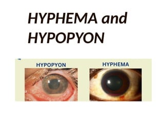
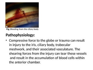
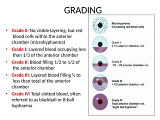
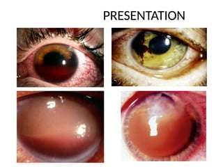
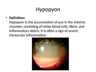
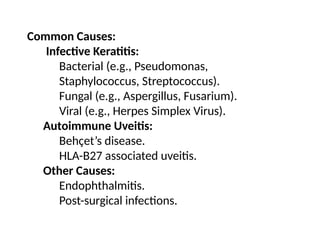
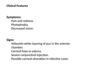
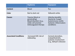
HYPHEMA and
HYPOPYON
Definition:
Blood in the anterior chamber of eye is called
Hyphema.
Hyphema
Pathophysiology:
• Compressive force to the globe or trauma can result
in injury to the iris, ciliary body, trabecular
meshwork, and their associated vasculature. The
shearing forces from the injury can tear these vessels
and result in the accumulation of blood cells within
the anterior chamber.
Fig; Bleeding from the ciliary body
• Grade 0: No visible layering, but red
blood cells within the anterior
chamber (microhyphaema)
• Grade I: Layered blood occupying less
than 1/3 of the anterior chamber
• Grade II: Blood filling 1/3 to 1/2 of
the anterior chamber
• Grade III: Layered blood filling ½ to
less than total of the anterior
chamber
• Grade IV: Total clotted blood, often
referred to as blackball or 8-ball
hyphaema
GRADING
PRESENTATION
PRESENTATION
• Symptoms:
Symptoms can be variable depending on the
etiology. Typically patients complain of blurry
vision, pain, headahce, photophobia, H/O
trauma.
• Signs:
Blood or clot or both in the AC, usually visible
without a slit lamp.
MANAGEMENT
TREATMENT
1.Sedation or complete bed rest with limited
activites.
2. Cycloplegics; Atropine 1% E/D
3. Anti inflamatoty
- Steroids, mild NSAIDs
4.Ocular hypotensive agents in case of IOP
5.Place shield or patch over eye
6. Rx of the cause
7.(surgically) A/C paracentesis with irrigation
and aspiration
Hypopyon
• Definition:
Hypopyon is the accumulation of pus in the anterior
chamber, consisting of white blood cells, fibrin, and
inflammatory debris. It is often a sign of severe
intraocular inflammation.
Common Causes:
Infective Keratitis:
Bacterial (e.g., Pseudomonas,
Staphylococcus, Streptococcus).
Fungal (e.g., Aspergillus, Fusarium).
Viral (e.g., Herpes Simplex Virus).
Autoimmune Uveitis:
Behçet’s disease.
HLA-B27 associated uveitis.
Other Causes:
Endophthalmitis.
Post-surgical infections.
Clinical Features
Symptoms:
Pain and redness.
Photophobia.
Decreased vision.
Signs:
Yellowish-white layering of pus in the anterior
chamber.
Corneal haze or edema.
Severe conjunctival injection.
Possible corneal ulceration in infective cases.
Hyphema Hypopyon
Content Blood Pus
Color Red to dark red Yellowish-white
Causes Trauma (blunt or
penetrating)
Blood disorders (e.g.,
sickle cell anemia)
Anticoagulant therapy
Infective keratitis
(bacterial, fungal)
Autoimmune uveitis
Systemic inflammatory
conditions
Associated Conditions Increased IOP, risk of
rebleeding
Corneal ulceration,
endophthalmitis,
systemic involvement
(e.g., Behçet’s disease)

hypopyon vs ................hyphema.pptx

  • 1.
  • 2.
    Definition: Blood in theanterior chamber of eye is called Hyphema. Hyphema
  • 3.
    Pathophysiology: • Compressive forceto the globe or trauma can result in injury to the iris, ciliary body, trabecular meshwork, and their associated vasculature. The shearing forces from the injury can tear these vessels and result in the accumulation of blood cells within the anterior chamber. Fig; Bleeding from the ciliary body
  • 4.
    • Grade 0:No visible layering, but red blood cells within the anterior chamber (microhyphaema) • Grade I: Layered blood occupying less than 1/3 of the anterior chamber • Grade II: Blood filling 1/3 to 1/2 of the anterior chamber • Grade III: Layered blood filling ½ to less than total of the anterior chamber • Grade IV: Total clotted blood, often referred to as blackball or 8-ball hyphaema GRADING
  • 5.
  • 6.
    PRESENTATION • Symptoms: Symptoms canbe variable depending on the etiology. Typically patients complain of blurry vision, pain, headahce, photophobia, H/O trauma. • Signs: Blood or clot or both in the AC, usually visible without a slit lamp.
  • 7.
  • 8.
    TREATMENT 1.Sedation or completebed rest with limited activites. 2. Cycloplegics; Atropine 1% E/D 3. Anti inflamatoty - Steroids, mild NSAIDs 4.Ocular hypotensive agents in case of IOP 5.Place shield or patch over eye 6. Rx of the cause 7.(surgically) A/C paracentesis with irrigation and aspiration
  • 9.
    Hypopyon • Definition: Hypopyon isthe accumulation of pus in the anterior chamber, consisting of white blood cells, fibrin, and inflammatory debris. It is often a sign of severe intraocular inflammation.
  • 10.
    Common Causes: Infective Keratitis: Bacterial(e.g., Pseudomonas, Staphylococcus, Streptococcus). Fungal (e.g., Aspergillus, Fusarium). Viral (e.g., Herpes Simplex Virus). Autoimmune Uveitis: Behçet’s disease. HLA-B27 associated uveitis. Other Causes: Endophthalmitis. Post-surgical infections.
  • 11.
    Clinical Features Symptoms: Pain andredness. Photophobia. Decreased vision. Signs: Yellowish-white layering of pus in the anterior chamber. Corneal haze or edema. Severe conjunctival injection. Possible corneal ulceration in infective cases.
  • 12.
    Hyphema Hypopyon Content BloodPus Color Red to dark red Yellowish-white Causes Trauma (blunt or penetrating) Blood disorders (e.g., sickle cell anemia) Anticoagulant therapy Infective keratitis (bacterial, fungal) Autoimmune uveitis Systemic inflammatory conditions Associated Conditions Increased IOP, risk of rebleeding Corneal ulceration, endophthalmitis, systemic involvement (e.g., Behçet’s disease)