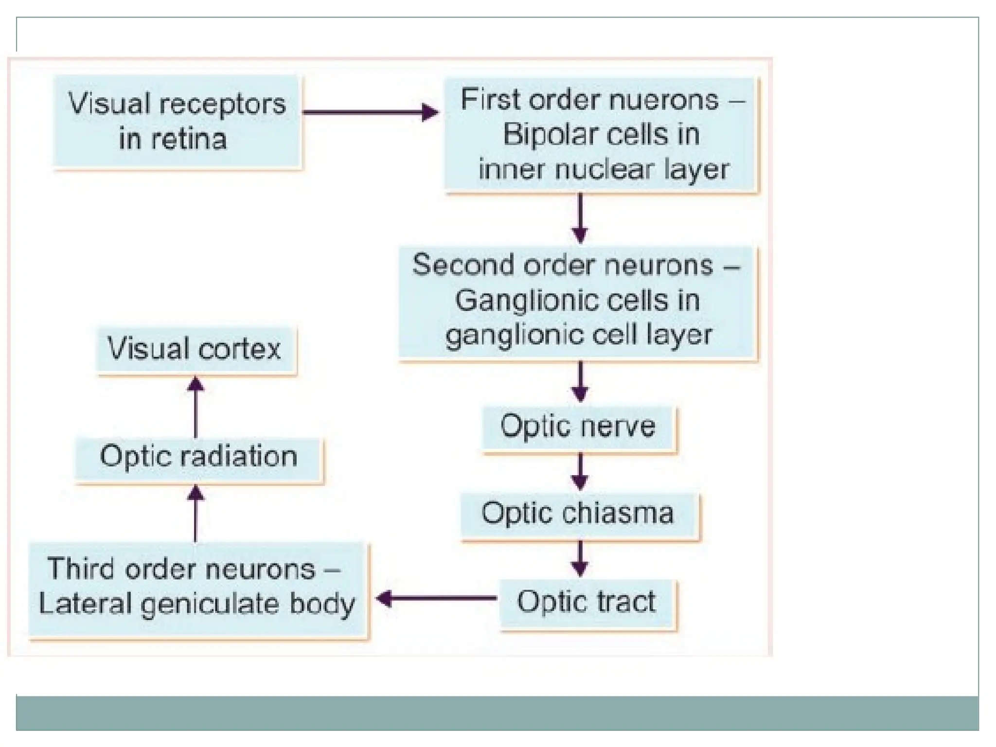
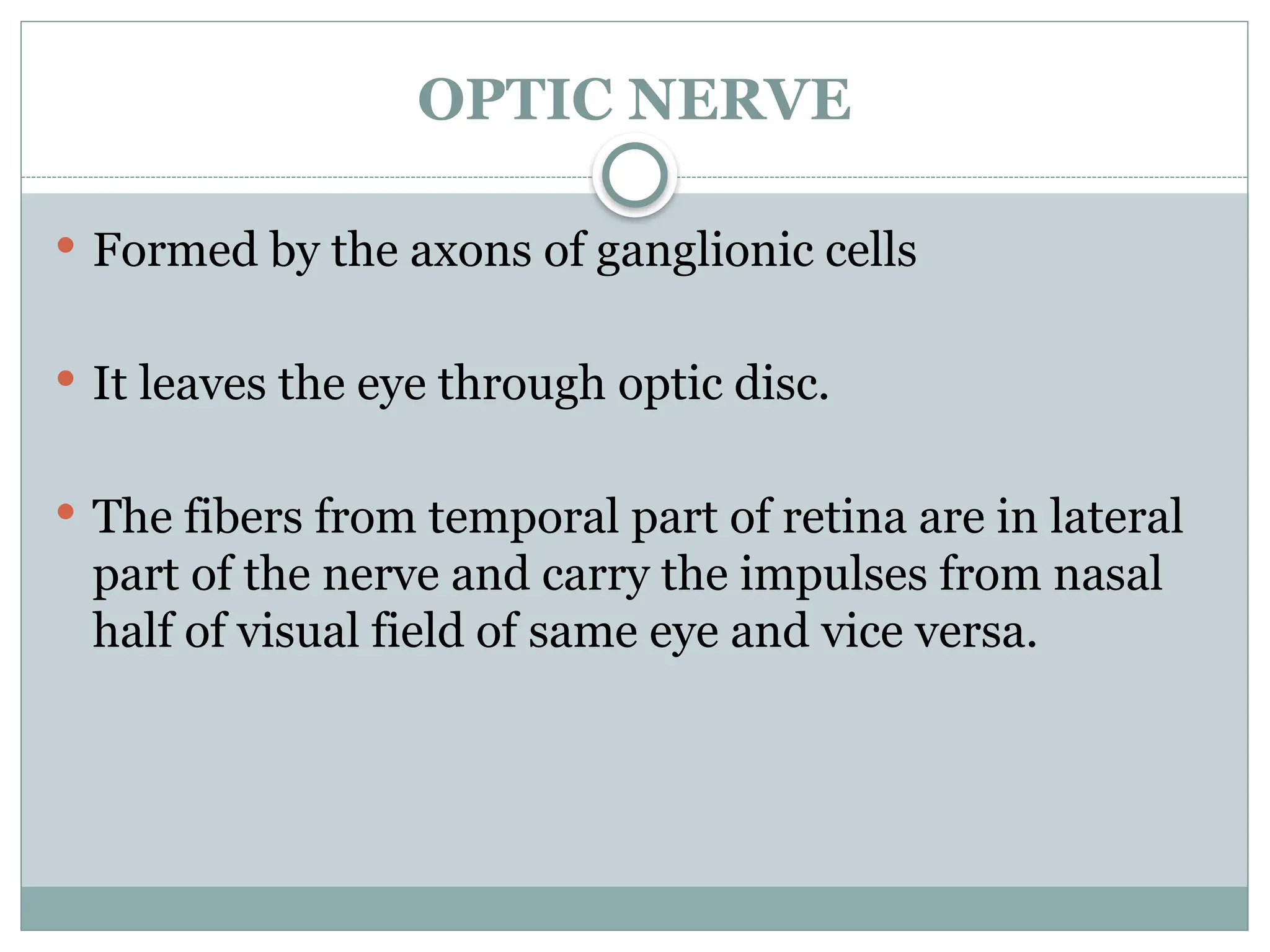
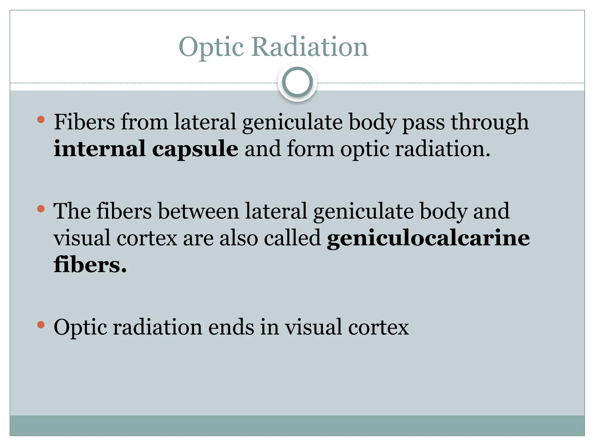
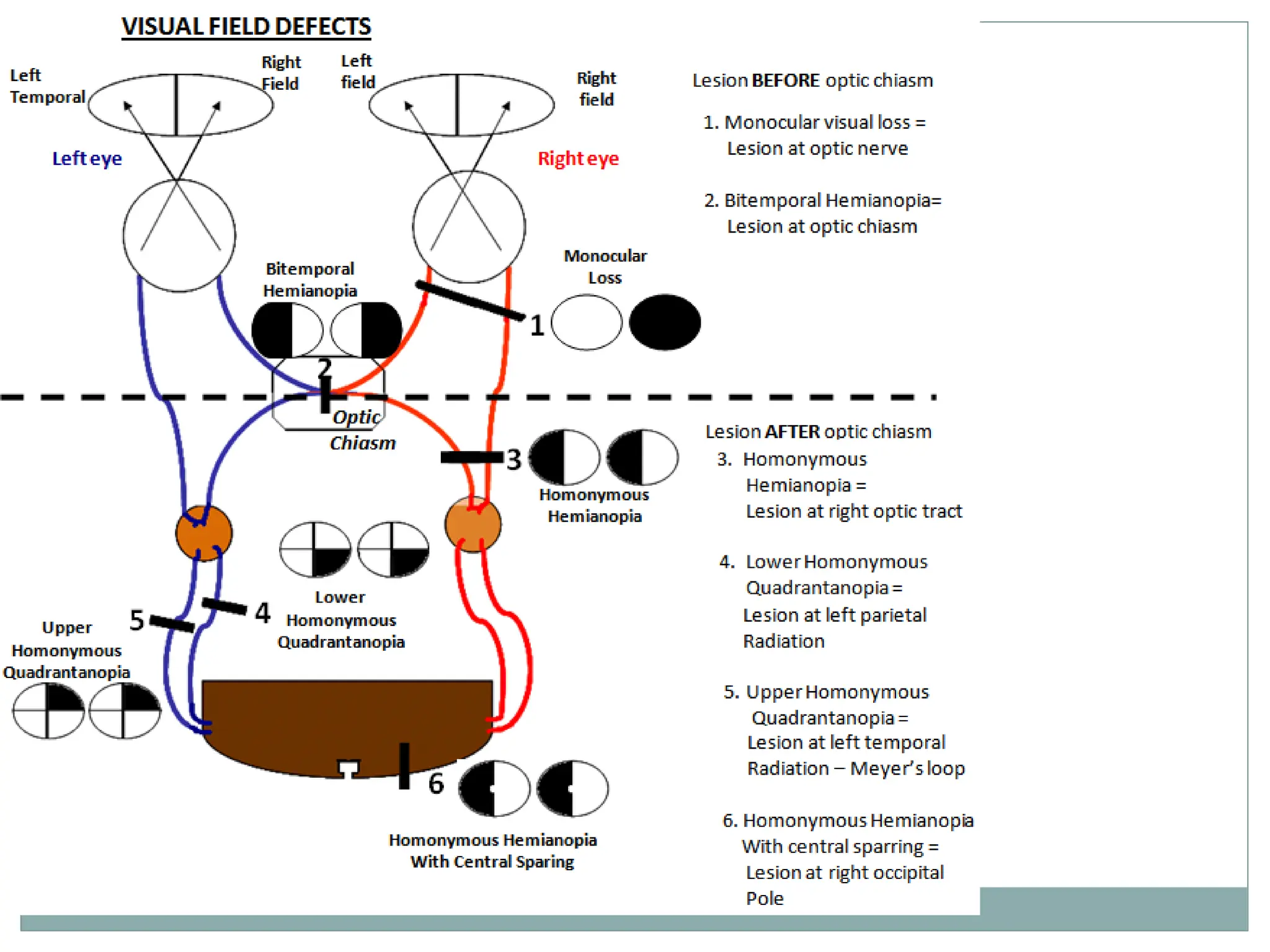
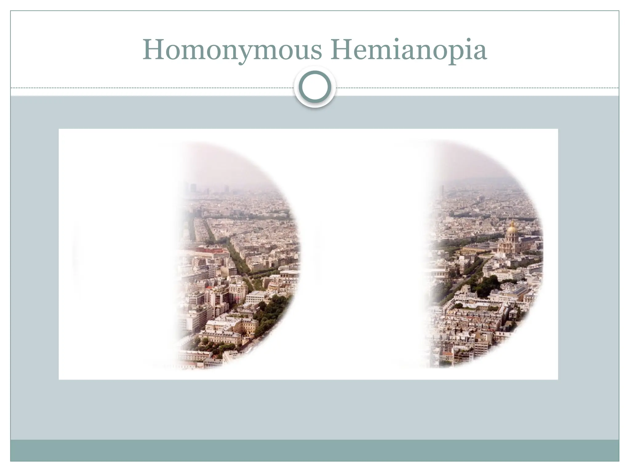
Visual Pathway
Interpret!
 30 year old patient Mr. XYZ , a known case of
Syphilis, presented to Department of
Ophthalmology, NMCH with complaints of partial
loss of vision.What type of visual field defect you
suspect ? Why?
What is Visual Pathway?
 Visual pathway or optic pathway is the
nervous pathway that transmits impulses
from retina to visual centre in cerebral
cortex.
VISUAL RECEPTORS
 Rods and cones
 They synapse with dendrites of bipolar cells of inner
nuclear layer of the retina.
FIRST ORDER NEURONS
 First order neurons (primary neurons) are bipolar
cells in the retina.
 Axons from the bipolar cells synapse with dendrites
of ganglionic cells.
SECOND ORDER NEURONS
 They are the ganglionic cells in ganglionic cell
layer of retina.
 Axons of the ganglionic cells form optic nerve.
 Optic nerve leaves the eye and terminates in lateral
geniculate body.
THIRD ORDER NEURONS
 They are in the lateral geniculate body.
 Fibres arising from LGB, reaches the visual cortex.
OPTIC NERVE
 Formed by the axons of ganglionic cells
 It leaves the eye through optic disc.
 The fibers from temporal part of retina are in lateral
part of the nerve and carry the impulses from nasal
half of visual field of same eye and vice versa.
„ OPTIC CHIASMA
 Medial fibers of each optic nerve cross the midline
and join the uncrossed lateral fibers of opposite side,
to form the optic tract.
Optic Tract
 Formed by uncrossed fibers of optic nerve on the
same side and crossed fibers of optic nerve from the
opposite side.
 The fibers turn around the peduncle to reach the
lateral geniculate body in thalamus.
LATERAL GENICULATE BODY
 Majority of the fibers of optic tract terminate in
lateral geniculate body, which forms the
subcortical center for visual sensation.
 From here, the geniculocalcarine tract or optic
radiation arises.
Optic Radiation
 Fibers from lateral geniculate body pass through
internal capsule and form optic radiation.
 The fibers between lateral geniculate body and
visual cortex are also called geniculocalcarine
fibers.
 Optic radiation ends in visual cortex
Visual Cortex
 Primary visual area (17) - Perception of visual
impulses
 Secondary visual area (18) - Interpretation of visual
impulses
 Occipital eye field (19) - Movement of eyes.
Visual Pathway lesions
 Hemianopia : Blindness over half the field of vision.
 Homonymous : Visual field loss on the same side of
both eyes.
Causes of Optic Nerve Lesion
 Optic atrophy
 Traumatic avulsion of the optic nerve
 Indirect optic neuropathy
 Acute optic neuritis
Lesions of Chiasma
 Suprasellar aneurysms
 Tumours of pituitary gland
 Third ventricular dilatation due to obstructive
hydrocephalus
 Atheroma of the carotids or posterior
communicating arteries
Optic tract lesions
 Syphilitic meningitis
 Tuberculosis and tumours of optic thalamus
 Aneurysms of superior cerebellar or posterior
cerebral arteries.
Lesion in Optic Radiation
 Vascular occlusions
 Primary and secondary tumours
 Trauma
Homonymous Hemianopia
Upper Homonymous Quadrantanopia
Lower Homonymous Quadrantanopia
INVESTIGATIONS
 FIELD TESTING
1. CONFRONTATION TEST
2. AMSLER GRID
3. PERIMETRY
 MRI (Magnetic Resonance Imaging)
 CT SCAN (Computed Tomography)
Interpret!
 30 year old patient Mr. XYZ , a known case of
Syphilis, presented to Department of
Ophthalmology, NMCH with complaints of partial
loss of vision.What type of visual field defect you
suspect ? Why?
 Homonymous hemianopia due to Optic tract lesion
Thank you 

visual pathway in..... human beings.pptx

  • 1.
  • 2.
    Interpret!  30 yearold patient Mr. XYZ , a known case of Syphilis, presented to Department of Ophthalmology, NMCH with complaints of partial loss of vision.What type of visual field defect you suspect ? Why?
  • 3.
    What is VisualPathway?  Visual pathway or optic pathway is the nervous pathway that transmits impulses from retina to visual centre in cerebral cortex.
  • 8.
    VISUAL RECEPTORS  Rodsand cones  They synapse with dendrites of bipolar cells of inner nuclear layer of the retina.
  • 9.
    FIRST ORDER NEURONS First order neurons (primary neurons) are bipolar cells in the retina.  Axons from the bipolar cells synapse with dendrites of ganglionic cells.
  • 10.
    SECOND ORDER NEURONS They are the ganglionic cells in ganglionic cell layer of retina.  Axons of the ganglionic cells form optic nerve.  Optic nerve leaves the eye and terminates in lateral geniculate body.
  • 11.
    THIRD ORDER NEURONS They are in the lateral geniculate body.  Fibres arising from LGB, reaches the visual cortex.
  • 12.
    OPTIC NERVE  Formedby the axons of ganglionic cells  It leaves the eye through optic disc.  The fibers from temporal part of retina are in lateral part of the nerve and carry the impulses from nasal half of visual field of same eye and vice versa.
  • 14.
    „ OPTIC CHIASMA Medial fibers of each optic nerve cross the midline and join the uncrossed lateral fibers of opposite side, to form the optic tract.
  • 15.
    Optic Tract  Formedby uncrossed fibers of optic nerve on the same side and crossed fibers of optic nerve from the opposite side.  The fibers turn around the peduncle to reach the lateral geniculate body in thalamus.
  • 16.
    LATERAL GENICULATE BODY Majority of the fibers of optic tract terminate in lateral geniculate body, which forms the subcortical center for visual sensation.  From here, the geniculocalcarine tract or optic radiation arises.
  • 17.
    Optic Radiation  Fibersfrom lateral geniculate body pass through internal capsule and form optic radiation.  The fibers between lateral geniculate body and visual cortex are also called geniculocalcarine fibers.  Optic radiation ends in visual cortex
  • 18.
    Visual Cortex  Primaryvisual area (17) - Perception of visual impulses  Secondary visual area (18) - Interpretation of visual impulses  Occipital eye field (19) - Movement of eyes.
  • 20.
    Visual Pathway lesions Hemianopia : Blindness over half the field of vision.  Homonymous : Visual field loss on the same side of both eyes.
  • 22.
    Causes of OpticNerve Lesion  Optic atrophy  Traumatic avulsion of the optic nerve  Indirect optic neuropathy  Acute optic neuritis
  • 23.
    Lesions of Chiasma Suprasellar aneurysms  Tumours of pituitary gland  Third ventricular dilatation due to obstructive hydrocephalus  Atheroma of the carotids or posterior communicating arteries
  • 24.
    Optic tract lesions Syphilitic meningitis  Tuberculosis and tumours of optic thalamus  Aneurysms of superior cerebellar or posterior cerebral arteries.
  • 25.
    Lesion in OpticRadiation  Vascular occlusions  Primary and secondary tumours  Trauma
  • 26.
  • 27.
  • 28.
  • 29.
    INVESTIGATIONS  FIELD TESTING 1.CONFRONTATION TEST 2. AMSLER GRID 3. PERIMETRY  MRI (Magnetic Resonance Imaging)  CT SCAN (Computed Tomography)
  • 30.
    Interpret!  30 yearold patient Mr. XYZ , a known case of Syphilis, presented to Department of Ophthalmology, NMCH with complaints of partial loss of vision.What type of visual field defect you suspect ? Why?  Homonymous hemianopia due to Optic tract lesion
  • 31.