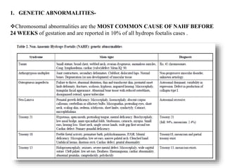
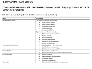
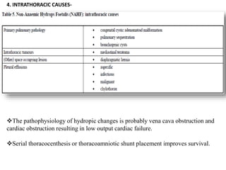
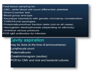
Hydrops fetalis, or fetal hydrops, is an abnormal accumulation of fluid in fetal compartments and body cavities. It is classified as immune or non-immune. Immune hydrops is caused by maternal-fetal blood group incompatibility while non-immune hydrops has various potential causes including genetic abnormalities, infections, heart defects, and tumors. Diagnosis involves ultrasound to detect fluid accumulation while treatment depends on the underlying etiology but may include fetal transfusions, medications, or early delivery. Prognosis varies significantly depending on the cause and gestational age at presentation and treatment.
2. Hydrops fetalis is a severe fetal condition characterized by an abnormal
and excessive accumulation of fluid within the fetal extravascular
compartments and body cavities. It is characterized by presence of atleast
two of the following features:
• EDEMA(collection of fluid in the subcutaneous plane) with width of
≥5mm classically seen as BUDDHA POSITION ON USG WITH
A HALO SIGN
• Ascites
• Pleural effusion
• Pericardial effusion
In addition, hydrops foetalis is frequently associated with polyhydramnios
and thickened placenta.
3. IMMUNE
HYDROPS
• 10-20%CASES
• Antigen incompatibility
between the mother and
fetus.
• There is an underlying well
defined cause.
• Prognosis-good
• Decreased incidence due to
widespread use of anti-D
NON
IMMUNE
HYDROPS
• 80-90%cases
• Occurs due to failure of
the interstitial fluid to
return into the venous
system.
• No identifiable circulating
antibody to any red cell
antigen
• Any other cause besides
immune
HYDROPS FETALIS IS BROADLY CLASSIFIED INTO-
Hydrops foetalis can be divided into Immune Hydrops Foetalis (12.7%) associated with antigen
antibody mediated red cell haemolysis, and Non-Immune Hydrops Foetalis (87.3%), associated
with a wide range of aetiological factors . With improved treatment and diagnosis of Rhesus Iso-
immunisation, nonimmune factors have become more frequent causes.
4.
5. RED CELL HAEMOLYSIS
FETAL ANEMIA AND
EXTRAMEDULLARY
HAEMATOPOIESIS
HEPATOMEGALY
SPLENOMEGALY
HIGH OUTPUT CARDIAC FAILURE
TISSUE ACIDOSIS AND FETAL ANOXIA
6. Characteristics of Hydrops foetalis
secondary to Rh alloimmunization-
• Foetus should be Rh positive
•ICT – positive
•Polyhydramnios
•Pale oedematous baby
•Pale and large placenta
•Buddha position with halo sign on usg
7. The basic mechanism for the formation of foetal hydrops is an IMBALANCE OF INTERSTITIAL
FLUID PRODUCTION AND THE LYMPHATIC RETURN.
8. The current classification of immune and nonimmune hydrops foetalis
is not sufficient to describe the complex pathophysiology leading to
heart failure and generalized oedema in the foetus and neonate and is
confusing since immunological phenomena play a role in both entities.
A more practical approach to describe the aetiology of hydrops
foetalis is based on the presence or absence of anaemia.
The distinction between anaemic and non-anaemic hydrops foetalis
(AHF and NAHF) allows for a more structured approach towards
diagnosis and therapy.
9.
10. ANAEMIC HYDROPS FOETALIS- AHF has two main groups
of causative factors as haematological disorders and intrauterine
infections
•Account for 10-27% cases
•The most common type of AHF (62.5%) is Rhesus
Alloimunization
• Intrauterine transfusion in anaemic hydrops foetalis
dramatically improves foetal and neonatal outcome
and reverses hydrops foetalis in 65%.
HAEMATOLOGICAL
DISORDERS
•Parvovirus B19, Toxoplasmosis, Adenovirus, Coxsackie
virus, Rubella, cytomegalovirus, leptospirosis and
congenital hepatitis and syphilis
•Serological tests in the affected hydropic foetus may
be negative due to the prozone phenomenon
•Symptoms include anaemia, hepatic dysfunction,
hypoproteinemia, and portal hypertension
INTRAUTERINE
INFECTIONS
11.
12. HYDROPS FOETALIS SECONDARY TO PARVOVIRUS B19
INFECTION-
Parvovirus B19, a small "parvo" single stranded DNA virus,
accounts for 27% of cases of AHF in anatomically normal foetuses.
It usually infects rapidly dividing cell lines, such as
erythroid progenitor cells
This virus has been shown to cause a congenital infection syndrome, manifested by RASH,
ANEMIA, HEPATOMEGALY, AND CARDIOMEGALY, and infection can lead to miscarriage or
nonimmune hydrops fetalis
Maternal viraemia reaches its peak approximately 1 week after infection and symptoms such as
erythema infectiosum, mild fever, arthralgia, and headache start approximately 17 days after
infection. Immediately following maternal viraemia the foetus can be infected.
The parvovirus B19 receptor, abundant in the placenta in early gestation, may provide a pathway to
the foetus. Viral infection of the foetus may persist until term or after birth, even when infection
occurs in early gestation. Transplacental transmission rate -33%
13. The interval between parvo B19 infection and development of AHF ranges from 2 to 6
weeks . SEVENTY PERCENT OF ALL CASES OF AHF ASSOCIATED WITH
PARVO B19 INFECTION OCCUR BETWEEN 20 AND 24 WEEKS OF
GESTATION .
DIAGNOSIS-
• Parvo B19 virus is diagnosed by detection of viral DNA by PCR on cordocentesis blood
samples and/or amniotic fluid
• Identification of specific IgM-antibodies
Intrauterine transfusion of red blood cells and/or platelets increases foetal survival
from 55 to 82% . Resolution of hydrops foetalis following successful intrauterine
transfusion may take up to 12 weeks.
Negative prognostic factors are maternal seroconversion during early gestation, low
gestational age at detection of AHF, number and results of intrauterine transfusion,
and prolonged duration from intrauterine transfusion to resolution of hydrops
foetalis.
The outcome following B19V infection is surprisingly good; spontaneous
resolution occurs in approximately one third of such incidents, and
approximately 85% of those who receive fetal transfusions survive. The virus is not
14. Non-Anaemic Hydrops Foetalis (NAHF)
GENETIC ABNORMALITIES
INBORN ERRORS OF METABOLISM
CONGENITAL HEART DEFECTS
INTRATHORACIC PATHOLOGY
Twin-twin transfusion syndrome (TTTS)
ENDOCRINAL PATHOLOGY
INTESTINAL ISCHAEMIA LEADING TO MECONIUM PERITONITIS
IDIOPATHIC
15.
16. 1. GENETIC ABNORMALITIES-
Chromosomal abnormalities are the MOST COMMON CAUSE OF NAHF BEFORE
24 WEEKS of gestation and are reported in 10% of all hydrops foetalis cases .
17. 2. INBORN ERRORS OF METABOLISM-
Inborn errors of metabolism lead to NAHF through obstruction of the venous bloodstream
by visceromegaly and local circulatory problems secondary to storage substances
(particularly in the reticuloendothelial system).
FEATURES-
•GENERALISED OEDEMA and EXTREME HEPATOSPLENOMEGALY
• ANAEMIA may result from hypersplenism and reduction of erythropoietic stem cells by
bone marrow infiltration with storage cells.
•HYPOPROTEINEMIA due to liver dysfunction may cause hydrops.
•Cardiomyopathy-rare
•In lysosomal storage disease, the placenta is pale and bulky and microscopy shows the
presence of lysosomal vacuolation.
• Especially cases of familial NAHF should be investigated for lysosomal storage disorders
as parental consanguinity is a known risk factor.
18. 3. CONGENITAL HEART DEFECTS-
CONGENITAL HEART DISEASE IS THE MOST COMMON CAUSE Of Hydrops Foetalis AFTER 24
WEEKS OF GESTATION.
19. • 13.8 to 40% of all NAHF cases.
• Congestive heart failure is the primary cause in these cases of NAHF
• Foetal echocardiography is a useful tool for diagnosis and monitoring progress of disease.
STRUCTURAL HEART DISEASE
• Sustained foetal supraventricular tachycardia is rare, but frequently leads to NAHF.
• Maternally administered Digoxin is the first-line treatment option to resolve foetal
hydrops due to cardiac arrhythmia.
• Placental oedema and poor placental function can impair transplacental delivery of drugs. In such
cases direct intraperitoneal or intravascular foetal therapy is a good alternative and more direct
option . The combination of transplacental Amiodarone and Digoxin therapy has been used with
success
• Sotalol is another effective treatment of foetal tachycardia with excellent placental transfer.
• After conversion to a normal sinus rhythm, total resolution of hydrops requires 4-6 weeks.
• The risk of recurrence is 48%
FOETAL TACHYARRHYTHMIA
• 8-11%
• The most common form is DCM(Dilated cardiomyopathy) is that seen in infants born of diabetic
mothers
• This form of cardiomyopathy will completely resolve in 6-12 months.
CARDIOMYOPATHY
• Causes- infectious agents or autoimmune diseases. The course of the illness is frequently
fulminant or fatal.
MYOCARDITIS
20. 4. INTRATHORACIC CAUSES-
The pathophysiology of hydropic changes is probably vena cava obstruction and
cardiac obstruction resulting in low output cardiac failure.
Serial thoracocenthesis or thoracoamniotic shunt placement improves survival.
21. 5.Twin-twin transfusion syndrome (TTTS)-
It complicates monochorionic twin gestation in 15% of cases, can be a cause of
NAHF.
TTTS presents during the second trimester with an oligo-polyhydramnion
sequence,i.e. the deepest vertical pool in the donor sac is <2 cm and in
the recipient’s sac >8 cm.
Staging has been described by Quintero et al. :
TTTS is caused by an unbalanced interfoetal transfusion via vascular anastomoses
in monochorionic twins. It is mediated by mostly > 1 arterio-venous anastomosis in
association with absent bi-directional superficial anastomoses.
Stage I: Bladder In Donor Visible
Stage II: Bladder In Donor Not Visible
Stage III: Pathologic Umbilical Artery Or Recipient Reverse Flow In Ductus Venosus Or
Abnormal Doppler Readings In The Donor (Absent/Reduced End-diastolic Flow, Pulsatile
Umbilical Venous Flow)
Stage IV: Hydrops Foetalis
Stage V: Demise Of One Or Both Twins
22. Foetal haemodynamics are disturbed by an increased expression of vasopressors
such as endothelin, renin, and angiotensin II and a decrease in vasodilators such as
nitric oxide (NO).
The recipient twin can develop signs of fluid overload and high output cardiac
failure, including cardiomegaly, ascites and pleural-pericardial effusions (hydrops
foetalis).
Hydropic changes may be seen in the recipient, donor, or both.
Hydrops foetalis has been described as early as 14 weeks of gestation . When
hydropic changes occur in the second trimester, prognosis is extremely poor.
Antenatal demise of the recipient twin in TTTS can be associated with subsequent
development of hydrops foetalis in the surviving twin donor. This phenomenon may
be due to ischaemia reperfusion injury of the previously poorly perfused twin.
Hydropic signs in the donor foetus may also be seen after selective laser
coagulation procedures (25% of cases),they are transient and caused by an increase in
foetal circulating volume and adaptation after occlusion of vascular anastomoses.
23. Treatment modalities for foetal hydrops in TTTS include
1. SERIALAMNIOREDUCTION (to reduce the risk of preterm labour by
reducing increased amniotic pressure)
2. SEPTOSTOMY (creating a hole in the intertwin membrane)
3. LASER ABLATION OF VASCULAR ANASTOMOSES .
4. In severe stage III/IV disease, SELECTIVE FETICIDE BY BIPOLAR
DIATHERMY of the umbilical cord has been performed to rescue one child .
Treatment can be based on the Quintero staging system: stage I/II: serial
amnioreduction, stage III: selective laser ablation, severe stage III/IV: selective
feticide.
Long-term sequelae of TTTS consist of –
psychomotor retardation and cerebral palsy.
24. 6.GASTROINTESTINAL AETIOLOGY: MECONIUM PERITONITIS-
MECONIUM PERITONITIS IS A STERILE CHEMICAL PERITONITIS induced by
meconium leaking into the peritoneal cavity following perforation of the bowel (most commonly
the ileum) proximal to a site of obstruction.
The perforation has closed by the time of birth, but the resulting adhesions between
bowel loops and omentum wall up the meconium collection forming pseudocysts which can be
detected by prenatal ultrasound.
Meconium peritonitis is rare before 20 weeks gestation.
25. 7. ENDOCRINE AETIOLOGY-
Several case reports suggest a possible relationship between foetal
hypothyroidism and pleural effusions.
Thyroid hormone may play a role in the regulation of lymphatic flow rate
and lung liquid clearance since a case of foetal chylothorax resulting in
NAHF resolved after treatment with thyroxin.
NAHF secondary to foetal tachycardia has been reported in foetal
hyperthyroidism due to thyroid-stimulating immunoglobulins in maternal
Graves disease and could be treated by administration of anti-thyroid
drugs to the mother.
26.
27.
28.
29.
30. THE FIRST SIGN OF FOETAL HYDROPS IN EARLY PREGNANCY (11-15
WEEKS OF GESTATION) IS OFTEN THE PRESENCE OF
GENERALIZED SKIN OEDEMA IN THE FOETAL HEAD AND NECK
AREAAT ULTRASOUND INVESTIGATION. Pleural effusions are rarely
diagnosed before 15 weeks of gestation.
Prenatal Diagnosis by Ultrasound Doppler investigations by ultrasound play an
important role in the diagnosis of foetal anaemia . Blood flow in anaemic foetuses
has a hyperdynamic pattern, which can be detected with Doppler velocimetry in
various foetal blood vessels. Velocity changes are thought to result from increased
cardiac output and decreased viscosity of foetal blood. Blood flow in the middle
cerebral artery (MCA) is the first to respond to foetal anaemia due to the early
response of the brain tissue to anaemia. Doppler ultrasonography has the advantage
of being noninvasive.
36. 1. Antenatal management should be directed towards making a diagnosis,
with the aim of identifying those in whom either prenatal or immediate post-natal
intervention may be
effective.
Diagnostic techniques include-
• Fetal ultrasonography
• Fetal echocardiography
•Examination of maternal blood for fetal erythrocytes (kleihauer-betke test),
•Amniocentesis and sampling of fetal blood.
In some cases, fetal intervention is effective
(e.g., fetal transfusion for anemia due to Parvovirus B19 infection, treatment of
fetal tachycardia).
In others, delivery corrects the underlying problem (e.g., chorioangioma of
placenta).
Very few hydropic infants survive if delivered before 30 weeks of gestation.
37. 2. Postnatal treatment includes
A. Vigorous resuscitation with interventions (e.g., thoracentesis, paracentesis)
to remove excess fluid .
B. Treatment of asphyxia, which is common in these infants.
C. Diagnosis of cause of hydrops, both for management of the patient and
counseling of the parents regarding risk of recurrence in future pregnancies.
This may require extensive diagnostic interventions, including careful post-
mortem examination, skin sample for karyotyping and full body radiographs in
those who do not survive.
D. Treatment of underlying cause
OUTCOME: Approximately 50% of fetuses with non-immune hydrops
fetalis die in utero, and about half of the liveborn infants survive.
Editor's Notes
Fluid accumulation in the fetus can result from congestive heart failure, obstructed lymphatic flow, or decreased plasma osmotic pressure. The fetus is particularly