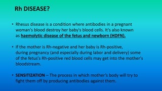
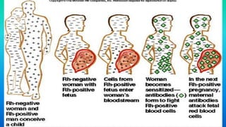
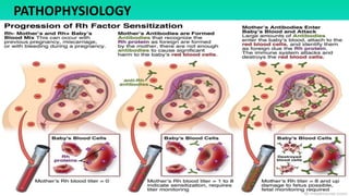
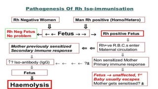
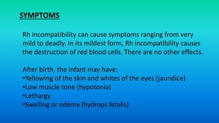
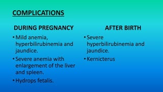
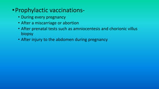
Rh incompatibility occurs when a pregnant woman has Rh-negative blood and her fetus has Rh-positive blood. During pregnancy or delivery, the fetus's Rh-positive blood can enter the mother's bloodstream and cause her to form antibodies. If she has another Rh-positive baby, those antibodies can cross the placenta and destroy the baby's red blood cells, causing anemia or even hydrops fetalis. To prevent Rh disease, Rh-negative mothers are given Rh immunoglobulin injections during and after pregnancy to prevent antibody formation. For affected pregnancies, fetal blood transfusions may be needed to treat severe anemia.
It is a composite graphical recording of cervical dilatation and descent of head against duration of labour in hours.
It also gives information about fetal and maternal condition that are all recorded on single sheet of paper.
Rh Incompatibility I Hemolytic Disease of the NewbornSwatilekha Das
Rh Incompatibility I Hemolytic Disease of the Newborn-
Hi All,
I am Swatilekha Das, B.Sc, M.Sc Nurse and working as Assistant Professor of Nursing in a Nursing college. I worked as Clinical Instructor, nursing educator, nursing trainer, Nursing Tutor at hospitals, nursing schools and colleges.
ABOUT THIS ppt-
In this ppt I discussed about definition of rh incompatibility, cause, pathophysiology, diagnostic tests, treatment and screening and prevention of Rh incompatibility.
To know about it check the ppt till end.
I hope you enjoy this ppt and if you do then please click on the like button and share the with your friends too . Don't Forget to follow to see more such ppt. Thank you for checking the ppt.
@All Rights Reserved..
Rh Incompatibility in Pregnancy. Rh incompatibility occurs when a pregnant woman whose blood type is Rh-negative is exposed to Rh-positive blood from her fetus, leading to the mother's development of Rh antibodies
It explains the mechanism of normal labour to medical and para-medical staff.It also puts light on principle movements underlying mechanism of normal labour with pictures.Thank You Like an share it to the maximum.
This topic contains detailed description about labour, its definition, date of onset of labour, calculations of date of delivery, causes of onset of labour, physiology of normal labour, and events, clinical course and management of each stages of labour.
Hydatidiform Mole (HM) is a rare mass or growth that forms inside the uterus at the beginning of a pregnancy. It is a type of gestational trophoblastic disease (GTD).
When a normal sperm cell fertilizes one of these oocytes, the resulting embryo has only one set of chromosomes. Because the embryo has no genes from the mother, the pregnancy cannot develop normally, resulting in a hydatidiform mole.
Rh incompatibility is a condition that occurs during pregnancy if
a woman hasRh-negative blood and
her baby has Rh-positive blood.
"Rh-negative" and "Rh-positive" refer to
whether your blood has Rh factor. Rh factor is a protein on red blood cells. If
you have Rh factor, you're Rh-positive. If you don't have it, you're
Rh-negative. Rh factor is inherited (passed from parents to children through
the genes). Most people are Rh-positive.
Whether you have Rh factor doesn't affect your general health.
However, it can cause problems during pregnancy.
HAEMOLYTIC DISEASE OF THE NEW BORN (HDN).pptxGroup271
Hemolytic anemia is a disorder in which red blood cells are destroyed faster than they can be made. The destruction of red blood cells is called hemolysis.
Red blood cells carry oxygen to all parts of your body. If you have a lower than normal amount of red blood cells, you have anemia. When you have anemia, your blood can’t bring enough oxygen to all your tissues and organs. Without enough oxygen, your body can’t work as well as it should.
Hemolytic anemia can be inherited or acquired:
Recomendações da OMS sobre cuidados maternos e neonatais para uma experiência pós-natal positiva.
Em consonância com os ODS – Objetivos do Desenvolvimento Sustentável e a Estratégia Global para a Saúde das Mulheres, Crianças e Adolescentes, e aplicando uma abordagem baseada nos direitos humanos, os esforços de cuidados pós-natais devem expandir-se para além da cobertura e da simples sobrevivência, de modo a incluir cuidados de qualidade.
Estas diretrizes visam melhorar a qualidade dos cuidados pós-natais essenciais e de rotina prestados às mulheres e aos recém-nascidos, com o objetivo final de melhorar a saúde e o bem-estar materno e neonatal.
Uma “experiência pós-natal positiva” é um resultado importante para todas as mulheres que dão à luz e para os seus recém-nascidos, estabelecendo as bases para a melhoria da saúde e do bem-estar a curto e longo prazo. Uma experiência pós-natal positiva é definida como aquela em que as mulheres, pessoas que gestam, os recém-nascidos, os casais, os pais, os cuidadores e as famílias recebem informação consistente, garantia e apoio de profissionais de saúde motivados; e onde um sistema de saúde flexível e com recursos reconheça as necessidades das mulheres e dos bebês e respeite o seu contexto cultural.
Estas diretrizes consolidadas apresentam algumas recomendações novas e já bem fundamentadas sobre cuidados pós-natais de rotina para mulheres e neonatos que recebem cuidados no pós-parto em unidades de saúde ou na comunidade, independentemente dos recursos disponíveis.
É fornecido um conjunto abrangente de recomendações para cuidados durante o período puerperal, com ênfase nos cuidados essenciais que todas as mulheres e recém-nascidos devem receber, e com a devida atenção à qualidade dos cuidados; isto é, a entrega e a experiência do cuidado recebido. Estas diretrizes atualizam e ampliam as recomendações da OMS de 2014 sobre cuidados pós-natais da mãe e do recém-nascido e complementam as atuais diretrizes da OMS sobre a gestão de complicações pós-natais.
O estabelecimento da amamentação e o manejo das principais intercorrências é contemplada.
Recomendamos muito.
Vamos discutir essas recomendações no nosso curso de pós-graduação em Aleitamento no Instituto Ciclos.
Esta publicação só está disponível em inglês até o momento.
Prof. Marcus Renato de Carvalho
www.agostodourado.com
- Video recording of this lecture in English language: https://youtu.be/lK81BzxMqdo
- Video recording of this lecture in Arabic language: https://youtu.be/Ve4P0COk9OI
- Link to download the book free: https://nephrotube.blogspot.com/p/nephrotube-nephrology-books.html
- Link to NephroTube website: www.NephroTube.com
- Link to NephroTube social media accounts: https://nephrotube.blogspot.com/p/join-nephrotube-on-social-media.html
Report Back from SGO 2024: What’s the Latest in Cervical Cancer?bkling
Are you curious about what’s new in cervical cancer research or unsure what the findings mean? Join Dr. Emily Ko, a gynecologic oncologist at Penn Medicine, to learn about the latest updates from the Society of Gynecologic Oncology (SGO) 2024 Annual Meeting on Women’s Cancer. Dr. Ko will discuss what the research presented at the conference means for you and answer your questions about the new developments.
HOT NEW PRODUCT! BIG SALES FAST SHIPPING NOW FROM CHINA!! EU KU DB BK substit...GL Anaacs
Contact us if you are interested:
Email / Skype : kefaya1771@gmail.com
Threema: PXHY5PDH
New BATCH Ku !!! MUCH IN DEMAND FAST SALE EVERY BATCH HAPPY GOOD EFFECT BIG BATCH !
Contact me on Threema or skype to start big business!!
Hot-sale products:
NEW HOT EUTYLONE WHITE CRYSTAL!!
5cl-adba precursor (semi finished )
5cl-adba raw materials
ADBB precursor (semi finished )
ADBB raw materials
APVP powder
5fadb/4f-adb
Jwh018 / Jwh210
Eutylone crystal
Protonitazene (hydrochloride) CAS: 119276-01-6
Flubrotizolam CAS: 57801-95-3
Metonitazene CAS: 14680-51-4
Payment terms: Western Union,MoneyGram,Bitcoin or USDT.
Deliver Time: Usually 7-15days
Shipping method: FedEx, TNT, DHL,UPS etc.Our deliveries are 100% safe, fast, reliable and discreet.
Samples will be sent for your evaluation!If you are interested in, please contact me, let's talk details.
We specializes in exporting high quality Research chemical, medical intermediate, Pharmaceutical chemicals and so on. Products are exported to USA, Canada, France, Korea, Japan,Russia, Southeast Asia and other countries.
The prostate is an exocrine gland of the male mammalian reproductive system
It is a walnut-sized gland that forms part of the male reproductive system and is located in front of the rectum and just below the urinary bladder
Function is to store and secrete a clear, slightly alkaline fluid that constitutes 10-30% of the volume of the seminal fluid that along with the spermatozoa, constitutes semen
A healthy human prostate measures (4cm-vertical, by 3cm-horizontal, 2cm ant-post ).
It surrounds the urethra just below the urinary bladder. It has anterior, median, posterior and two lateral lobes
It’s work is regulated by androgens which are responsible for male sex characteristics
Generalised disease of the prostate due to hormonal derangement which leads to non malignant enlargement of the gland (increase in the number of epithelial cells and stromal tissue)to cause compression of the urethra leading to symptoms (LUTS
NVBDCP.pptx Nation vector borne disease control programSapna Thakur
NVBDCP was launched in 2003-2004 . Vector-Borne Disease: Disease that results from an infection transmitted to humans and other animals by blood-feeding arthropods, such as mosquitoes, ticks, and fleas. Examples of vector-borne diseases include Dengue fever, West Nile Virus, Lyme disease, and malaria.
New Directions in Targeted Therapeutic Approaches for Older Adults With Mantl...i3 Health
i3 Health is pleased to make the speaker slides from this activity available for use as a non-accredited self-study or teaching resource.
This slide deck presented by Dr. Kami Maddocks, Professor-Clinical in the Division of Hematology and
Associate Division Director for Ambulatory Operations
The Ohio State University Comprehensive Cancer Center, will provide insight into new directions in targeted therapeutic approaches for older adults with mantle cell lymphoma.
STATEMENT OF NEED
Mantle cell lymphoma (MCL) is a rare, aggressive B-cell non-Hodgkin lymphoma (NHL) accounting for 5% to 7% of all lymphomas. Its prognosis ranges from indolent disease that does not require treatment for years to very aggressive disease, which is associated with poor survival (Silkenstedt et al, 2021). Typically, MCL is diagnosed at advanced stage and in older patients who cannot tolerate intensive therapy (NCCN, 2022). Although recent advances have slightly increased remission rates, recurrence and relapse remain very common, leading to a median overall survival between 3 and 6 years (LLS, 2021). Though there are several effective options, progress is still needed towards establishing an accepted frontline approach for MCL (Castellino et al, 2022). Treatment selection and management of MCL are complicated by the heterogeneity of prognosis, advanced age and comorbidities of patients, and lack of an established standard approach for treatment, making it vital that clinicians be familiar with the latest research and advances in this area. In this activity chaired by Michael Wang, MD, Professor in the Department of Lymphoma & Myeloma at MD Anderson Cancer Center, expert faculty will discuss prognostic factors informing treatment, the promising results of recent trials in new therapeutic approaches, and the implications of treatment resistance in therapeutic selection for MCL.
Target Audience
Hematology/oncology fellows, attending faculty, and other health care professionals involved in the treatment of patients with mantle cell lymphoma (MCL).
Learning Objectives
1.) Identify clinical and biological prognostic factors that can guide treatment decision making for older adults with MCL
2.) Evaluate emerging data on targeted therapeutic approaches for treatment-naive and relapsed/refractory MCL and their applicability to older adults
3.) Assess mechanisms of resistance to targeted therapies for MCL and their implications for treatment selection
Pulmonary Thromboembolism - etilogy, types, medical- Surgical and nursing man...VarunMahajani
Disruption of blood supply to lung alveoli due to blockage of one or more pulmonary blood vessels is called as Pulmonary thromboembolism. In this presentation we will discuss its causes, types and its management in depth.
2. • Proteins (antigens) occurring only on surface of
RBC’s
• Rh + if proteins present
• Rh – if proteins absent
• A+, A-, B+, B-, AB+, AB-, O+, O-
• Most important for pregnancy
• Inheritance is Autosomal Dominant
• 15% Caucasian population are Rh-
Rh FACTOR
3. INTRODUCTION
• During Pregnancy, the mother and the fetus have different Rh
protein factors, this condition is called Rh incompatibility.
• Like our blood type, we inherit our Rh factor type from our
parents. Most people are Rh-positive, but a small percentage of
people are Rh-negative
• Rh factor doesn’t directly affect the health.
• However, Rh factor becomes important during pregnancy.
4. • Rhesus disease is a condition where antibodies in a pregnant
woman's blood destroy her baby's blood cells. It's also known
as haemolytic disease of the fetus and newborn (HDFN).
• If the mother is Rh-negative and her baby is Rh-positive,
during pregnancy (and especially during labor and delivery) some
of the fetus's Rh-positive red blood cells may get into the mother's
bloodstream.
• SENSITIZATION – The process in which mother’s body will try to
fight them off by producing antibodies against them.
Rh DISEASE?
9. CAUSES
•Ectopic pregnancy
•Partial molar
pregnancy
•Blighted ovum
•Antepartum bleeding
•External version
•Platelet transfusion
•Placenta previa
•Placental abruption
•Abdominal/pelvic trauma
•In utero fetal death
•Any invasive obstetric
procedure (eg, amniocentesis)
•Lack of prenatal care
•Postpartum (Rh+baby)
•Spontaneous abortion
10. Rh incompatibility can cause symptoms ranging from very
mild to deadly. In its mildest form, Rh incompatibility causes
the destruction of red blood cells. There are no other effects.
After birth, the infant may have:
•Yellowing of the skin and whites of the eyes (jaundice)
•Low muscle tone (hypotonia)
•Lethargy
•Swelling or edema (hydrops fetalis)
SYMPTOMS
12. Diagnosis
• MCA doppler
• Cell free DNA
• Kleihauer test
• Amniocentesis and liley graph
• Direct coomb’s test
13. SCREENING TESTS
• ABO & Rh Ab at 1st prenatal visit
• At 28 weeks
• Postpartum Bleeding
• Antepartum bleeding and before
giving any immune globulin
• Neonatal bloods ABO, Rh
GOLD STANDARD TESTS
• Indirect Coombs:
mix Rh(D)+ cells with maternal serum
anti-Rh(D) Ab will adhere
RBC’s then washed & suspended in
Coombs serum
RBC’s coated with Ab will be
agglutinated
• Direct Coombs:
mix infant’s RBC’s with Coombs serum
maternal Ab present if cells agglutinate
14.
15.
16. Non Reliable Parameters:
• Placental thickness
• Umbilical vein diameter
• Hepatic size
• Splenic size
• Polyhydramnios
Visualization of walls of fetal bowel from small amounts
intra abdominal fluid may be 1st sign of impending
hydrops
U/S reliable for hydrops (ascites, pleural effusions, skin
edema) – Hgb < 70
Ultrasound Parameters
18. Management
• Anti D immunoglobulin
• Fetal blood transfusion (fetal Hct <30%)
• Phototherapy
19. • Routes of administration-
• Into umbilical vein at the point of cord insertion
• Into intrahepatic vein
• Into peritoneal cavity
• Into fetal heart
• Transfused blood-
• RhD negative
• Crossmatched with a maternal sample
• Densely packed (Hb around 30g/L)
• White cell depleted and irradiated
• Screened for infection including CMV
20. •Prophylactic vaccinations-
• During every pregnancy
• After a miscarriage or abortion
• After prenatal tests such as amniocentesis and chorionic villus
biopsy
• After injury to the abdomen during pregnancy
Editor's Notes
Rh incompatibility, also known as Rh disease, is a condition that occurs when a woman with Rh-negative blood type is exposed to Rh-positive blood cells, leading to the development of Rh antibodies.
Rh incompatibility can occur by 2 main mechanisms. The most common type occurs when an Rh-negative pregnant mother is exposed to Rh-positive fetal red blood cells secondary to fetomaternal hemorrhage during the course of pregnancy from spontaneous or induced abortion, trauma,[1] invasive obstetric procedures, or normal delivery. Rh incompatibility can also occur when an Rh-negative female receives an Rh-positive blood transfusion. In part, this is the reason that blood banks prefer using blood type "O negative" or "type O, Rh negative," as the universal donor type in emergency situations when there is no time to type and crossmatch blood.
The most common cause of Rh incompatibility is exposure from an Rh-negative mother by Rh-positive fetal blood during pregnancy or delivery. As a consequence, blood from the fetal circulation may leak into the maternal circulation, and, after a significant exposure, sensitization occurs leading to maternal antibody production against the foreign Rh antigen.
Once produced, maternal Rh immunoglobulin G (IgG) antibodies may cross freely from the placenta to the fetal circulation, where they form antigen-antibody complexes with Rh-positive fetal erythrocytes and eventually are destroyed, resulting in a fetal alloimmune-induced hemolytic anemia.[2] Although the Rh blood group systems consist of several antigens (eg, D, C, c, E, e), the D antigen is the most immunogenic; therefore, it most commonly is involved in Rh incompatibility.
This means that if blood cells from your baby cross your bloodstream, which can happen during pregnancy, labor, and delivery, your immune system will make antibodies against your baby’s red blood cells. Antibodies are parts of your body’s immune system. They destroy foreign substances.
If you have an Rh-negative blood type, you’re considered sensitized to your baby once your body has made these antibodies. This means that your body might send these antibodies across the placenta to attack your baby’s red blood cells. Your placenta is the organ that connects you and your baby.
This is a disease that destroys an unborn baby's blood cells, potentially resulting in newborns being born with jaundice (yellowing of the skin and eyes) and anemia. In some cases the results could be brain damage, heart failure, and even death. But today, doctors are able to detect and treat Rh disease in the fetus, so about 95 percent of babies with severe Rh disease survive.
Sensitization - Amniocentesis
Chorionic villus sampling (CVS)
Bleeding during pregnancy
Manual rotation of a baby in a breech presentation before labor
Blunt trauma to the abdomen during pregnancy
It also is possible to develop antibodies after a miscarriage, an ectopic pregnancy, or an induced abortion. If an Rh-negative woman becomes pregnant after one of these events, she does not receive treatment, and the fetus is Rh positive, the fetus may be at risk of Rh-related problems.
Once sensitized, it takes approximately one month for Rh antibodies in the maternal circulation to equilibrate in the fetal circulation. In 90% of cases, sensitization occurs during delivery.
The amount of fetal blood necessary to produce Rh incompatibility varies. In one study, less than 1 mL of Rh-positive blood was shown to sensitize volunteers with Rh-negative blood. Conversely, other studies have suggested that 30% of persons with Rh-negative blood never develop Rh incompatibility, even when challenged with large volumes of Rh-positive blood. Once sensitized, it takes approximately one month for Rh antibodies in the maternal circulation to equilibrate in the fetal circulation. In 90% of cases, sensitization occurs during delivery. Therefore, most firstborn infants with Rh-positive blood type are not affected because the short period from first exposure of Rh-positive fetal erythrocytes to the birth of the infant is insufficient to produce a significant maternal IgG antibody response.
The risk and severity of sensitization response increases with each subsequent pregnancy involving a fetus with Rh-positive blood. In women who are prone to Rh incompatibility, the second pregnancy with an Rh-positive fetus often produces a mildly anemic infant, whereas succeeding pregnancies produce more seriously affected infants who ultimately may die in utero from massive antibody-induced hemolytic anemia.
Risk of sensitization depends largely upon the following 3 factors:
Volume of transplacental hemorrhage
Extent of the maternal immune response
Concurrent presence of ABO incompatibility
The incidence of Rh incompatibility in the Rh-negative mother who is also ABO incompatible is reduced dramatically to 1-2% and is believed to occur because the mother's serum contains antibodies against the ABO blood group of the fetus. The few fetal red blood cells that are mixed with the maternal circulation are destroyed before Rh sensitization can proceed to a significant extent. Fortunately, ABO incompatibility usually does not cause serious sequela.
Rh incompatibility is only of medical concern for females who are pregnant or plan to have children in the future. Rh-positive antibodies circulating in the bloodstream of an Rh-negative woman otherwise have no adverse effects.
partial molar pregnancy is a variation of a molar pregnancy, an abnormal pregnancy in which an embryo (the fertilized egg) either develops incompletely, or doesn’t develop at all. Instead, a cluster of grape-like cysts (known as a hydatidiform mole) grows in the uterus.
A blighted ovum occurs when a fertilized egg implants in the uterus but doesn't develop into an embryo. It is also referred to as an anembryonic (no embryo) pregnancy and is a leading cause of early pregnancy failure or miscarriage. Often it occurs so early that you don't even know you are pregnant.
A blighted ovum causes about one out of two miscarriages in the first trimester of pregnancy. A miscarriage is when a pregnancy ends on its own within the first 20 weeks.
Placenta previa (pluh-SEN-tuh PREH-vee-uh) occurs when a baby's placenta partially or totally covers the opening in the mother's cervix — the lower end of the uterus that connects to the top of the vagina. Placenta previa can cause severe bleeding before or during delivery.
Direct Coombs test
The direct Coombs test finds antibodies attached to your red blood cells. The antibodies may be those your body made because of disease or those you get in a blood transfusion.
The direct Coombs test also may be done on a newborn baby with Rh-positive blood whose mother has Rh-negative blood. The test shows whether the mother has made antibodies and if the antibodies have moved through the placenta to her baby.
Indirect Coombs test
The indirect Coombs test finds certain antibodies that are in the liquid part of your blood (serum). These antibodies can attack red blood cells but are not attached to your red blood cells. The indirect Coombs test is commonly done to find antibodies in a recipient's or donor's blood before a transfusion.
A test to determine whether a woman has Rh-positive or Rh-negative blood (Rh antibody titer) is done early in pregnancy. If she is Rh-negative, steps can be taken to protect the baby.
During pregnancy:
Mild anemia, hyperbilirubinemia, and jaundice. The placenta helps rid some of the bilirubin, but not all.
Severe anemia with enlargement of the liver and spleen. When these organs and the bone marrow cannot compensate for the fast destruction of red blood cells, severe anemia results and other organs are affected.
Hydrops fetalis. This occurs as the baby's organs are unable to handle the anemia. The heart begins to fail and large amounts of fluid build up in the baby's tissues and organs. A fetus with hydrops is at great risk of being stillborn.
After birth:
Severe hyperbilirubinemia and jaundice. The baby's liver is unable to handle the large amount of bilirubin that results from red blood cell breakdown. The baby's liver is enlarged and anemia continues.
Kernicterus. Kernicterus is the most severe form of hyperbilirubinemia and results from the buildup of bilirubin in the brain. This can cause seizures, brain damage, deafness, and death.