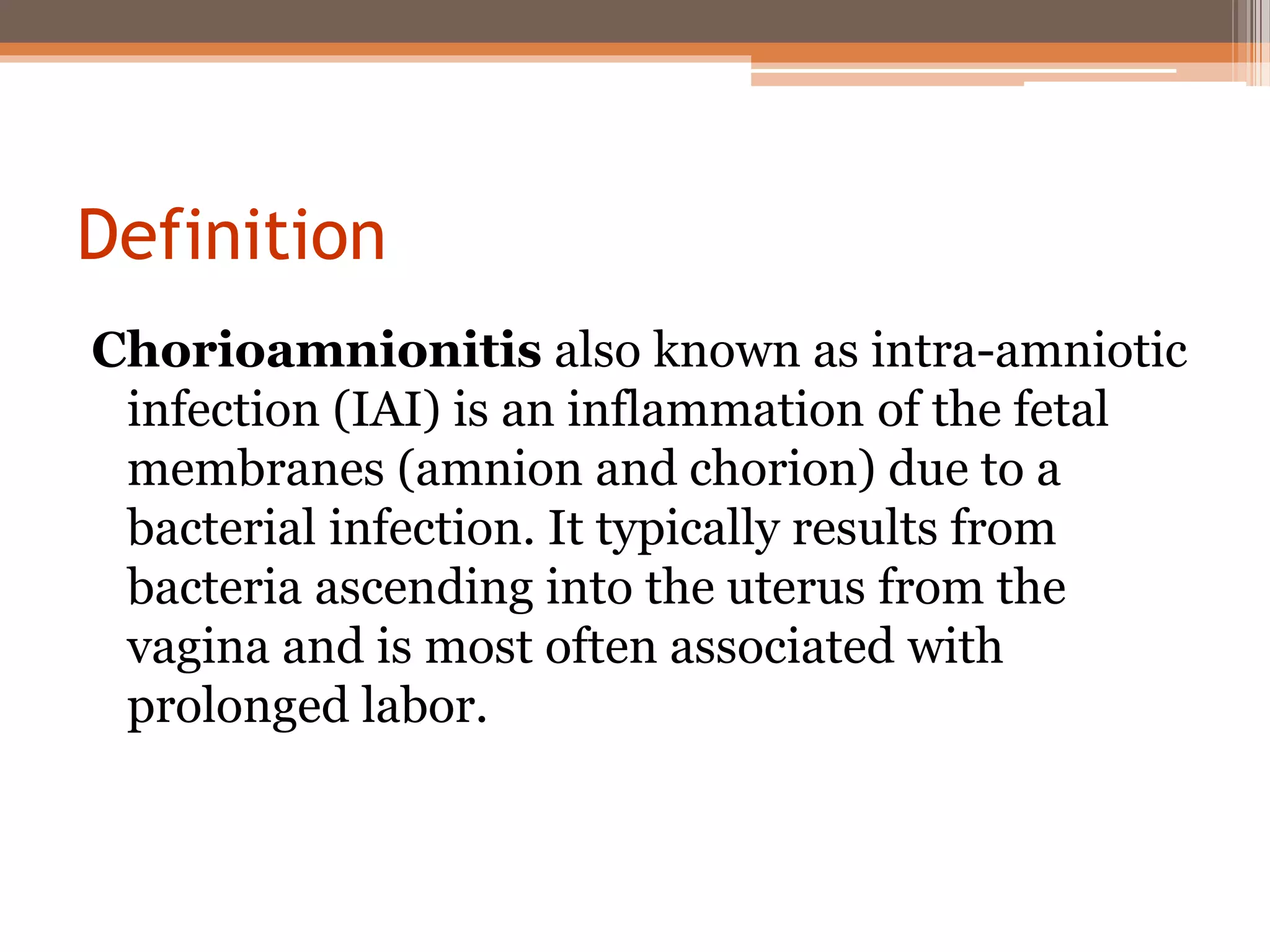
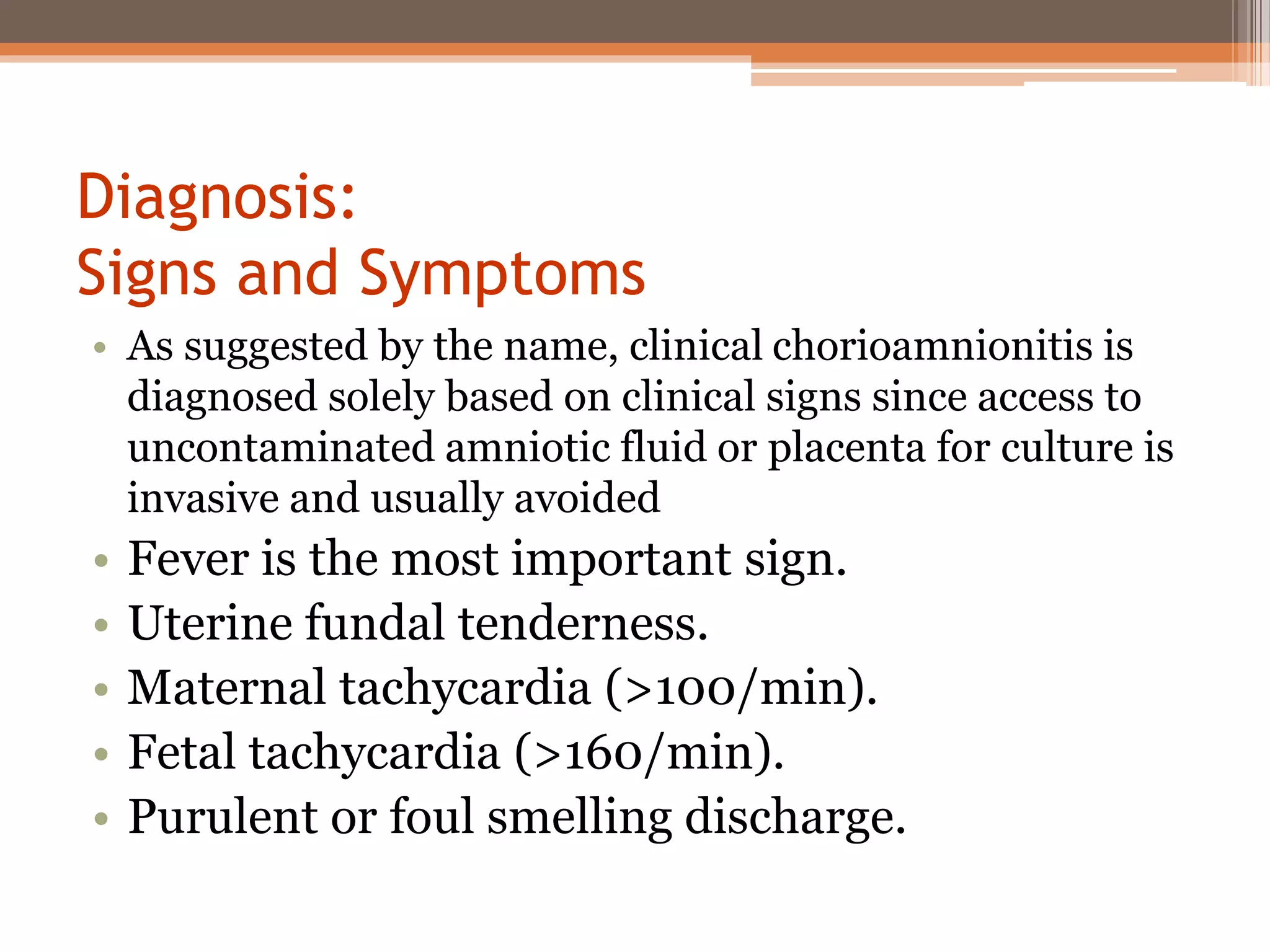
Chorioamnionitis and premature
rupture of membranes (PROM).
Supervised By:
Dr. Sahar Hisham
Done By:
Ali Hussein Rahman
Safa Oday Hatem
Chorioamnionitis
Definition
Chorioamnionitis also known as intra-amniotic
infection (IAI) is an inflammation of the fetal
membranes (amnion and chorion) due to a
bacterial infection. It typically results from
bacteria ascending into the uterus from the
vagina and is most often associated with
prolonged labor.
Epidemiology
• 1–4% of all births in the US are complicated by
chorioamnionitis.
• Chorioamnionitis, complicates as many as 40–70%
of preterm births with premature membrane
rupture or spontaneous labor, and 1–13% of term
births.
• 12% of primary cesarean births at term involve
clinical chorioamnionitis, with the most common
indication for cesarean in these cases being failure
to progress usually after membrane rupture.
Risk Factors
1. Longer duration of membrane rupture.
2. Prolonged labor.
3. Nulliparity.
4. African American ethnicity.
5. Internal monitoring of labor.
6. Multiple vaginal exams.
7. Meconium-stained amniotic fluid.
8. Smoking, alcohol or drug abuse.
9. Immune-compromised states.
10. Epidural anesthesia.
11. Colonization with group B streptococcus.
12. Bacterial vaginosis.
13. Sexually transmissible genital infections and vaginal
colonization with ureaplasma.
Pathogenesis
Routes of infection:
1. Most commonly by retrograde or ascending
infection from the lower genital tract (cervix and
vagina).
Less commonly:
2. Hematogenous/transplacental passage.
3. Iatrogenic infection complicating amniocentesis
or chorionic villous sampling.
4. Anterograde infection from the peritoneum via
the fallopian tubes has also been postulated.
Organisms causing chorioamnionitis
• Chorioamnionitis is a polymicrobial infection
over 65% of positive amniotic fluid cultures
involve two or more organisms. The genital
mycoplasmas, Ureaplasma
urealyticum and Mycoplasma hominis (genital
mycoplasmas), constitute the most frequent
microbes
• Other common isolates in women with
chorioamnionitis include anaerobes such
as Gardnerella vaginalis(25%) and bacteroides
(30%), as well as aerobes including Group B
streptococcus (GBS, 15%) and gram-negative
rods including Escherichia coli (8%).
Diagnosis:
Signs and Symptoms
• As suggested by the name, clinical chorioamnionitis is
diagnosed solely based on clinical signs since access to
uncontaminated amniotic fluid or placenta for culture is
invasive and usually avoided
• Fever is the most important sign.
• Uterine fundal tenderness.
• Maternal tachycardia (>100/min).
• Fetal tachycardia (>160/min).
• Purulent or foul smelling discharge.
• Chorioamnionitis that is subclinical by definition
does not present the above clinical signs but may
manifest as:
• Preterm labor or, more commonly.
• Preterm premature rupture of membranes
(PPROM).
• Premature ROM at term (membrane rupture at
≥37 weeks gestation but prior to onset of uterine
contractions).
• In the absence of other etiologies the
combination of 3 clinical criteria provides a
highly accurate diagnosis of chorioamnionitis.
The presence of risk factors of chorioamnionitis,
especially membrane rupture, further
strengthens the diagnosis.
Laboratory tests:
• i) CBC
• Maternal leucocytosis in approximately 70–90% of cases
of clinical chorioamnionitis. However, isolated
leucocytosis in the absence of other signs or symptoms is
of limited value since it may be induced by several other
conditions including labor and steroid use.
• ii) Other blood tests
• like C-reactive protein (CRP)
iii) Amniotic fluid testing:
• Amniocentesis and culture
Differential diagnosis
• Pyelonephritis
• Influenza
• Appendicitis
• Pneumonia
• Thrombophlebitis
• Round ligament pain
• Colitis
• Connective tissue disorders
• Placental abruption.
Management
• Antibiotics
• Intravenous administration of ampicillin every 6
hours and gentamicin every 8–24 hours until
delivery is the typical regimen.
• If cesarean delivery is performed, clindamycin every
8 hours (or metronidazole) is often added for
anaerobic coverage.
• Optimal treatment should also include
administration of a single intravenous additional
dose of antibiotics after delivery (<5% failure rate)
further oral antibiotic treatment is not beneficial in
most cases.
• i) Supportive measures
• antipyretics (acetaminophen) This is particularly
important in:
•
1)the intrapartum period since fetal acidosis in the
setting of fever has been associated with a marked
increase in the incidence of neonatal
encephalopathy.
2)reducing fetal tachycardia thereby avoiding the
tendency to perform cesarean for a non-reassuring
fetal status.
Complications of chorioamnionitis
• Maternal complications
• Cesarean delivery
• Endomyometritis
• Wound infection
• Pelvic abscess
• Bacteremia
• Postpartum hemorrhage
• Septic shock
• Disseminated intravascular coagululation
• Adult respiratory distress syndrome
• Maternal death
Fetal complications
• Fetal death
• Neonatal sepsis
• Perinatal death,
• Asphyxia,
• Early onset neonatal sepsis,
• Septic shock,
• Pneumonia,
• Intraventricular hemorrhage (IVH),
• Cerebral white matter damage, and
• Cerebral palsy.
prevention
• Prophylactic antibiotics, typically ampicillin and
erythromycin, have been demonstrated in large
clinical trials and systematic reviews to confer
benefits including reduction in a primary
composite of neonatal death, chronic lung
disease or major cerebral abnormality on
ultrasound.
Premature rupture of membranes
(PROM)
• Fetal membranes consist of 2 layers:
• 1. chorion (outer).
• 2. Amnion (inner).
• PROM is spontaneous rupture of membrane any
time beyond 22nd week of pregnancy but before
onset of labour.
• Incidence: 10% of all pregnancies.
Types of PROM
• 1. Term: is rupture of membranes beyond 37th
weeks of gestation but before the onset of labour.
• Incidence is: 8% of all pregnancies.
• 2. Preterm: rupture of membranes before 37
completed weeks of gestation.
• Incidence is: 2 to 3 % of all pregnancies.
Etiology:
• In majority, causes not known.
• Possible causes:
1. . increases friability of the membranes.
2. Decreased tensile strength of membranes.
3. Polyhydramnios.
4. Cervical incompetence.
5. Multiple pregnancies.
6. Infections: e.g. chorioamnionitis, UTI and lower
genital tract infections.
7. Cervical length <2.5 cm.
8. Prior preterm labour.
9. Low BMI (<19 kg/m2).
Diagnosis
• History.
• Patient complains of discharge of clear fluid (liquor)
vaginally.
• Examination:
• Speculum: shows liquor draining through cervical
os.
• Differential diagnosis:
• 1. Hydrorrhoea gravidarum: a state where periodic
watery discharge occurs probably due to successive
decidual glandular secretion.
• 2. incontinence of urine.
• 3. PROM.
Investigations
• Examination of collected fluid from posterior
fornix:
• A. Fern test, crystallization of liquor when dried
on a slide.
• B. Litmus test or Nitrazine paper test for
detection of pH (6 to 6.2)
Hazards of PROM:
• Maternal hazards:
1. Preterm labor.
2. Increased risk of infection.
• Fetal:
1. Cord prolapse.
2. Intrauterine infection.
3. Fetal pulmonary hypoplasia.
4. Neonatal sepsis.
5. Respiratory distress syndrome.
6. Intraventricular hemorrhage.
7. Necrotizing enterocolitis. (NEC).
Management
• Depends on:
• 1. Gestational age of the fetus.
• 2. whether the patient is in labour or not.
• 3. Any evidence of sepsis.
• 4. Prospect of fetal survival in that institution, if
delivery occurs.
Management
• If there is: amnionitis, placental abruption, fetal
death or distress, labour process then a prompt
effective delivery should be done with broad
spectrum intrapartum antibiotics and admit the
baby to the nursery intensive care unit if needed.
• If the above conditions are absent:
• 1. GA < 34 weeks: management aims to continue
for fetal maturity so transfere the patient with
the fetus in utero to a center equipped with
NICU.
• 2. GA 34 weeks and above: wait for spontaneous
onset of labour for 24-48 hrs. if fails then
induction of labour with oxytocin or C/S (for
obstetric reasons).

Chorioamnionitis and PROM - Nahrain University

  • 1.
    Chorioamnionitis and premature ruptureof membranes (PROM). Supervised By: Dr. Sahar Hisham Done By: Ali Hussein Rahman Safa Oday Hatem
  • 2.
  • 3.
    Definition Chorioamnionitis also knownas intra-amniotic infection (IAI) is an inflammation of the fetal membranes (amnion and chorion) due to a bacterial infection. It typically results from bacteria ascending into the uterus from the vagina and is most often associated with prolonged labor.
  • 4.
    Epidemiology • 1–4% ofall births in the US are complicated by chorioamnionitis. • Chorioamnionitis, complicates as many as 40–70% of preterm births with premature membrane rupture or spontaneous labor, and 1–13% of term births. • 12% of primary cesarean births at term involve clinical chorioamnionitis, with the most common indication for cesarean in these cases being failure to progress usually after membrane rupture.
  • 5.
    Risk Factors 1. Longerduration of membrane rupture. 2. Prolonged labor. 3. Nulliparity. 4. African American ethnicity. 5. Internal monitoring of labor. 6. Multiple vaginal exams. 7. Meconium-stained amniotic fluid. 8. Smoking, alcohol or drug abuse. 9. Immune-compromised states. 10. Epidural anesthesia. 11. Colonization with group B streptococcus. 12. Bacterial vaginosis. 13. Sexually transmissible genital infections and vaginal colonization with ureaplasma.
  • 6.
    Pathogenesis Routes of infection: 1.Most commonly by retrograde or ascending infection from the lower genital tract (cervix and vagina). Less commonly: 2. Hematogenous/transplacental passage. 3. Iatrogenic infection complicating amniocentesis or chorionic villous sampling. 4. Anterograde infection from the peritoneum via the fallopian tubes has also been postulated.
  • 8.
    Organisms causing chorioamnionitis •Chorioamnionitis is a polymicrobial infection over 65% of positive amniotic fluid cultures involve two or more organisms. The genital mycoplasmas, Ureaplasma urealyticum and Mycoplasma hominis (genital mycoplasmas), constitute the most frequent microbes
  • 9.
    • Other commonisolates in women with chorioamnionitis include anaerobes such as Gardnerella vaginalis(25%) and bacteroides (30%), as well as aerobes including Group B streptococcus (GBS, 15%) and gram-negative rods including Escherichia coli (8%).
  • 11.
    Diagnosis: Signs and Symptoms •As suggested by the name, clinical chorioamnionitis is diagnosed solely based on clinical signs since access to uncontaminated amniotic fluid or placenta for culture is invasive and usually avoided • Fever is the most important sign. • Uterine fundal tenderness. • Maternal tachycardia (>100/min). • Fetal tachycardia (>160/min). • Purulent or foul smelling discharge.
  • 12.
    • Chorioamnionitis thatis subclinical by definition does not present the above clinical signs but may manifest as: • Preterm labor or, more commonly. • Preterm premature rupture of membranes (PPROM). • Premature ROM at term (membrane rupture at ≥37 weeks gestation but prior to onset of uterine contractions).
  • 13.
    • In theabsence of other etiologies the combination of 3 clinical criteria provides a highly accurate diagnosis of chorioamnionitis. The presence of risk factors of chorioamnionitis, especially membrane rupture, further strengthens the diagnosis.
  • 14.
    Laboratory tests: • i)CBC • Maternal leucocytosis in approximately 70–90% of cases of clinical chorioamnionitis. However, isolated leucocytosis in the absence of other signs or symptoms is of limited value since it may be induced by several other conditions including labor and steroid use. • ii) Other blood tests • like C-reactive protein (CRP) iii) Amniotic fluid testing: • Amniocentesis and culture
  • 15.
    Differential diagnosis • Pyelonephritis •Influenza • Appendicitis • Pneumonia • Thrombophlebitis • Round ligament pain • Colitis • Connective tissue disorders • Placental abruption.
  • 16.
    Management • Antibiotics • Intravenousadministration of ampicillin every 6 hours and gentamicin every 8–24 hours until delivery is the typical regimen. • If cesarean delivery is performed, clindamycin every 8 hours (or metronidazole) is often added for anaerobic coverage. • Optimal treatment should also include administration of a single intravenous additional dose of antibiotics after delivery (<5% failure rate) further oral antibiotic treatment is not beneficial in most cases.
  • 17.
    • i) Supportivemeasures • antipyretics (acetaminophen) This is particularly important in: • 1)the intrapartum period since fetal acidosis in the setting of fever has been associated with a marked increase in the incidence of neonatal encephalopathy. 2)reducing fetal tachycardia thereby avoiding the tendency to perform cesarean for a non-reassuring fetal status.
  • 18.
    Complications of chorioamnionitis •Maternal complications • Cesarean delivery • Endomyometritis • Wound infection • Pelvic abscess • Bacteremia • Postpartum hemorrhage • Septic shock • Disseminated intravascular coagululation • Adult respiratory distress syndrome • Maternal death
  • 19.
    Fetal complications • Fetaldeath • Neonatal sepsis • Perinatal death, • Asphyxia, • Early onset neonatal sepsis, • Septic shock, • Pneumonia, • Intraventricular hemorrhage (IVH), • Cerebral white matter damage, and • Cerebral palsy.
  • 20.
    prevention • Prophylactic antibiotics,typically ampicillin and erythromycin, have been demonstrated in large clinical trials and systematic reviews to confer benefits including reduction in a primary composite of neonatal death, chronic lung disease or major cerebral abnormality on ultrasound.
  • 21.
    Premature rupture ofmembranes (PROM)
  • 22.
    • Fetal membranesconsist of 2 layers: • 1. chorion (outer). • 2. Amnion (inner). • PROM is spontaneous rupture of membrane any time beyond 22nd week of pregnancy but before onset of labour. • Incidence: 10% of all pregnancies.
  • 23.
    Types of PROM •1. Term: is rupture of membranes beyond 37th weeks of gestation but before the onset of labour. • Incidence is: 8% of all pregnancies. • 2. Preterm: rupture of membranes before 37 completed weeks of gestation. • Incidence is: 2 to 3 % of all pregnancies.
  • 24.
    Etiology: • In majority,causes not known. • Possible causes: 1. . increases friability of the membranes. 2. Decreased tensile strength of membranes. 3. Polyhydramnios. 4. Cervical incompetence. 5. Multiple pregnancies. 6. Infections: e.g. chorioamnionitis, UTI and lower genital tract infections. 7. Cervical length <2.5 cm. 8. Prior preterm labour. 9. Low BMI (<19 kg/m2).
  • 25.
    Diagnosis • History. • Patientcomplains of discharge of clear fluid (liquor) vaginally. • Examination: • Speculum: shows liquor draining through cervical os. • Differential diagnosis: • 1. Hydrorrhoea gravidarum: a state where periodic watery discharge occurs probably due to successive decidual glandular secretion. • 2. incontinence of urine. • 3. PROM.
  • 26.
    Investigations • Examination ofcollected fluid from posterior fornix: • A. Fern test, crystallization of liquor when dried on a slide. • B. Litmus test or Nitrazine paper test for detection of pH (6 to 6.2)
  • 27.
    Hazards of PROM: •Maternal hazards: 1. Preterm labor. 2. Increased risk of infection. • Fetal: 1. Cord prolapse. 2. Intrauterine infection. 3. Fetal pulmonary hypoplasia. 4. Neonatal sepsis. 5. Respiratory distress syndrome. 6. Intraventricular hemorrhage. 7. Necrotizing enterocolitis. (NEC).
  • 28.
    Management • Depends on: •1. Gestational age of the fetus. • 2. whether the patient is in labour or not. • 3. Any evidence of sepsis. • 4. Prospect of fetal survival in that institution, if delivery occurs.
  • 29.
    Management • If thereis: amnionitis, placental abruption, fetal death or distress, labour process then a prompt effective delivery should be done with broad spectrum intrapartum antibiotics and admit the baby to the nursery intensive care unit if needed.
  • 30.
    • If theabove conditions are absent: • 1. GA < 34 weeks: management aims to continue for fetal maturity so transfere the patient with the fetus in utero to a center equipped with NICU. • 2. GA 34 weeks and above: wait for spontaneous onset of labour for 24-48 hrs. if fails then induction of labour with oxytocin or C/S (for obstetric reasons).