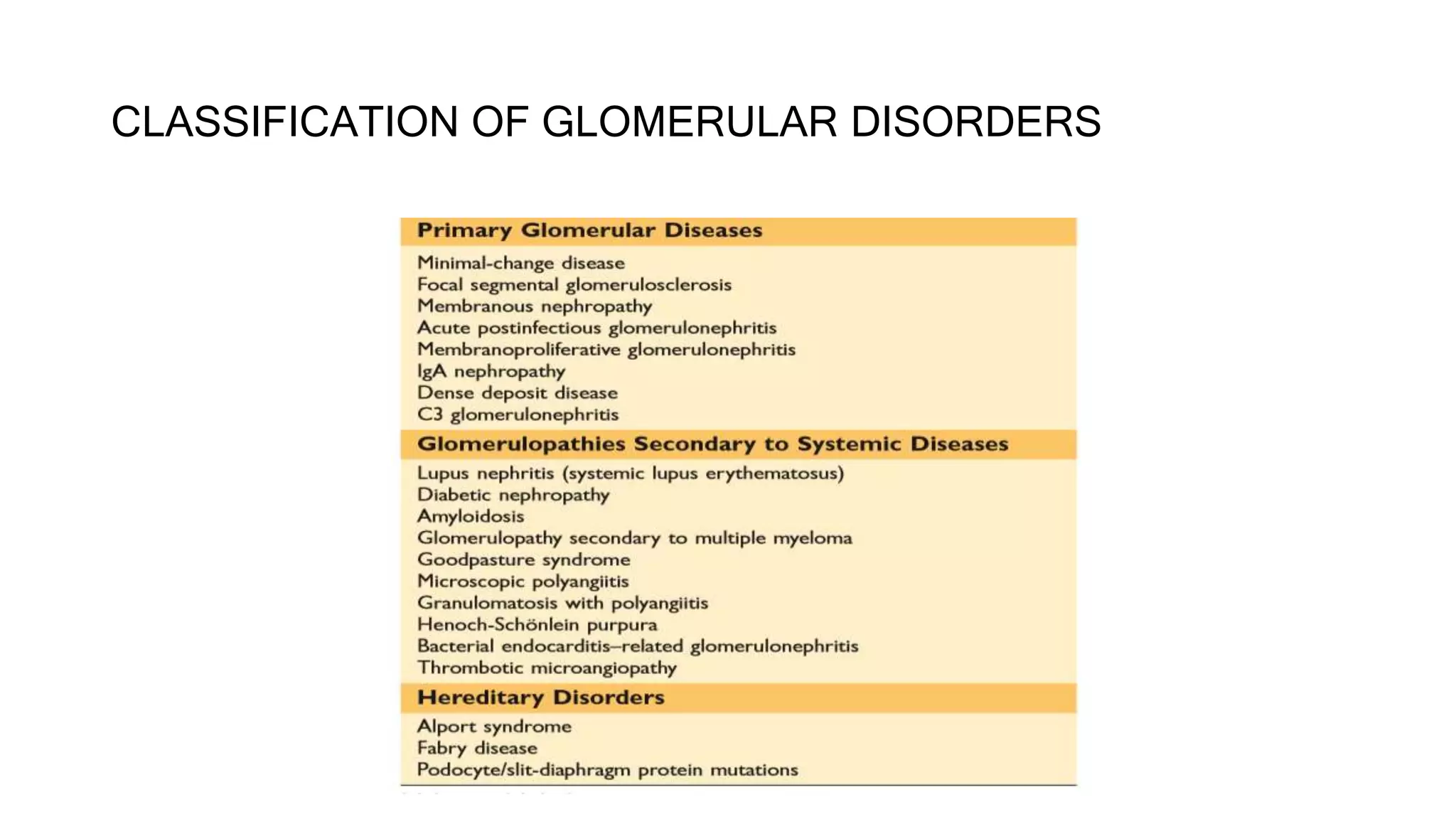
ACUTE NEPHRITIC
SYNDROMES
GUIDED BY
DR.M.MAYYA
ASST. PROFESSOR
DEPT OF NEPHROLOGY
KIMS HUBBALLI
PRESENTED BY
DR.SURYAKANTH
POST GRADUATE STUDENT
KIMS HUBBALLI
GLOMERULUS
Pathogenisis
There are many forms of glomerular disease with pathogenesis variably linked to the
presence of
• Genetic mutations,
• Infection,
• Toxin exposure,
• Autoimmunity,
• Atherosclerosis,
• Hypertension,
• Emboli,
• Thrombosis
• Diabetes mellitus.
• Even after careful study, however, the cause often remains unknown, and the lesion
is called idiopathic.
IMMUNE MECHANISMS
• Immune mechanisms underlie most types of primary glomerular
diseases and many of the secondary glomerular diseases.
• Two mechanisms of antibody deposition in the glomerulus have been
established:
• (1) Deposition of circulating antigen- antibody complexes in the
glomerular capillary wall or mesangium, and
• (2) Antibodies reacting in situ within the glomerulus.
IMMUNE MECHANISMS
Progression of glomerular disease
• Persistent glomerulonephritis that worsens renal function is always
accompanied by
1. Interstitial nephritis,
2. Renal fibrosis, and
3. Tubular atrophy.
CLASSIFICATION OF GLOMERULAR DISORDERS
•ACUTE NEPHRITIC SYNDROMES
Introduction
Acute nephritic syndromes classically present with the following:
• Hypertension
• Hematuria
• Red blood cell casts
• Subnephrotic proteinuria (1- 2 g/24 h )
• Fluid retention
• Rise in serum creatinine
• Reduction in GFR
ACUTE NEPHRITIC SYNDROMES
SPECRTUM OF PRESENTATION
Causes according to age
CHILDREN AND ADOLESCENTS
• Post-streptococcal
glomerulonephritis
• MPGN
• Cresentric GN
• IgA nephropathy
• Lupus Nephritis
• Hemolytic uremic syndrome
• Henoch-Schönlein purpura
ADULTS
• IgA Nephropathy
• Membranoproliferative GN
• SLE or lupus nephritis
• ANCA associated
• Rapidly progressive (crescentic)
glomerulonephritis
• Hepatitis B or C
• Infective endocarditis
• Abdominal abscesses
• Viral diseases such as mononucleosis, measles,
mumps
INFECTIOUS CAUSES OF
ACUTE NEPHRITIS
• Infectious syndromes
• Specific bacterial diseases
• Mycobacterial, rickettsial,
mycoplasmal, chlamydial, and
spirochetal diseases
• Fungal infections
• Viruses
• Parasitic infestations
NON-INFECTIOUS CAUSES
OF ACUTE NEPHRITIS
Immune mediated glomerulonephritis
• Immune complex GN
• IgAN,HSP,LN…
• ANTI-GBM GN
• GPS,Anti-GBM
• ANCA associated GN
• MP,WG,CSS
• MPGN
SITE OF INJURY IN ACUTE
NEPHRITIC SYNDROME
CLINICAL APPROACH
CLINICAL APPROACH
(POST-INFECTIOUS)
On the background of Acute
Nephritis features….
• 5 and 12 years(90%) and >40(10%)
• boys > girls
• Pharyngitis(1-3)
• Impetigo(2-6)
• may be severe enough to appear as RPGN
• Constitutional symptoms - as many as 50% of
cases
IVDU
Cardiac
murmur
Dental
abscess.
Osteomyelitis,
deep seated
abscesses
Treated
hydrocephalus
PSGN Endocarditis ShuntAbscess
RAPIDLY PROGRESSIVE
GLOMERULONEPHRITIS
IMMUNE COMPLEX GN ANTI-GBM GN ANCA ASSOCIATED GN
IMMUNE COMPLEX GN
On the background of Acute
Nephritis features….
• 20s and 30s
• men > women
• asymptomatic
microscopic
hematuria most
often seen in
adults
• No vasulitis
• Asso with
systemic
diseases
• <20 years of
age(4-7)
• Macroscopic
haematuria -
time of
infections
• Purpuric
rash,
arthritis,
abdominal
pain
(Systemic
vasulitis)
• Young women
in 20 and 30s
• Arthralgia,
photosensitive
skin rash,
pleurisy, and
pericarditis
• 5 - 12 years
• boys > girls
• Acute Strept
infection(1-6
weeks before
presentation)
IgA
Nephropathy
HSP
Lupus
Nephritis
Acute PSGN MPGN
ANTI-GBM GN
On the background of Acute
Nephritis features….
Young men(late 20s)
both sexes
(50s and 70s)
With lung
haemorrhage
GPS
20s:Young men;
50s and 60s:both sexes
Without lung
haemorrhage
Anti-GBM GN
• ANCA ASSOCIATED GN
• With the background of
Acute Nephritis
features….
• 50s/60s
• men = women
• Weight loss,
malaise, URT/LRT
symptoms,
arthritis, palpable
purpura
• no Asthma
• 50s/60s
• men = women
• Weight loss,
malaise, URT/LRT
symptoms,
arthritis, palpable
purpura
• no Asthma
• 20-40
• Males>female
s
• Asthma
• Neuropthy
Microscopic
Polyangitis
Granulomatosis with
polyangitis CSS
LABORATORY EVALUATIONS
DARK URINE
DIPSTICK FOR
HEMENEGATIVE
DRUGS
FOOD DYES
MELANIN
POSITIVE
GROSS
HEMATURIA
MICROSCOPIC
HEMATURIA
TRAUMA
PRESENT
TRAUMA
ABSENT
IMAGING OF
ABDOMEN
AND PELVIS
CYSTOSCOPY
PAINFUL PAINLESS
URINE
CALCIUM
URINE CULTURE
RENAL CALCULI
U.CA
FAMILIAL
NEPHRITIS
HSP
EUMORPHIC
RBCs
DYSMORPHIC
RBCs
U.CA
R/O MEATAL
STENOSIS
W/U FOR
GLOMERULO
NEPHRITIS
NO
RBCs
HB,MB
Photomicrograph of urine sediment
with a
red cell cast
Phase contrast micrograph
showing dysmorphic red cells
in urine sediment
Scanning electron micrograph
showing dysmorphic red cells
in urine sediment
INVESTIGATIONS
POST-INFECTIOUS GN
• Anti
streptolysin
titre
• anti-DNAse B
• Throat swab or
skin swab
• 2D ECHO
• TEE
• USG
• VP shunt
evidence
PSGN Endocarditis ShuntAbscess
OTHER SPECIFIC
INVESTIGATIONS
IMMUNE COMPLEX GN
Glomerular immune complex
localisatIon with granular capillary
wall and/or mesangeal staining
Raised serum
IgA in 50% of
cases,
antinuclear
antibody/
anti-ds DNA,
anticardiolipin
antibody
antistreptolysi
n titre
IF STUDIES
AND
COMPLEMENT
LEVELS
IgA
Nephropathy
Lupus
Nephritis Acute PSGN MPGN
OTHER SPECIFIC
INVESTIGATIONS
ANTI-GBM GN
Circulating Anti-GBM antibodies
with linear GBM staining for IgG
• With evidence of lung
haemorrhage
GPS
• Without lung
haemorrhage
Anti-GBM GN
OTHER SPECIFIC
INVESTIGATIONS
ANCA ASSOCIATED GN
Circulating ANCA with paucity
of glomerular
immunoglobulin staining
Normal Complement
Vasculitis with no
granuloma
MPO
ANCA
Granulomas
RP3
Eosinophilia
and
granuloma
MPO
Microscopic
Polyangitis
Granulomatosis
polyangitis
CSS
OTHER SPECIFIC
INVESTIGATIONS
1. ACUTE POST-STREPTOCOCCAL
GLOMERULONEPHRITIS
• AGN that follows an infection with a nephritogenic strain of group A beta
hemolytic streptococci.
• The classic example of the acute nephritic syndrome.
• Skin and throat infections with particular M types of streptococci (nephritogenic
strains) antedate glomerular disease;
• M types 47, 49, 55, 2, 60, and 57 are seen following impetigo and
• M types 1, 2, 4, 3, 25, 49, and 12 with pharyngitis.
Nelson Textbook of Pediatrics, 7th Edition
Streptococcal infection of the throat or skin ( impetigo)
Poststreptococcal glomerulonephritis due to impetigo develops 2–6 weeks
after skin infection and 1–3 weeks after streptococcal pharyngitis
LM AND EM OF PSGN
IF OF PSGN
PSGN
Renal biopsy shows
1. Hypercellularity of mesangial
and endothelial cells,
2. Glomerular infiltrates of
polymorphonuclear
leukocytes,
3. Granular subendothelial
immune deposits of IgG, IgM,
C3, C4, and C5–9, and
subepithelial deposits (which
appear as “humps).
Epidemiology
• Peak incidence - age 5-12 y/o, uncommon <3y/o.
• Male : female ratio is 2 : 1.
Clinical course
• Spontaneous improvement typically begins within 1 wk
• Resolution of edema in 5-10 days
• Hypertension in 3-4 wk
• Proteinuria -- normalize by 4-6 wk
• Urinalysis may be abnormal (persistent microscopic hematuria) upto a year
Antibodies to streptococcal antigen(s)
• Anti-streptolysin O titer (ASOT) >333 TOD Units
• COMMONLY elevated after a pharyngeal infection but
• RARELY increases after streptococcal skin infections
• Anti deoxyribonuclease (DNase) B antibodies
• best single antibody titer to document cutaneous streptococcal infection
• Anti-hyaluronidase antibodies
• Anti-streptokinase antibodies
IndIcatIons for renal BIopsy
• Severe acute renal failure requiring dialysis.
• Features suggesting non post-infectious agn as the cause of acute nephritis.
• Absence of the latent period between streptococcal infection and acute
glomerulonephritis
• Normal complement levels initially in early course
• Persistence of low C3 beyond 6-12 wks
• Delayed resolution
• Oliguria > 2 weeks
• Azotaemia > 3 weeks
• Gross haematuria > 3 weeks
• Persistent proteinuria > 6 months
Management
• Treatment is supportive
• control of hypertension, edema, and dialysis as needed.
• Strict monitoring – I/O/weight/BP
• Penicillin V for 10 days to eliminate β - haemolytic streptococcal infection (give
erythromycin if penicillin is contraindicated) patients,
• Diet – no added salt to diet, Protein restriction is unnecessary.
• No role for immunosuppressive therapy(even in the setting of crescents)
Management…
FOLLOW UP:
• Recheck serum complement at 6-8 wks.
• Annually check BP, renal function test and urinalysis every 1-3 month for 1 yr
then yearly.
PROGNOSIS
• Short term outcome: Excellent, mortality <0.5%.
• Long term outcome: 1.8% (1-3%) of children develop chronic kidney disease
• Complete resolution  within 3–6 weeks
• Complete recovery
• >95% of children
• 60% of adult.
2. infection-related glomerulonephritis
• IRGN is an immune-mediated renal injury, which is caused due to
non-renal infections, which are most commonly of bacterial origin.
• male:female ratio ranging from 1.4:1 to 3:1.
• In the pediatric age group, the sites of infection are usually the throat
or skin; however,
• the adult population shows a much-varied site of preceding infection
including upper respiratory tract, skin, lung, heart, urinary tract,
teeth/oral mucosa, bone, and deep-seated visceral, or somatic
abscesses.
LM AND IF in IRGN
• The most common LM finding on renal biopsy is diffuse proliferative and
exudative GN with abundant intracapillary neutrophils.
• On IF, C3 dominant or codominant glomerular staining is most commonly seen.
• It may show various patterns, which have been typically described as “starry sky
pattern” or the “garland pattern” or the mesangial pattern depending on
whether the capillary wall or mesangium is stained
Irgn…
• Renal biopsy plays an important role in the assessment of prognosis
and underlying GN and should be considered early, especially in
patients with an initial requirement of RRT or severe
proteinuria/persistent proteinuria.
3. SUBACUTE BACTERIAL ENDOCARDITIS
• Endocarditis-associated glomerulonephritis is typically a complication of SBE,
particularly in patients who remain untreated for a long time, have negative
blood cultures, or have right-sided endocarditis.
• Common comorbidities are valvular heart disease, intravenous drug use,
hepatitis C, and diabetes mellitus.
Grossly, the kidneys in SBE have subcapsular
hemorrhages with a “flea-bitten” appearance,
Renal biopsy reveals focal proliferation around
foci of necrosis associated with abundant
mesangial, subendothelial, and subepithelial
infiltrates.
4. LUPUS NEPHRITIS
• Lupus nephritis is a common and serious complication of systemic lupus
erythematosus (SLE) and most severe in African-American female adolescents.
• Thirty to 50% of patients will have clinical manifestations of renal disease at the
time of diagnosis, and 60% of adults and 80% of children develop renal
abnormalities at some point in the course of their disease.
Classification of lupus nephritis
Lupus nephritis class I
• Minimal Mesangial Immune Deposits
Lupus nephritis class II
• Mesangial Proliferation.
• Patients with ISN/RPS class I and II biopsies have an excellent renal
prognosis and need no therapy directed to the kidney.
Lupus nephritis class III
Focal Nephritis
TREATMENT : Short course of high-dose corticosteroid therapy or a brief
course of other immunosuppressive agents.
Lupus nephritis class Iv
• Global, Diffuse Proliferative Lesions
TREATMENT : inducing a remission with administration of highdose steroids
and either cyclophosphamide or mycophenolate mofetil for 2–6 months,
followed by maintenance therapy with lower doses of the same,
Lupus nephritis class V
• Thickening of GBM with subepithelial deposits.
• TREATMENT: Therapy with inhibitors of the renin-angiotensin system
also may attenuate the proteinuria.
Lupus nephritis class VI
• ISN/RPS class VI, advanced sclerosing LN or end-stage LN, is reserved for
biopsies with more than 90% of the glomeruli sclerotic and no residual activity.
6.ANTIGLOMERULAR BASEMENT MEMBRANE
DISEASE
• Patients who develop autoantibodies directed against glomerular basement
antigens frequently develop a glomerulonephritis termed antiglomerular basement
membrane (anti-GBM) disease.
• When they present with lung hemorrhage and glomerulonephritis, they have a
pulmonary- renal syndrome called Goodpasture’s syndrome.
• The target epitopes for this autoimmune disease lie in the quaternary structure
of a3 NC1 domain of collagen IV.
Goodpasture’s syndrome
• Goodpasture’s syndrome appears in two age groups: in young men in
their late twenties and in men and women in their sixties and
seventies.
• Disease in the younger age group is usually explosive, with
hemoptysis, a sudden fall in hemoglobin, fever, dyspnea, and
hematuria.
• Presentation with oliguria is often associated with a particularly bad
outcome.
ANTI-GBM GN
ANTI-GBM GN…
• The performance of an urgent kidney biopsy is important in suspected cases of
Goodpasture’s syndrome to confirm the diagnosis and assess prognosis.
• Renal biopsy - focal or segmental necrosis that later, with aggressive destruction
of the capillaries by cellular proliferation, leads to crescent formation in
Bowman’s space .
• As these lesions progress, there is concomitant interstitial nephritis with fibrosis
and tubular atrophy.
5. IgA NEPHROPATHY
• It is the most common form of glomerulonephritis
worldwide.
• IgA nephropathy, characterized by mesangial deposits of
IgAcontaining immune complexes.
• Recurrent asymptomatic hematuria is the most common
clinical presentation.
• It commonly affects children and young adults and has
a variable course.
LM IN IGAN
EM AND IF IN IGAN
IgAN… Clinical course
• Clues to an increased risk of progression(loss of renal function)
• Onset in old age
• Heavy proteinuria
• Hypertension
• Extent of glomerulosclerosis on biopsy
• Absence of episodes of macroscopic hematuria,
• Male
• The hematuria typically lasts for several days and then subsides, only to return every few months.
The subsequent course is highly variable
• Many patients maintain normal renal function for decades.
Prognosis
• IgA nephropathy is a benign disease for the majority of patients, and 5–30% of
patients may go into a complete remission, with others having hematuria but
well preserved renal function.
• In the minority of patients who have progressive disease, progression is slow, with
renal failure seen in only 25–30% of patients with IgA nephropathy over 20–
25 years.
IgA Nephropathy- TREATMENT
• Angiotensin-converting enzyme (ACE) inhibitors in patients with
proteinuria or declining renal function.
• When presenting as RPGN, patients typically receive:
• Steroids
• Cytotoxic agents
7. ANCA SMALL-VESSEL VASCULITIS
• Granulomatosis with Polyangiitis
• Microscopic Polyangiitis
• Churg-Strauss Syndrome
7.1Granulomatosis with Polyangitis
• Clinical features
Fever,
Purulent rhinorrhea,
Nasal ulcers,
Sinus pain,
Polyarthralgias/arthritis,
Cough,
Hemoptysis,
 Shortness of breath,
 Microscopic hematuria, and
proteinuria;
Chest x-ray often reveals nodules and
persistent infiltrates, sometimes with
cavities.
Biopsy of involved tissue will show a small-
vessel vasculitis and adjacent noncaseating
granulomas
Management of granulomatosis with
polyangitis
• Treatment :
• Induction of remission :
• 1 Rituximab Plus Prednisone
• 2.Cyclophopshamide And Prednsione
• Maintainence of remission :
• 1.Azathioprine (2mg/kg/day orally)
• 2. Methotrexate (20-25mg/wk PO/IM)
• 3.Rituximab (500mg /week)
7.2 Microscopic Polyangitis
• Clinically, these patients look somewhat similar to those with granulomatosis
with polyangitis, except they rarely have significant lung disease or destructive
sinusitis.
• The distinction is made on biopsy, where the vasculitis in microscopic polyangiitis
is without granulomas.
• Some patients will also have injury limited to the capillaries and venules.
• The 5-year survival rate for patients with treated microscopic polyangiitis
is 74%.
• Treatment is same as Granulomatosis With Polyangitis
• Life-threatening disease should be treated with the combination of
prednisone and daily cyclophosphamide or rituximab.
Management of microscopic polyangitis
7.3 Churg-Strauss Syndrome
Small-vessel vasculitis is associated with
• Peripheral eosinophilia,
• Cutaneous purpura,
• Mononeuritis,
• Asthma, and
• Allergic rhinitis
Lung inflammation, including fleeting cough and pulmonary infiltrates are
common
Renal biopsy : Small-vessel vasculitis and focal segmental necrotizing
glomerulonephritis.
Management of css
• The prognosis of untreated CSS is poor,with a reported 5-year survival of
25%.
• Glucocorticoids alone appear to be effective in many patients.
• In patients who present with fulminant multisystem disease, particularly
cardiac involvement, the treatment of choice is a combined regimen of
daily cyclophosphamideand prednisone followed by azathioprine or
methotrexate
• Mepoluzimab is FDA approved for the treatment of severe eosinophilic
asthma and may particularly have a role in thesettingof relapsing resistant
asthma in eosinophilic granulomatosis.
8.MPGN
• MPGN is sometimes called mesangiocapillary glomerulonephritis or lobar
glomerulonephritis.
• It is an immune-mediated glomerulonephritis characterized by thickening of the
GBM with mesangioproliferative changes;
Types of MPGN
LM OF MPGN
IF IN MPGN
C3 GLOMERULOPATHIES
(DENSE DEPOSIT DISEASE AND C3
GLOMERULONEPHRITIS)
• EPIDEMIOLOGY
• Dense deposit disease accounts for about 25% of MPGN in children but is much
less common in adults.
• The large majority of patients are children between the ages of 8 and 16.
• It is estimated to affect 2 to 3 persons per million.
DDD
• PATHOLOGY
• The term dense deposit disease emphasizes the pathognomonic feature
of discontinuous electron-dense bands within the GBM .
• These are accompanied by spherical to irregular mesangial dense
deposits and occasional subendothelial and subepithelial deposits,
some of which resemble the “humps” seen in postinfectious GN
Em in ddd
IF IN DDD
CLINICAL FEATURES
• Patients with dense deposit disease may have hematuria, proteinuria,
or both.
• They may have a nephrotic or acute nephritic syndrome.
• At least a third of patients have all of the components of the
nephrotic syndrome on presentation.
• Kidney dysfunction occurs in at least half of cases and is more
common among adults than in children.
• When present at the outset of disease, kidney dysfunction portends a
poor prognosis.
Management
• Predictors of outcome
• Renal dysfunction – Serum Creatinine, GFR
• Proteinuria
• DDD- older age at diagnosis - an independent predictor of ESRD
• C3GN may have a more benign course than patients with DDD
Treatment
• Targeted therapies have not proven to be universally beneficial –
• Heterogeneity of the C3 glomerulopathies
• Control of alternative complement pathway activity
• Replacement of factor H - viable option
• Patients with genetic defects of inhibitory proteins of the alternative
complement pathway - replacement of factors
SUMMARY OF PRIMARY
GLOMERULAR DISEASES
REFERENCES
• Harrisons Principles of Internal Medicine – 20th Edition
• Brenner and Rector – The Kidney – 9th Edition
• Nelson Textbook of Pediatrics, 7th Edition
• Robins and Cotran pathological basis of disease - 9th edition
• CMDT-2019
•Thank you one and all

Glomerulonephritis - Acute Nephritic Syndromes

  • 1.
    ACUTE NEPHRITIC SYNDROMES GUIDED BY DR.M.MAYYA ASST.PROFESSOR DEPT OF NEPHROLOGY KIMS HUBBALLI PRESENTED BY DR.SURYAKANTH POST GRADUATE STUDENT KIMS HUBBALLI
  • 2.
  • 3.
    Pathogenisis There are manyforms of glomerular disease with pathogenesis variably linked to the presence of • Genetic mutations, • Infection, • Toxin exposure, • Autoimmunity, • Atherosclerosis, • Hypertension, • Emboli, • Thrombosis • Diabetes mellitus. • Even after careful study, however, the cause often remains unknown, and the lesion is called idiopathic.
  • 4.
    IMMUNE MECHANISMS • Immunemechanisms underlie most types of primary glomerular diseases and many of the secondary glomerular diseases. • Two mechanisms of antibody deposition in the glomerulus have been established: • (1) Deposition of circulating antigen- antibody complexes in the glomerular capillary wall or mesangium, and • (2) Antibodies reacting in situ within the glomerulus.
  • 5.
  • 7.
    Progression of glomerulardisease • Persistent glomerulonephritis that worsens renal function is always accompanied by 1. Interstitial nephritis, 2. Renal fibrosis, and 3. Tubular atrophy.
  • 8.
  • 9.
  • 10.
    Introduction Acute nephritic syndromesclassically present with the following: • Hypertension • Hematuria • Red blood cell casts • Subnephrotic proteinuria (1- 2 g/24 h ) • Fluid retention • Rise in serum creatinine • Reduction in GFR
  • 11.
  • 12.
  • 13.
    Causes according toage CHILDREN AND ADOLESCENTS • Post-streptococcal glomerulonephritis • MPGN • Cresentric GN • IgA nephropathy • Lupus Nephritis • Hemolytic uremic syndrome • Henoch-Schönlein purpura ADULTS • IgA Nephropathy • Membranoproliferative GN • SLE or lupus nephritis • ANCA associated • Rapidly progressive (crescentic) glomerulonephritis • Hepatitis B or C • Infective endocarditis • Abdominal abscesses • Viral diseases such as mononucleosis, measles, mumps
  • 14.
    INFECTIOUS CAUSES OF ACUTENEPHRITIS • Infectious syndromes • Specific bacterial diseases • Mycobacterial, rickettsial, mycoplasmal, chlamydial, and spirochetal diseases • Fungal infections • Viruses • Parasitic infestations NON-INFECTIOUS CAUSES OF ACUTE NEPHRITIS Immune mediated glomerulonephritis • Immune complex GN • IgAN,HSP,LN… • ANTI-GBM GN • GPS,Anti-GBM • ANCA associated GN • MP,WG,CSS • MPGN
  • 15.
    SITE OF INJURYIN ACUTE NEPHRITIC SYNDROME
  • 16.
  • 17.
    CLINICAL APPROACH (POST-INFECTIOUS) On thebackground of Acute Nephritis features…. • 5 and 12 years(90%) and >40(10%) • boys > girls • Pharyngitis(1-3) • Impetigo(2-6) • may be severe enough to appear as RPGN • Constitutional symptoms - as many as 50% of cases IVDU Cardiac murmur Dental abscess. Osteomyelitis, deep seated abscesses Treated hydrocephalus PSGN Endocarditis ShuntAbscess
  • 18.
  • 19.
    IMMUNE COMPLEX GN Onthe background of Acute Nephritis features…. • 20s and 30s • men > women • asymptomatic microscopic hematuria most often seen in adults • No vasulitis • Asso with systemic diseases • <20 years of age(4-7) • Macroscopic haematuria - time of infections • Purpuric rash, arthritis, abdominal pain (Systemic vasulitis) • Young women in 20 and 30s • Arthralgia, photosensitive skin rash, pleurisy, and pericarditis • 5 - 12 years • boys > girls • Acute Strept infection(1-6 weeks before presentation) IgA Nephropathy HSP Lupus Nephritis Acute PSGN MPGN
  • 20.
    ANTI-GBM GN On thebackground of Acute Nephritis features…. Young men(late 20s) both sexes (50s and 70s) With lung haemorrhage GPS 20s:Young men; 50s and 60s:both sexes Without lung haemorrhage Anti-GBM GN
  • 21.
    • ANCA ASSOCIATEDGN • With the background of Acute Nephritis features…. • 50s/60s • men = women • Weight loss, malaise, URT/LRT symptoms, arthritis, palpable purpura • no Asthma • 50s/60s • men = women • Weight loss, malaise, URT/LRT symptoms, arthritis, palpable purpura • no Asthma • 20-40 • Males>female s • Asthma • Neuropthy Microscopic Polyangitis Granulomatosis with polyangitis CSS
  • 22.
  • 23.
    DARK URINE DIPSTICK FOR HEMENEGATIVE DRUGS FOODDYES MELANIN POSITIVE GROSS HEMATURIA MICROSCOPIC HEMATURIA TRAUMA PRESENT TRAUMA ABSENT IMAGING OF ABDOMEN AND PELVIS CYSTOSCOPY PAINFUL PAINLESS URINE CALCIUM URINE CULTURE RENAL CALCULI U.CA FAMILIAL NEPHRITIS HSP EUMORPHIC RBCs DYSMORPHIC RBCs U.CA R/O MEATAL STENOSIS W/U FOR GLOMERULO NEPHRITIS NO RBCs HB,MB
  • 24.
    Photomicrograph of urinesediment with a red cell cast Phase contrast micrograph showing dysmorphic red cells in urine sediment Scanning electron micrograph showing dysmorphic red cells in urine sediment
  • 26.
  • 27.
    POST-INFECTIOUS GN • Anti streptolysin titre •anti-DNAse B • Throat swab or skin swab • 2D ECHO • TEE • USG • VP shunt evidence PSGN Endocarditis ShuntAbscess OTHER SPECIFIC INVESTIGATIONS
  • 28.
    IMMUNE COMPLEX GN Glomerularimmune complex localisatIon with granular capillary wall and/or mesangeal staining Raised serum IgA in 50% of cases, antinuclear antibody/ anti-ds DNA, anticardiolipin antibody antistreptolysi n titre IF STUDIES AND COMPLEMENT LEVELS IgA Nephropathy Lupus Nephritis Acute PSGN MPGN OTHER SPECIFIC INVESTIGATIONS
  • 29.
    ANTI-GBM GN Circulating Anti-GBMantibodies with linear GBM staining for IgG • With evidence of lung haemorrhage GPS • Without lung haemorrhage Anti-GBM GN OTHER SPECIFIC INVESTIGATIONS
  • 30.
    ANCA ASSOCIATED GN CirculatingANCA with paucity of glomerular immunoglobulin staining Normal Complement Vasculitis with no granuloma MPO ANCA Granulomas RP3 Eosinophilia and granuloma MPO Microscopic Polyangitis Granulomatosis polyangitis CSS OTHER SPECIFIC INVESTIGATIONS
  • 31.
    1. ACUTE POST-STREPTOCOCCAL GLOMERULONEPHRITIS •AGN that follows an infection with a nephritogenic strain of group A beta hemolytic streptococci. • The classic example of the acute nephritic syndrome. • Skin and throat infections with particular M types of streptococci (nephritogenic strains) antedate glomerular disease; • M types 47, 49, 55, 2, 60, and 57 are seen following impetigo and • M types 1, 2, 4, 3, 25, 49, and 12 with pharyngitis. Nelson Textbook of Pediatrics, 7th Edition
  • 32.
    Streptococcal infection ofthe throat or skin ( impetigo) Poststreptococcal glomerulonephritis due to impetigo develops 2–6 weeks after skin infection and 1–3 weeks after streptococcal pharyngitis
  • 33.
    LM AND EMOF PSGN
  • 34.
  • 35.
    PSGN Renal biopsy shows 1.Hypercellularity of mesangial and endothelial cells, 2. Glomerular infiltrates of polymorphonuclear leukocytes, 3. Granular subendothelial immune deposits of IgG, IgM, C3, C4, and C5–9, and subepithelial deposits (which appear as “humps).
  • 36.
    Epidemiology • Peak incidence- age 5-12 y/o, uncommon <3y/o. • Male : female ratio is 2 : 1. Clinical course • Spontaneous improvement typically begins within 1 wk • Resolution of edema in 5-10 days • Hypertension in 3-4 wk • Proteinuria -- normalize by 4-6 wk • Urinalysis may be abnormal (persistent microscopic hematuria) upto a year
  • 37.
    Antibodies to streptococcalantigen(s) • Anti-streptolysin O titer (ASOT) >333 TOD Units • COMMONLY elevated after a pharyngeal infection but • RARELY increases after streptococcal skin infections • Anti deoxyribonuclease (DNase) B antibodies • best single antibody titer to document cutaneous streptococcal infection • Anti-hyaluronidase antibodies • Anti-streptokinase antibodies
  • 38.
    IndIcatIons for renalBIopsy • Severe acute renal failure requiring dialysis. • Features suggesting non post-infectious agn as the cause of acute nephritis. • Absence of the latent period between streptococcal infection and acute glomerulonephritis • Normal complement levels initially in early course • Persistence of low C3 beyond 6-12 wks • Delayed resolution • Oliguria > 2 weeks • Azotaemia > 3 weeks • Gross haematuria > 3 weeks • Persistent proteinuria > 6 months
  • 39.
    Management • Treatment issupportive • control of hypertension, edema, and dialysis as needed. • Strict monitoring – I/O/weight/BP • Penicillin V for 10 days to eliminate β - haemolytic streptococcal infection (give erythromycin if penicillin is contraindicated) patients, • Diet – no added salt to diet, Protein restriction is unnecessary. • No role for immunosuppressive therapy(even in the setting of crescents)
  • 40.
    Management… FOLLOW UP: • Recheckserum complement at 6-8 wks. • Annually check BP, renal function test and urinalysis every 1-3 month for 1 yr then yearly.
  • 41.
    PROGNOSIS • Short termoutcome: Excellent, mortality <0.5%. • Long term outcome: 1.8% (1-3%) of children develop chronic kidney disease • Complete resolution  within 3–6 weeks • Complete recovery • >95% of children • 60% of adult.
  • 42.
    2. infection-related glomerulonephritis •IRGN is an immune-mediated renal injury, which is caused due to non-renal infections, which are most commonly of bacterial origin. • male:female ratio ranging from 1.4:1 to 3:1. • In the pediatric age group, the sites of infection are usually the throat or skin; however, • the adult population shows a much-varied site of preceding infection including upper respiratory tract, skin, lung, heart, urinary tract, teeth/oral mucosa, bone, and deep-seated visceral, or somatic abscesses.
  • 43.
    LM AND IFin IRGN • The most common LM finding on renal biopsy is diffuse proliferative and exudative GN with abundant intracapillary neutrophils. • On IF, C3 dominant or codominant glomerular staining is most commonly seen. • It may show various patterns, which have been typically described as “starry sky pattern” or the “garland pattern” or the mesangial pattern depending on whether the capillary wall or mesangium is stained
  • 44.
    Irgn… • Renal biopsyplays an important role in the assessment of prognosis and underlying GN and should be considered early, especially in patients with an initial requirement of RRT or severe proteinuria/persistent proteinuria.
  • 45.
    3. SUBACUTE BACTERIALENDOCARDITIS • Endocarditis-associated glomerulonephritis is typically a complication of SBE, particularly in patients who remain untreated for a long time, have negative blood cultures, or have right-sided endocarditis. • Common comorbidities are valvular heart disease, intravenous drug use, hepatitis C, and diabetes mellitus. Grossly, the kidneys in SBE have subcapsular hemorrhages with a “flea-bitten” appearance, Renal biopsy reveals focal proliferation around foci of necrosis associated with abundant mesangial, subendothelial, and subepithelial infiltrates.
  • 46.
    4. LUPUS NEPHRITIS •Lupus nephritis is a common and serious complication of systemic lupus erythematosus (SLE) and most severe in African-American female adolescents. • Thirty to 50% of patients will have clinical manifestations of renal disease at the time of diagnosis, and 60% of adults and 80% of children develop renal abnormalities at some point in the course of their disease.
  • 48.
  • 49.
    Lupus nephritis classI • Minimal Mesangial Immune Deposits
  • 50.
    Lupus nephritis classII • Mesangial Proliferation. • Patients with ISN/RPS class I and II biopsies have an excellent renal prognosis and need no therapy directed to the kidney.
  • 51.
    Lupus nephritis classIII Focal Nephritis TREATMENT : Short course of high-dose corticosteroid therapy or a brief course of other immunosuppressive agents.
  • 52.
    Lupus nephritis classIv • Global, Diffuse Proliferative Lesions TREATMENT : inducing a remission with administration of highdose steroids and either cyclophosphamide or mycophenolate mofetil for 2–6 months, followed by maintenance therapy with lower doses of the same,
  • 53.
    Lupus nephritis classV • Thickening of GBM with subepithelial deposits. • TREATMENT: Therapy with inhibitors of the renin-angiotensin system also may attenuate the proteinuria.
  • 54.
    Lupus nephritis classVI • ISN/RPS class VI, advanced sclerosing LN or end-stage LN, is reserved for biopsies with more than 90% of the glomeruli sclerotic and no residual activity.
  • 55.
    6.ANTIGLOMERULAR BASEMENT MEMBRANE DISEASE •Patients who develop autoantibodies directed against glomerular basement antigens frequently develop a glomerulonephritis termed antiglomerular basement membrane (anti-GBM) disease. • When they present with lung hemorrhage and glomerulonephritis, they have a pulmonary- renal syndrome called Goodpasture’s syndrome. • The target epitopes for this autoimmune disease lie in the quaternary structure of a3 NC1 domain of collagen IV.
  • 56.
    Goodpasture’s syndrome • Goodpasture’ssyndrome appears in two age groups: in young men in their late twenties and in men and women in their sixties and seventies. • Disease in the younger age group is usually explosive, with hemoptysis, a sudden fall in hemoglobin, fever, dyspnea, and hematuria. • Presentation with oliguria is often associated with a particularly bad outcome.
  • 57.
  • 58.
    ANTI-GBM GN… • Theperformance of an urgent kidney biopsy is important in suspected cases of Goodpasture’s syndrome to confirm the diagnosis and assess prognosis. • Renal biopsy - focal or segmental necrosis that later, with aggressive destruction of the capillaries by cellular proliferation, leads to crescent formation in Bowman’s space . • As these lesions progress, there is concomitant interstitial nephritis with fibrosis and tubular atrophy.
  • 60.
    5. IgA NEPHROPATHY •It is the most common form of glomerulonephritis worldwide. • IgA nephropathy, characterized by mesangial deposits of IgAcontaining immune complexes. • Recurrent asymptomatic hematuria is the most common clinical presentation. • It commonly affects children and young adults and has a variable course.
  • 61.
  • 62.
    EM AND IFIN IGAN
  • 63.
    IgAN… Clinical course •Clues to an increased risk of progression(loss of renal function) • Onset in old age • Heavy proteinuria • Hypertension • Extent of glomerulosclerosis on biopsy • Absence of episodes of macroscopic hematuria, • Male • The hematuria typically lasts for several days and then subsides, only to return every few months. The subsequent course is highly variable • Many patients maintain normal renal function for decades.
  • 64.
    Prognosis • IgA nephropathyis a benign disease for the majority of patients, and 5–30% of patients may go into a complete remission, with others having hematuria but well preserved renal function. • In the minority of patients who have progressive disease, progression is slow, with renal failure seen in only 25–30% of patients with IgA nephropathy over 20– 25 years.
  • 65.
    IgA Nephropathy- TREATMENT •Angiotensin-converting enzyme (ACE) inhibitors in patients with proteinuria or declining renal function. • When presenting as RPGN, patients typically receive: • Steroids • Cytotoxic agents
  • 66.
    7. ANCA SMALL-VESSELVASCULITIS • Granulomatosis with Polyangiitis • Microscopic Polyangiitis • Churg-Strauss Syndrome
  • 67.
    7.1Granulomatosis with Polyangitis •Clinical features Fever, Purulent rhinorrhea, Nasal ulcers, Sinus pain, Polyarthralgias/arthritis, Cough, Hemoptysis,  Shortness of breath,  Microscopic hematuria, and proteinuria; Chest x-ray often reveals nodules and persistent infiltrates, sometimes with cavities. Biopsy of involved tissue will show a small- vessel vasculitis and adjacent noncaseating granulomas
  • 69.
    Management of granulomatosiswith polyangitis • Treatment : • Induction of remission : • 1 Rituximab Plus Prednisone • 2.Cyclophopshamide And Prednsione • Maintainence of remission : • 1.Azathioprine (2mg/kg/day orally) • 2. Methotrexate (20-25mg/wk PO/IM) • 3.Rituximab (500mg /week)
  • 70.
    7.2 Microscopic Polyangitis •Clinically, these patients look somewhat similar to those with granulomatosis with polyangitis, except they rarely have significant lung disease or destructive sinusitis. • The distinction is made on biopsy, where the vasculitis in microscopic polyangiitis is without granulomas. • Some patients will also have injury limited to the capillaries and venules.
  • 72.
    • The 5-yearsurvival rate for patients with treated microscopic polyangiitis is 74%. • Treatment is same as Granulomatosis With Polyangitis • Life-threatening disease should be treated with the combination of prednisone and daily cyclophosphamide or rituximab. Management of microscopic polyangitis
  • 73.
    7.3 Churg-Strauss Syndrome Small-vesselvasculitis is associated with • Peripheral eosinophilia, • Cutaneous purpura, • Mononeuritis, • Asthma, and • Allergic rhinitis Lung inflammation, including fleeting cough and pulmonary infiltrates are common Renal biopsy : Small-vessel vasculitis and focal segmental necrotizing glomerulonephritis.
  • 74.
    Management of css •The prognosis of untreated CSS is poor,with a reported 5-year survival of 25%. • Glucocorticoids alone appear to be effective in many patients. • In patients who present with fulminant multisystem disease, particularly cardiac involvement, the treatment of choice is a combined regimen of daily cyclophosphamideand prednisone followed by azathioprine or methotrexate • Mepoluzimab is FDA approved for the treatment of severe eosinophilic asthma and may particularly have a role in thesettingof relapsing resistant asthma in eosinophilic granulomatosis.
  • 75.
    8.MPGN • MPGN issometimes called mesangiocapillary glomerulonephritis or lobar glomerulonephritis. • It is an immune-mediated glomerulonephritis characterized by thickening of the GBM with mesangioproliferative changes;
  • 76.
  • 77.
  • 78.
  • 79.
    C3 GLOMERULOPATHIES (DENSE DEPOSITDISEASE AND C3 GLOMERULONEPHRITIS) • EPIDEMIOLOGY • Dense deposit disease accounts for about 25% of MPGN in children but is much less common in adults. • The large majority of patients are children between the ages of 8 and 16. • It is estimated to affect 2 to 3 persons per million.
  • 80.
    DDD • PATHOLOGY • Theterm dense deposit disease emphasizes the pathognomonic feature of discontinuous electron-dense bands within the GBM . • These are accompanied by spherical to irregular mesangial dense deposits and occasional subendothelial and subepithelial deposits, some of which resemble the “humps” seen in postinfectious GN
  • 81.
  • 82.
  • 83.
    CLINICAL FEATURES • Patientswith dense deposit disease may have hematuria, proteinuria, or both. • They may have a nephrotic or acute nephritic syndrome. • At least a third of patients have all of the components of the nephrotic syndrome on presentation. • Kidney dysfunction occurs in at least half of cases and is more common among adults than in children. • When present at the outset of disease, kidney dysfunction portends a poor prognosis.
  • 84.
    Management • Predictors ofoutcome • Renal dysfunction – Serum Creatinine, GFR • Proteinuria • DDD- older age at diagnosis - an independent predictor of ESRD • C3GN may have a more benign course than patients with DDD
  • 85.
    Treatment • Targeted therapieshave not proven to be universally beneficial – • Heterogeneity of the C3 glomerulopathies • Control of alternative complement pathway activity • Replacement of factor H - viable option • Patients with genetic defects of inhibitory proteins of the alternative complement pathway - replacement of factors
  • 86.
  • 88.
    REFERENCES • Harrisons Principlesof Internal Medicine – 20th Edition • Brenner and Rector – The Kidney – 9th Edition • Nelson Textbook of Pediatrics, 7th Edition • Robins and Cotran pathological basis of disease - 9th edition • CMDT-2019
  • 89.