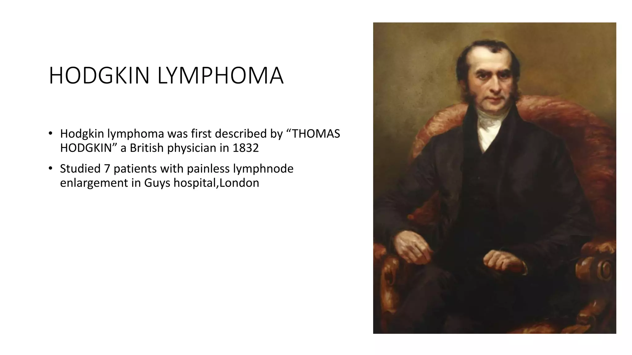
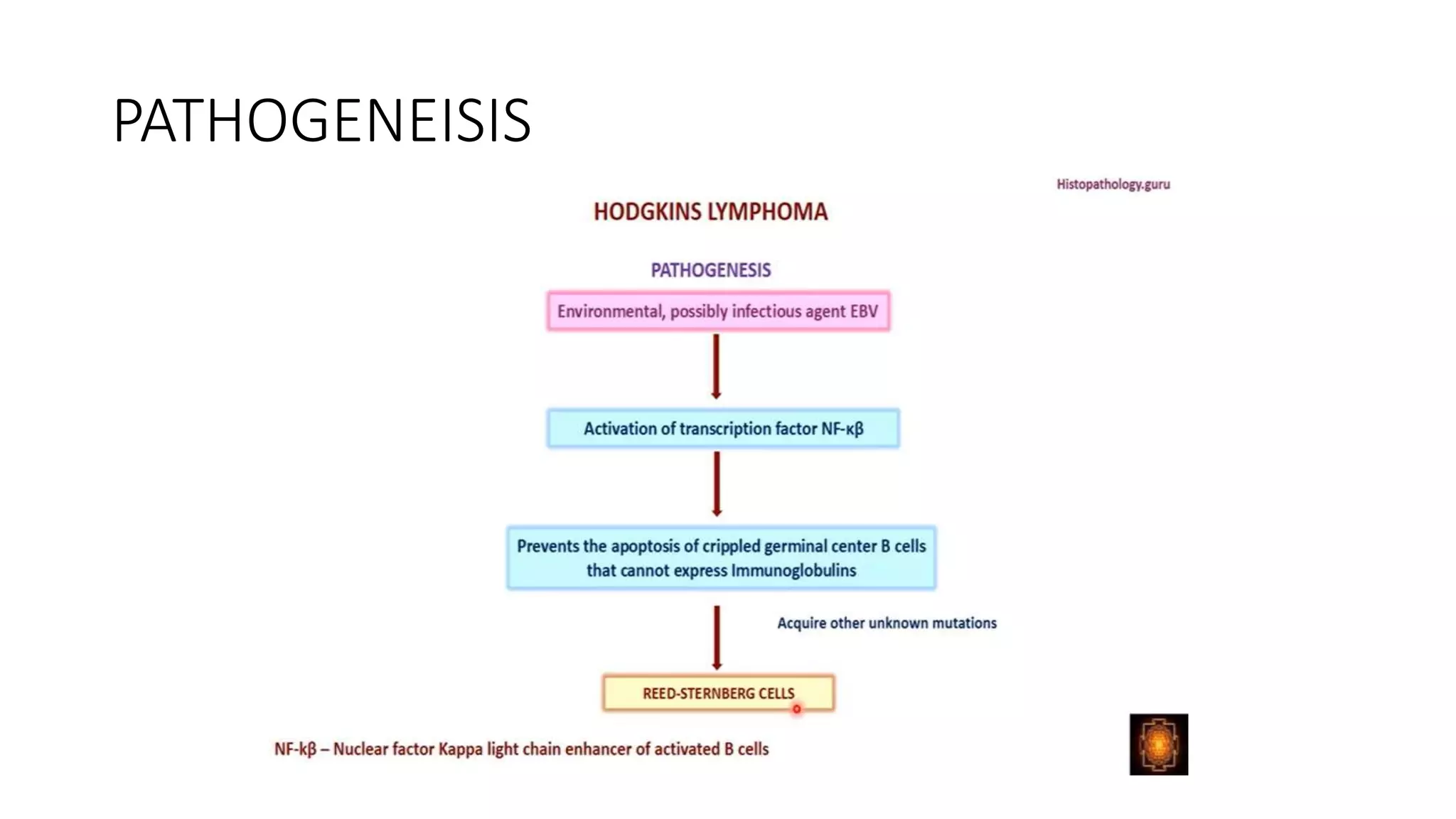
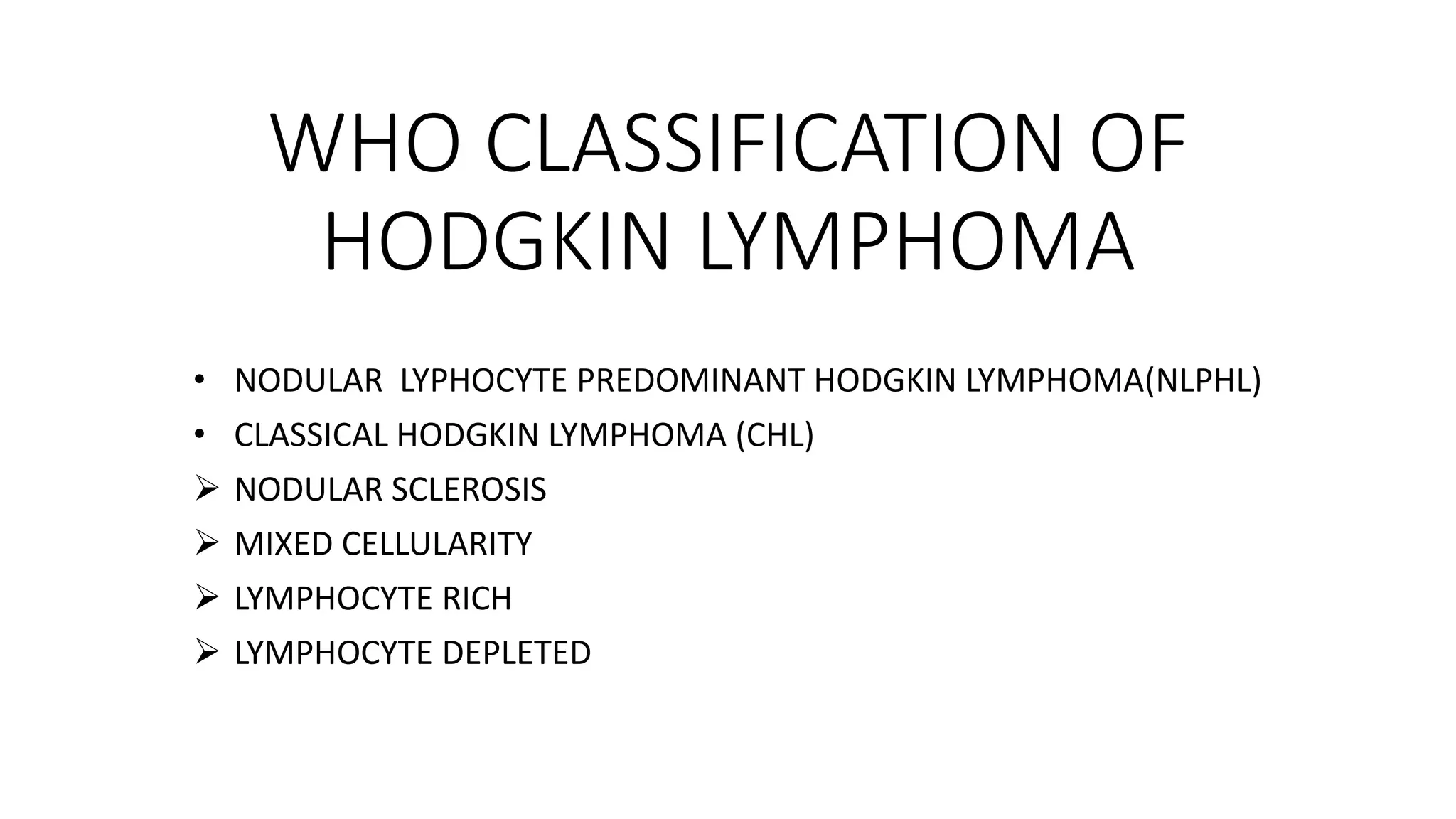
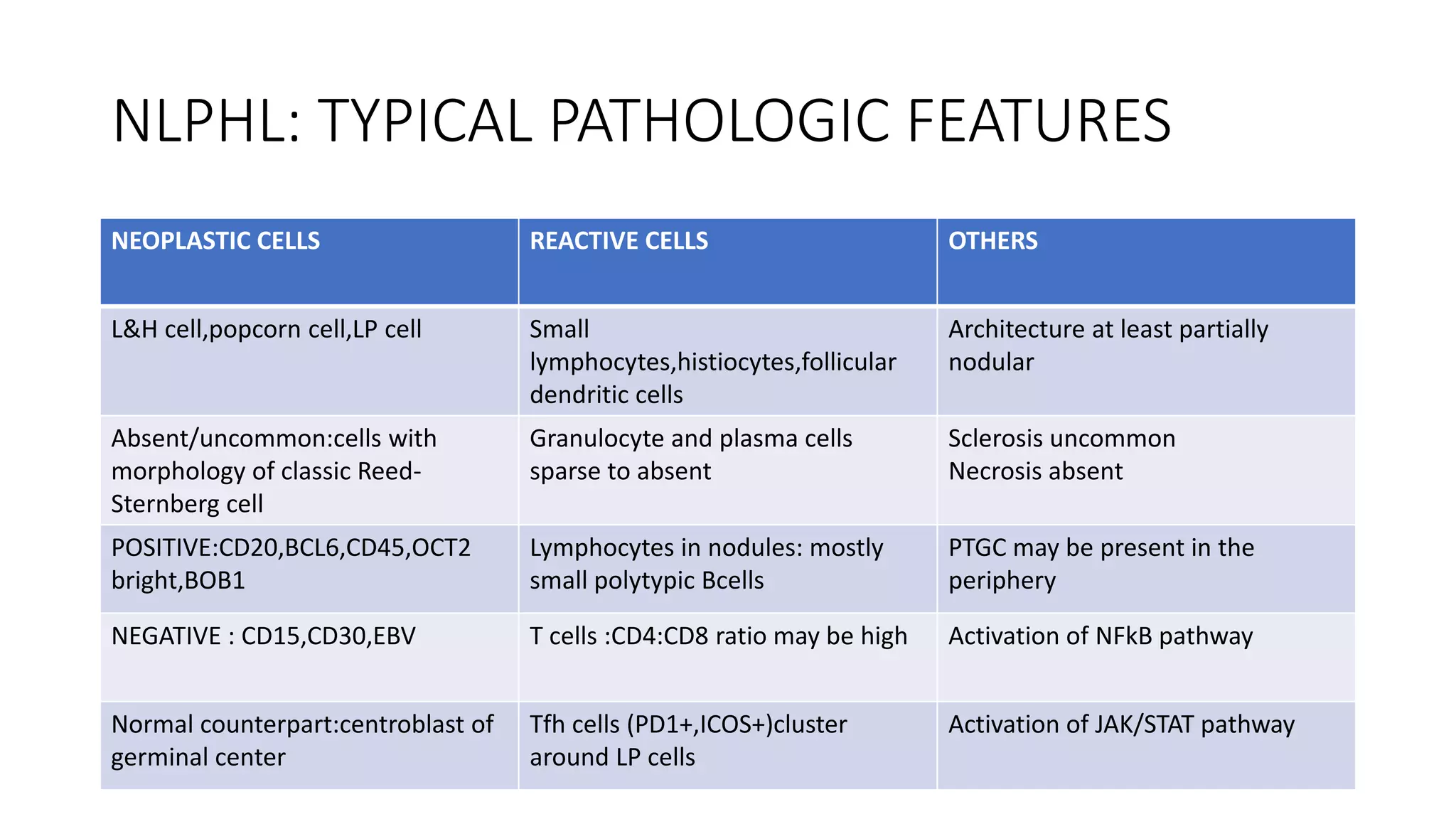
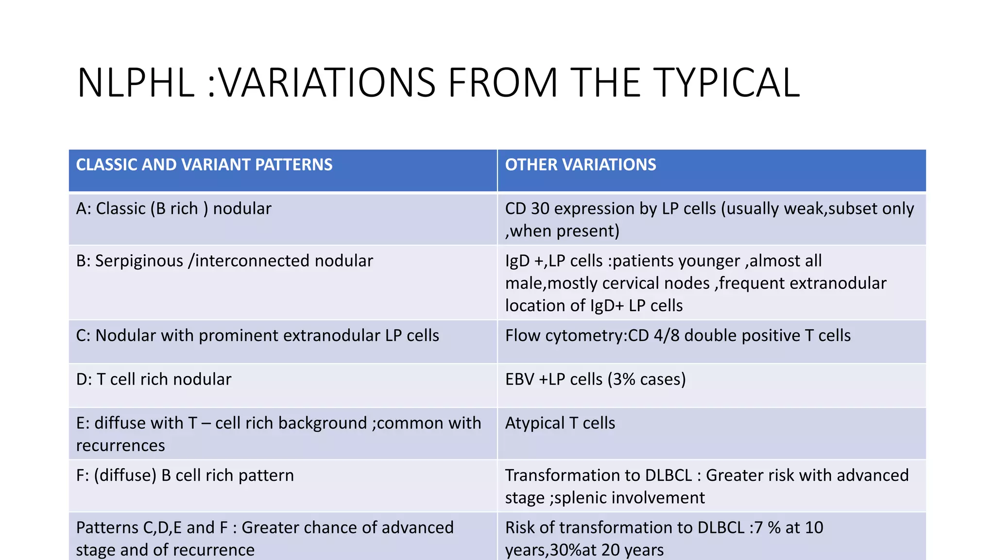
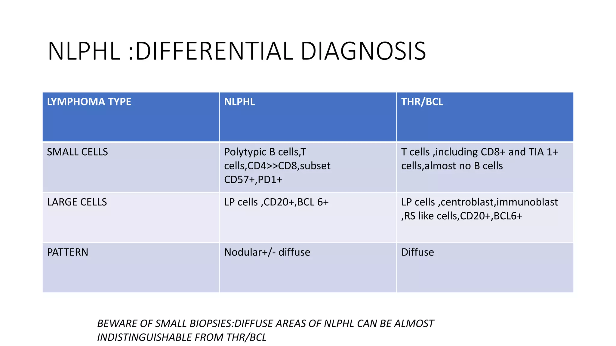
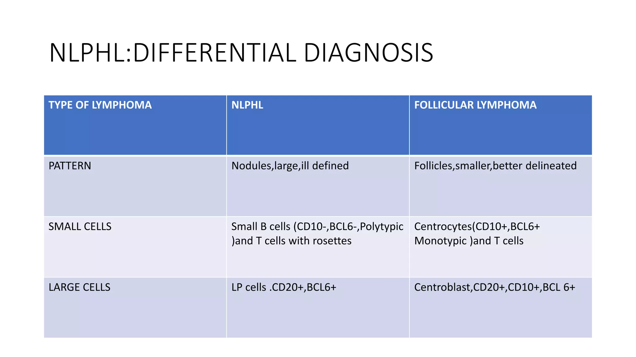
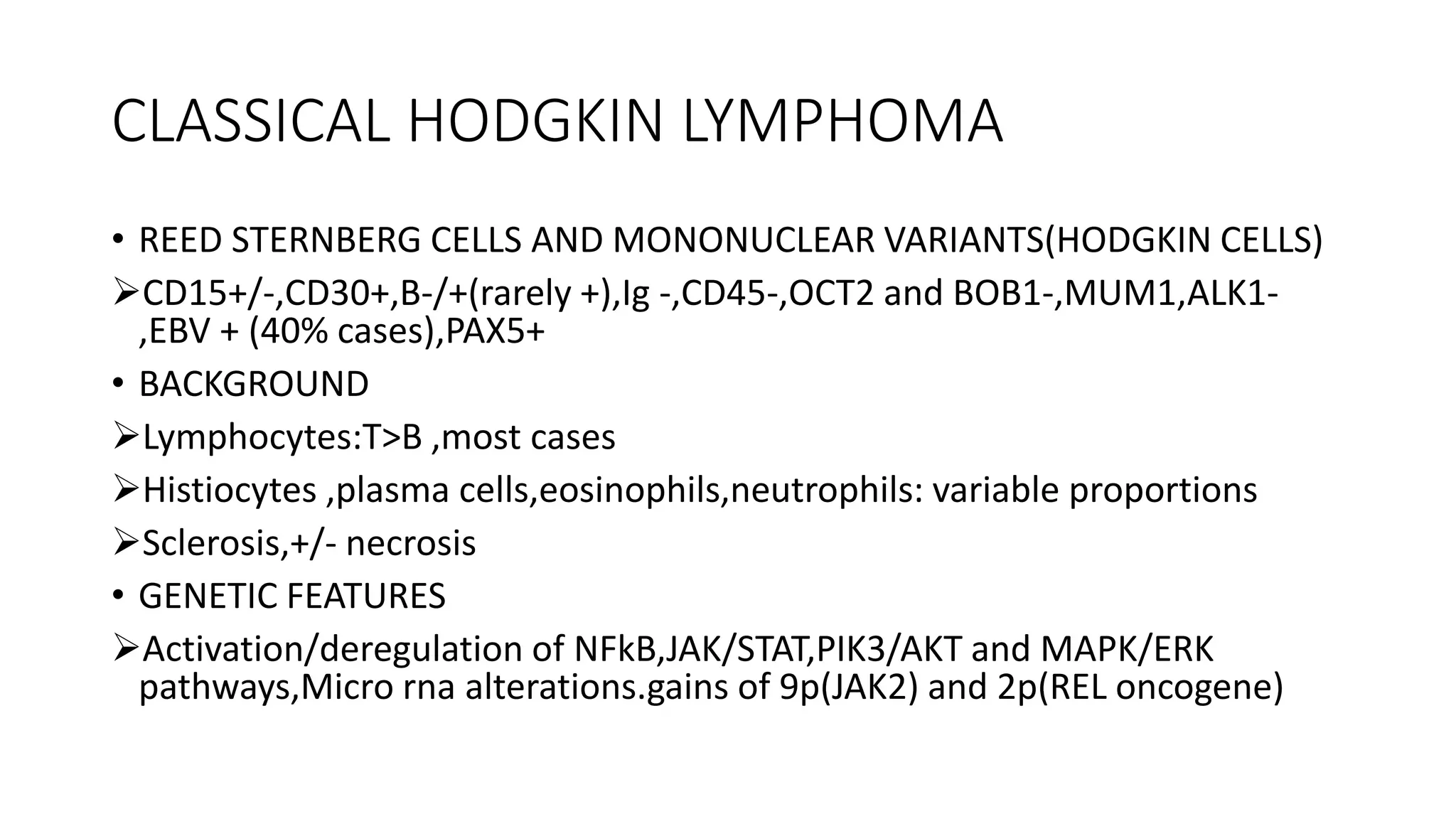
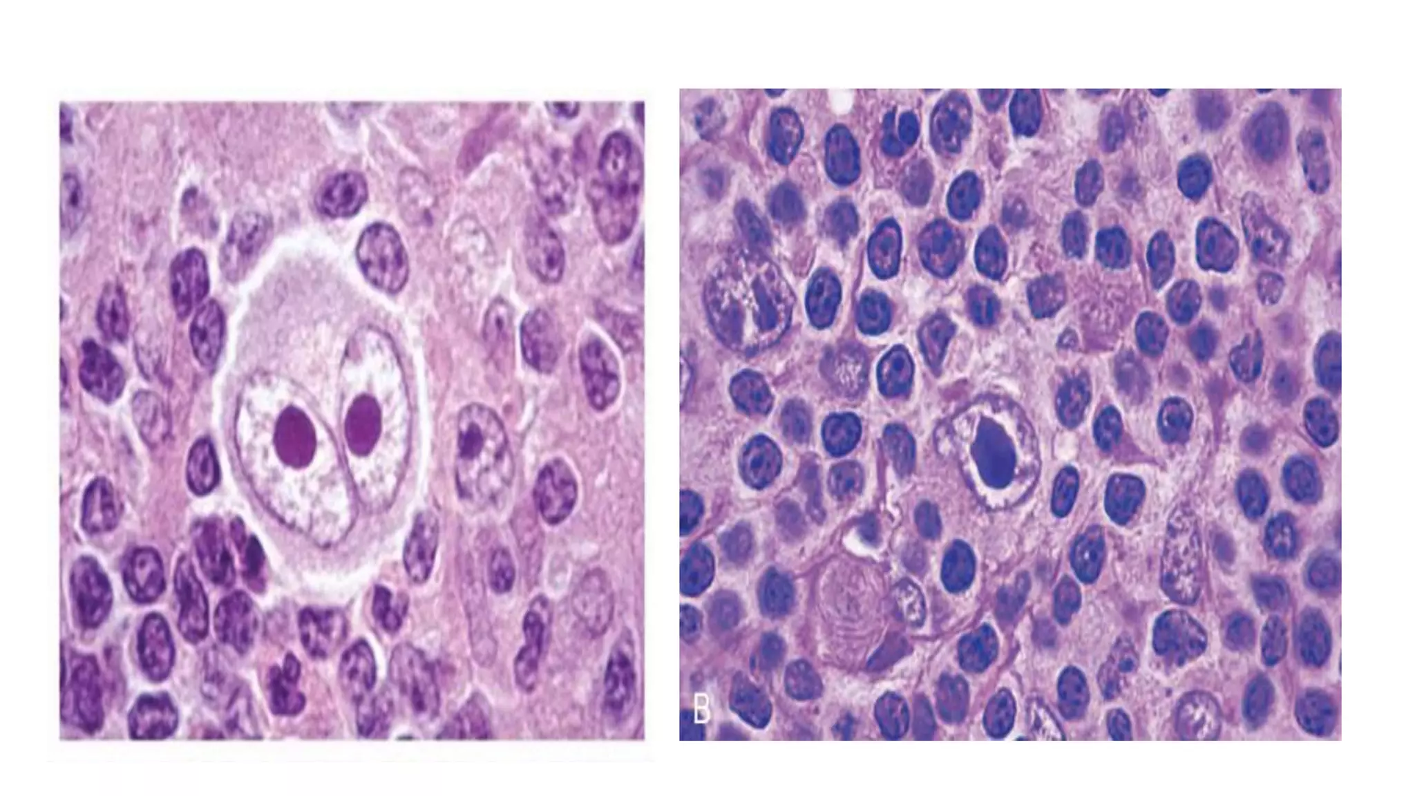
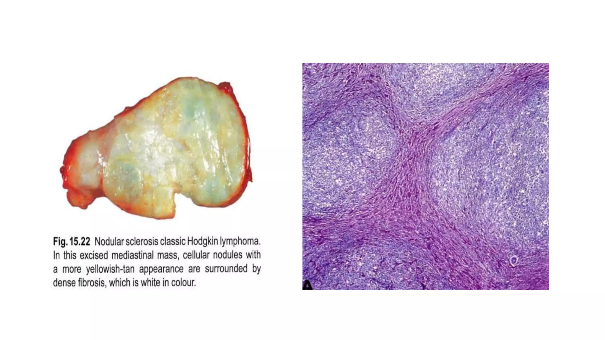
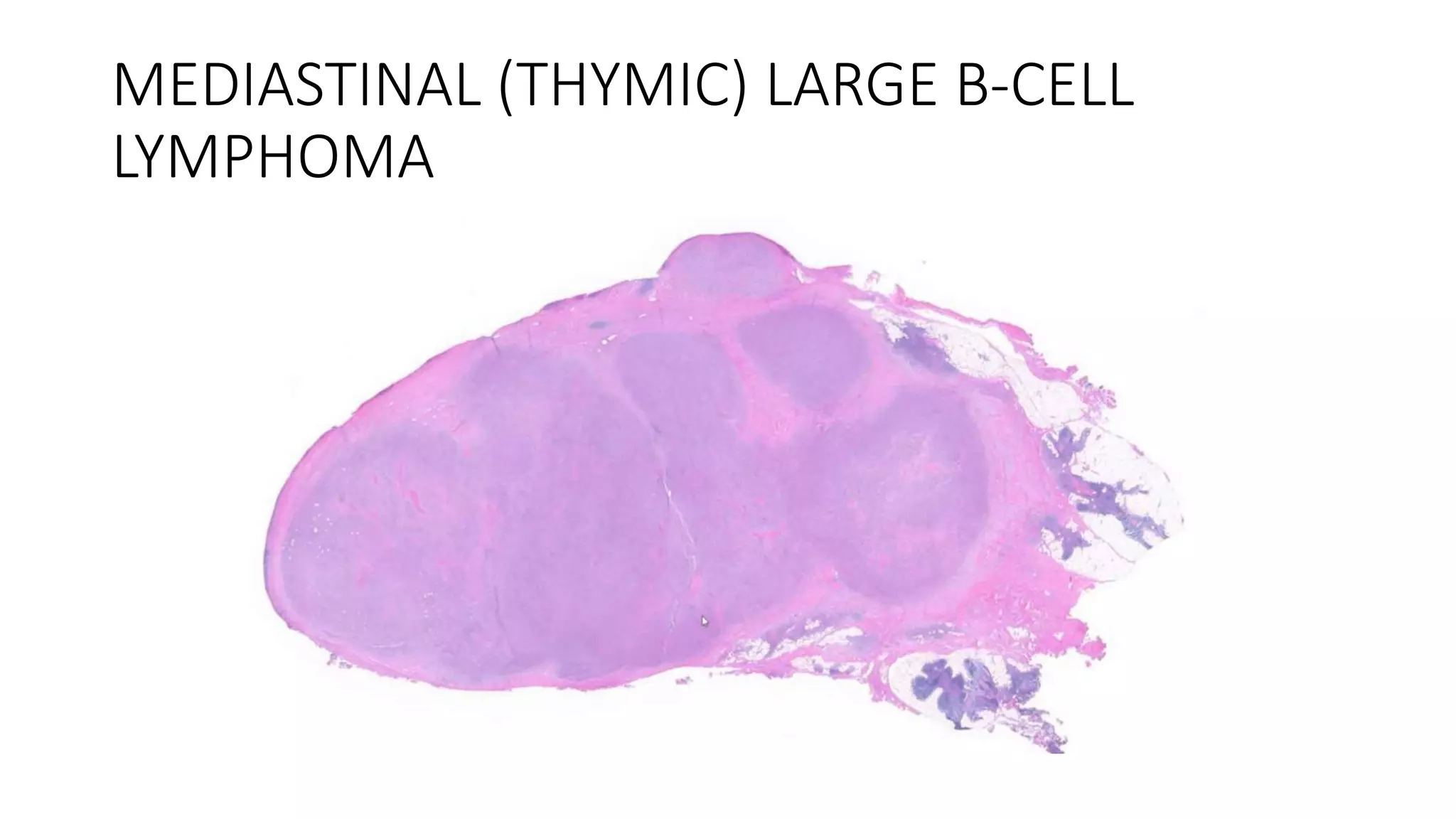
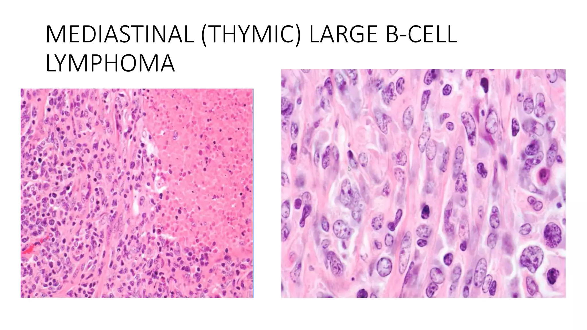
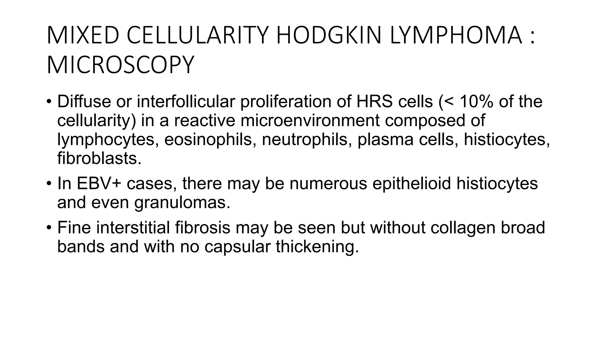
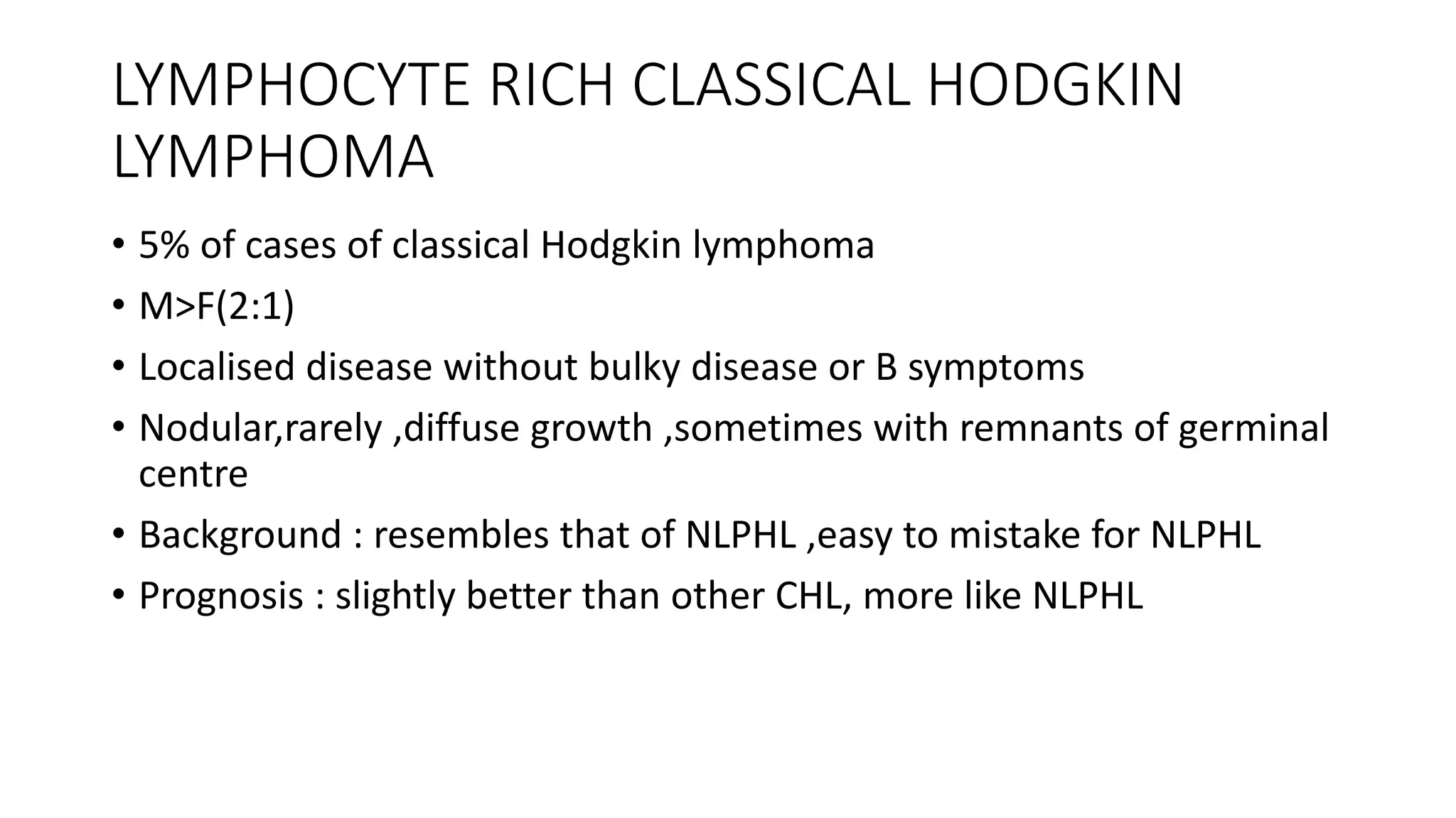
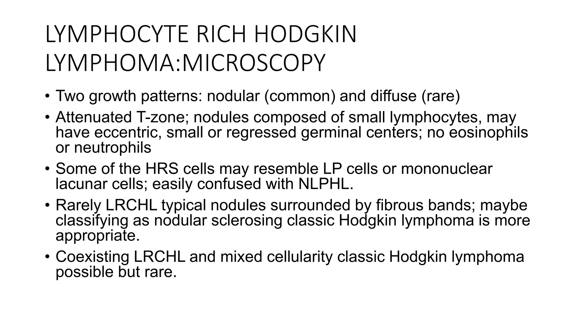
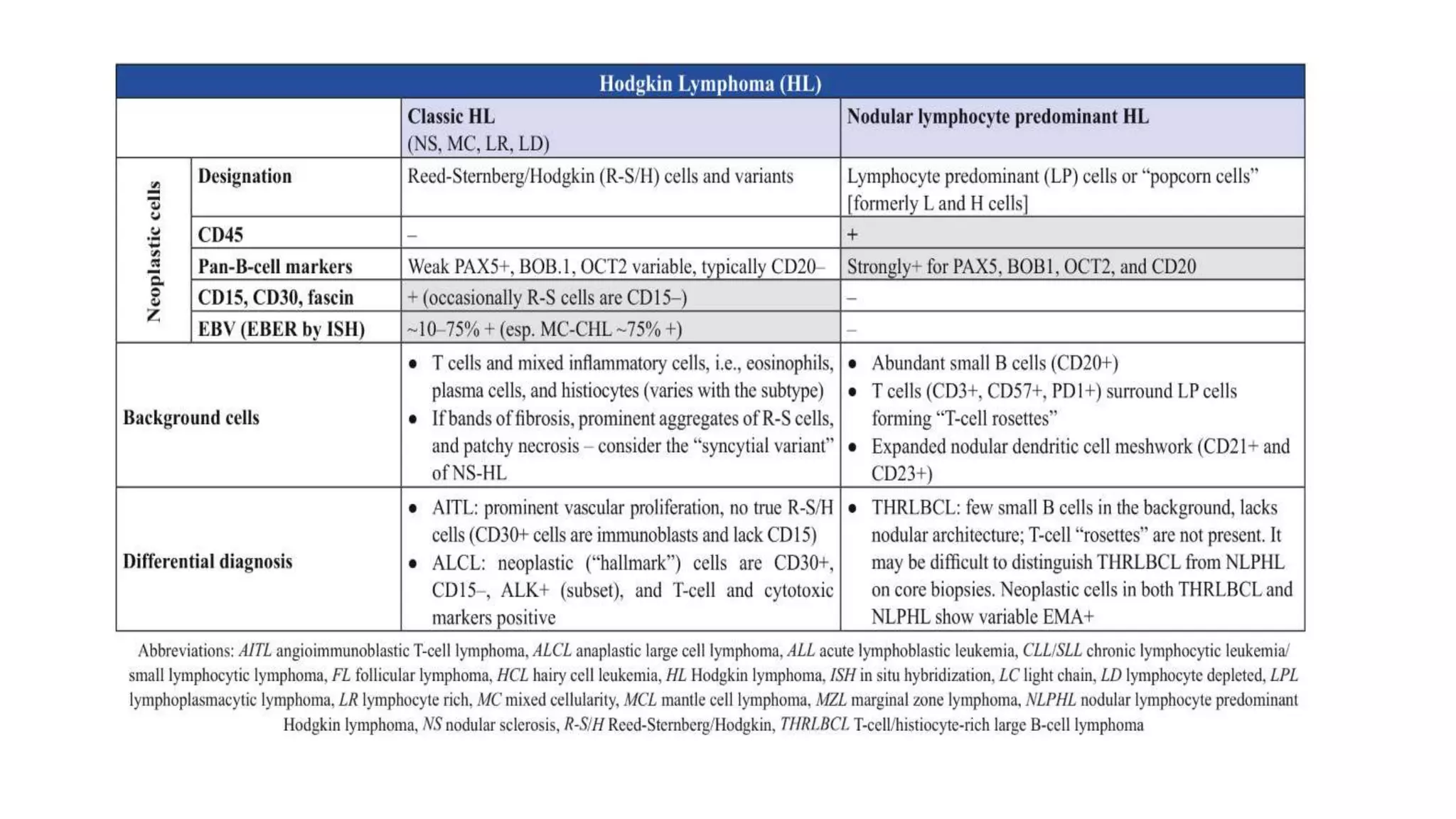
6. NORMAL DISTRIBUTION OF LYMPHOCYTE
SUBSETS
• CORTEX – B cells ,few T cells in GC,FDC, tingible body macrophages
• PARACORTEX – T cells,few Bcells, IDC,
immunoblast,histiocytes,plasmacytoid monocytes
• MEDULLARY CORDS – Small lymphocytes,plasma cells and
immunoblast
• SINUSES – sinus lining cells and histiocytes
9. HODGKIN LYMPHOMA
• Hodgkin lymphoma was first described by “THOMAS
HODGKIN” a British physician in 1832
• Studied 7 patients with painless lymphnode
enlargement in Guys hospital,London
10. • Disease primarily arises in lymph nodes from B-cells and secondarily
involves the extra nodal sites
• it is a disease characterized by heterogenous cellularity comprising of
majority of non neoplastic cells with minority of neoplastic cells called
REED-STERNBERG cells or HODGKIN CELLS
11. WHO DEFINITION
• They are composed of large dysplastic mononuclear and
multinucleated cells surrounded by a variable mixture of mature
non―neoplastic inflammatory cells.
• Abundant band―like and/or more diffuse collagen fibrosis may be
present.
• The neoplastic cells are often ringed by T cells in a rosette―like
manner.
21. NODULAR LYMPHOCYTE PREDOMINANT
HODGKIN LYMPHOMA
• 10% of all Hodgkin lymphoma
• M:F=3:1
• Wide range ,with median age in 30-40 years,unimodal age
distribution
• Localised disease involving peripheral nodes (cervical,axillary,inguinal)
• Relapse: common
• Indolent disease with excellent survival in most
• Transformation to DLBCL in minority
22. NLPHL: TYPICAL PATHOLOGIC FEATURES
NEOPLASTIC CELLS REACTIVE CELLS OTHERS
L&H cell,popcorn cell,LP cell Small
lymphocytes,histiocytes,follicular
dendritic cells
Architecture at least partially
nodular
Absent/uncommon:cells with
morphology of classic Reed-
Sternberg cell
Granulocyte and plasma cells
sparse to absent
Sclerosis uncommon
Necrosis absent
POSITIVE:CD20,BCL6,CD45,OCT2
bright,BOB1
Lymphocytes in nodules: mostly
small polytypic Bcells
PTGC may be present in the
periphery
NEGATIVE : CD15,CD30,EBV T cells :CD4:CD8 ratio may be high Activation of NFkB pathway
Normal counterpart:centroblast of
germinal center
Tfh cells (PD1+,ICOS+)cluster
around LP cells
Activation of JAK/STAT pathway
23. Lymph node showing vaguely nodular small
lymphocyte proliferation
Large neoplastic cells with folded /irregular contoured nuclei and
small nucleoli in a background of small lymphocyte and
histiocyte
25. NLPHL :VARIATIONS FROM THE TYPICAL
CLASSIC AND VARIANT PATTERNS OTHER VARIATIONS
A: Classic (B rich ) nodular CD 30 expression by LP cells (usually weak,subset only
,when present)
B: Serpiginous /interconnected nodular IgD +,LP cells :patients younger ,almost all
male,mostly cervical nodes ,frequent extranodular
location of IgD+ LP cells
C: Nodular with prominent extranodular LP cells Flow cytometry:CD 4/8 double positive T cells
D: T cell rich nodular EBV +LP cells (3% cases)
E: diffuse with T – cell rich background ;common with
recurrences
Atypical T cells
F: (diffuse) B cell rich pattern Transformation to DLBCL : Greater risk with advanced
stage ;splenic involvement
Patterns C,D,E and F : Greater chance of advanced
stage and of recurrence
Risk of transformation to DLBCL :7 % at 10
years,30%at 20 years
26. NLPHL:DIFFERENTIAL DIAGNOSIS
• Reactive hyperplasia: progressive transformation of GC
• Classic Hodgkin lymphoma: especially lymphocyte rich CHL
• T-cell/histiocyte rich large B-cell lymphoma
• Follicular lymphoma
• Peripheral T cell lymphoma: High CD4:CD8 ratio(common in NLPHL)
• T lymphoblastic lymphoma : CD4/CD8 double positive T
cells(common in NLPHL)
27.
28. NLPHL :DIFFERENTIAL DIAGNOSIS
LYMPHOMA TYPE NLPHL THR/BCL
SMALL CELLS Polytypic B cells,T
cells,CD4>>CD8,subset
CD57+,PD1+
T cells ,including CD8+ and TIA 1+
cells,almost no B cells
LARGE CELLS LP cells ,CD20+,BCL 6+ LP cells ,centroblast,immunoblast
,RS like cells,CD20+,BCL6+
PATTERN Nodular+/- diffuse Diffuse
BEWARE OF SMALL BIOPSIES:DIFFUSE AREAS OF NLPHL CAN BE ALMOST
INDISTINGUISHABLE FROM THR/BCL
30. NLPHL:DIFFERENTIAL DIAGNOSIS
TYPE OF LYMPHOMA NLPHL FOLLICULAR LYMPHOMA
PATTERN Nodules,large,ill defined Follicles,smaller,better delineated
SMALL CELLS Small B cells (CD10-,BCL6-,Polytypic
)and T cells with rosettes
Centrocytes(CD10+,BCL6+
Monotypic )and T cells
LARGE CELLS LP cells .CD20+,BCL6+ Centroblast,CD20+,CD10+,BCL 6+
32. CLASSICAL HODGKIN LYMPHOMA
• 90% OF HODGKIN LYPHOMA
• Bimodal age distribution,peaks among young adults & elderly adults
• Usually stage I/II , occasional widespread
• B symptoms in around 40 %
• Aggressive but curable
• 5-year survival in 5%,70 years age
• Cure in >85% or more ,now
33. CLASSICAL HODGKIN LYMPHOMA
• REED STERNBERG CELLS AND MONONUCLEAR VARIANTS(HODGKIN CELLS)
CD15+/-,CD30+,B-/+(rarely +),Ig -,CD45-,OCT2 and BOB1-,MUM1,ALK1-
,EBV + (40% cases),PAX5+
• BACKGROUND
Lymphocytes:T>B ,most cases
Histiocytes ,plasma cells,eosinophils,neutrophils: variable proportions
Sclerosis,+/- necrosis
• GENETIC FEATURES
Activation/deregulation of NFkB,JAK/STAT,PIK3/AKT and MAPK/ERK
pathways,Micro rna alterations.gains of 9p(JAK2) and 2p(REL oncogene)
36. NODULAR SCLEROSIS CLASSICAL HODGKIN
LYMPHOMA
• Adolescents and young adults ;F>M
• Usually, supradiaphragmatic disease (stage I/II)
• Mediastinum often involved
• EBV: minority of cases (10-25%)
37. NODULAR SCLEROSIS:MICROSCOPY
• Nodular growth pattern with broad fibroblast poor birefringent
collagen bands surrounding at least one nodule
• Usually confined within thickened lymphonodular capsule
• Highly variable numbers of HRS cells, small lymphocytes and
other inflammatory cells; often numerous eosinophils,
histiocytes and neutrophils; occasional foamy macrophages
• Mitoses uncommon
• Lacunar cells
40. DIFFERENTIAL DIAGNOSIS
1. Primary mediastinal (thymic) Large B-cell lymphoma
COMMON FEATURES:
Usually young adults with slight female preponderance
B cell origin
Ig negative
CD30 expression(NSCHL>MLBCL)
RS like cells in some MLBCL
Sclerosis
Similar gene expression
Activation of NFkB and JAK/STAT
CIITA translocation in subsets (15% on CHL)
41. NSCHL VERSUS MLBCL
FEATURE NS HODGKIN LYMPHOMA MLBCL
REACTIVE CELLS Reactive cells more numerous
,eosinophils common
Fewer reactive cells
overall;granulocyte uncommon
NEOPLASTIC CELLS Lacunar cells,diagnostic RS cells Oval /lobated cells with pale
cytoplasm
FIBROSIS Fibrous bands Packeting sclerosis are more
common
NECROSIS May have neutrophils Granulocyte usually absent
NEOPLASTIC
CELLS,IMMUNOPHENOTYPE
CD15+/-,CD30+,PAX5 +,CD20-
/+,CD45-,OCT2/BOB1 -
Diffuse strong CD20+,CD45+,CD30-
/+,OCT2/BOB1+,CD23+/-
PCR IGH PCR usually negative IGH PCR clonal
VC SYNDROME uncommon common
45. MIXED CELLULARITY CLASSICAL HODGKIN
LYMPHOMA
• 20-25% of CHL, more frequently in developing countries and HIV+
patients
• Any age affected ,most common type in older adults ,M;F =2:1
• Type of Hodgkin most strongly associated with EBV :75% of cases
EBV+
46. MIXED CELLULARITY HODGKIN LYMPHOMA :
MICROSCOPY
• Diffuse or interfollicular proliferation of HRS cells (< 10% of the
cellularity) in a reactive microenvironment composed of
lymphocytes, eosinophils, neutrophils, plasma cells, histiocytes,
fibroblasts.
• In EBV+ cases, there may be numerous epithelioid histiocytes
and even granulomas.
• Fine interstitial fibrosis may be seen but without collagen broad
bands and with no capsular thickening.
49. EBV positive in mixed cellularity CHL .EBER (CISH ) strong staining signal in the nuclei of
the neoplastic cells
50. LYMPHOCYTE RICH CLASSICAL HODGKIN
LYMPHOMA
• 5% of cases of classical Hodgkin lymphoma
• M>F(2:1)
• Localised disease without bulky disease or B symptoms
• Nodular,rarely ,diffuse growth ,sometimes with remnants of germinal
centre
• Background : resembles that of NLPHL ,easy to mistake for NLPHL
• Prognosis : slightly better than other CHL, more like NLPHL
51. LYMPHOCYTE RICH HODGKIN
LYMPHOMA:MICROSCOPY
• Two growth patterns: nodular (common) and diffuse (rare)
• Attenuated T-zone; nodules composed of small lymphocytes, may
have eccentric, small or regressed germinal centers; no eosinophils
or neutrophils
• Some of the HRS cells may resemble LP cells or mononuclear
lacunar cells; easily confused with NLPHL.
• Rarely LRCHL typical nodules surrounded by fibrous bands; maybe
classifying as nodular sclerosing classic Hodgkin lymphoma is more
appropriate.
• Coexisting LRCHL and mixed cellularity classic Hodgkin lymphoma
possible but rare.
52.
53.
54. LYMPHOCYTE –DEPLETED CLASSICAL
HODGKIN LYMPHOMA
• <2% of cases of CHL
• Seen in HIV+ patients ,developing countries ;M>F
• Widespread disease ,peripheral and/or internal lymphadenopathy
• Immunophenotye as for other CHL:EBV often +
• Clonal IGH may be found by PCR
• With optimum treatment, in HIV- patients,prognosis is nearly as good
as other CHL patients with same stage of disease
55. LYMPHOCYTE DEPLETED HODGKIN
LYMPHOMA :MICROSCOPY
• Relative predominance of HRS cells and the scarcity of background
lymphocytes in relation to the neoplastic cells.
• 2 patterns:
• Diffuse fibrosis:
• Prominent fibroblastic proliferation (nonbirefringent fibrillary stroma) without well
formed fibrous bands
• Numerous histiocytes
• Scattered Reed-Sternberg cells
• Scant lymphocytes
• Lack of plasma cells or eosinophils
• Reticular:
• Rich in Reed-Sternberg cells (often sheets) with anaplastic, pleomorphic or
sarcomatous features
• Scant background small lymphocytes
• Capsular and perinodal infiltration is common
56.
57. CHL:DIFFERENTIAL DIAGNOSIS
• Reactive lymphoid hyperplasia
Non specific reactive hyperplasia
Viral lymphadenitis
Granulomatous inflammation
• Non Hodgkin lymphoma
T cell/histiocyte rich B cell lymphoma
EBV+DLBCL,NOS
Peripheral T cell lymphoma
Non lymphoid malignancies
64. • With current treatment protocols, tumor stage rather
than histologic type is the most important prognostic
variable.
• The cure rate of patients with stages I and IIA is close
to 90%. Even with advanced disease (stages IVA and
IVB), disease-free survival at 5 years is 60% to 70%.
65. MANAGEMENT
• CHL is now curable in > 85% of cases.
• Modern polychemotherapy protocols such as ABVD (i.e. doxorubicin,
bleomycin, vinblastine, and dacarbazine) and escalated BEACOPP
(bleomycin,etoposide, doxorubicin, cyclophosphamide, vincristine,
procarbazine, and prednisone)
• Stage―adapted treatment
• In advanced stages, the International Prognostic Score (IPS) is used
• Novel targeted treatment approaches: CD30―directed antibody-drug
conjugate brentuximab vedotin
• AntiPD1 antibodies