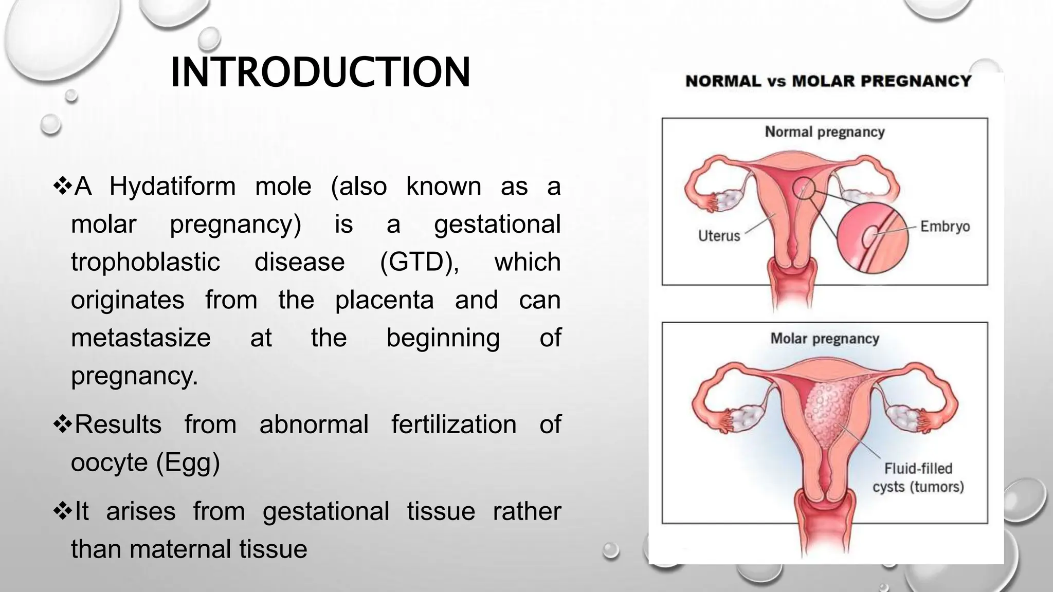
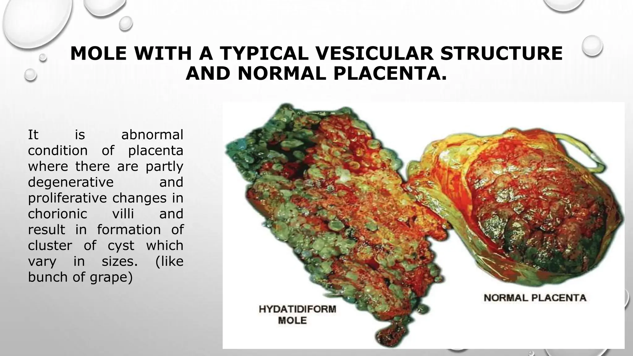
3. GESTATIONAL TROPHOBLASTIC DISEASE
(GTD)
• HYDATIFORM MOLE – BENIGN –PREMALIGNANT
CAN BECOME MALIGNANT AND INVASIVE
• GESTATIONAL CHORIOCARCINOMA –EXTREMELY
MALIGNANT AND INVASIVE
• PLACENTAL SITE TROPHOBLASTIC TUMOUR
4. INTRODUCTION
A Hydatiform mole (also known as a
molar pregnancy) is a gestational
trophoblastic disease (GTD), which
originates from the placenta and can
metastasize at the beginning of
pregnancy.
Results from abnormal fertilization of
oocyte (Egg)
It arises from gestational tissue rather
than maternal tissue
6. MOLE WITH A TYPICAL VESICULAR STRUCTURE
AND NORMAL PLACENTA.
It is abnormal
condition of placenta
where there are partly
degenerative and
proliferative changes in
chorionic villi and
result in formation of
cluster of cyst which
vary in sizes. (like
bunch of grape)
7. TYPES OF HYDATIDIFORM MOLE
Two types of HM
1.Complete Hydatidiform Mole
2.Incomplete or partial Hydatidiform Mole
9. Etiology Complete Hydatidiform Mole (CHM)
• Complete moles are usually DIPLOID &
tend to cause higher levels of the human
chorionic gonadotropin (HCG) which is
one of the main clinical features of this
process.
• In complete moles, the karyotype is
46XX, 90% of the time and 46XY 10% of
the time.
• Fetus is absent
• It occurs when enucleated egg is
fertilized by two sperms or haploid sperm
• Only paternal DNA is expressed CHD Specimen
11. Etiology Incomplete OR Partial Hydatidiform mole
(PHM)
• In partial moles, the karyotype is
90% of the time triploid and
either 69,XXX or 69,XXY.
• In partial moles, both maternal
and paternal DNA is expressed.
• There is formation of fetus but will
not survive
• Normal sperm fertilizes haploid
ovum duplicates or when two
sperms fertilizes a haploid ovum
13. INCIDENCE
• Asia (~1 in 500)
• The Middle east and Africa (~1 in 1000)
• In Europe and north America (~1 in 1500)
14. RISK FACTORS
• Extremes of maternal age:
• Greater than 35 years old carries a five to ten-fold increased risk
• Early teenage years, usually less than 20 years old
• Previous molar pregnancy increases the risk in future pregnancies
• Women with previous spontaneous abortions or infertilities
• Dietary factors including patients that have diets deficient in carotene
(vitamin A precursor) and animal fats
• Smoking
16. Clinical Features
Dark Brown to bright red Vaginal bleeding( prune juice
appearance) in the first trimester.
Hyperemesis (severe nausea and vomiting), which is due to
the high level of the HCG hormone circulating in the
bloodstream.
Some patients also endorse passage of vaginal tissue
described as grape-like clusters or vesicles.
Advance stage possibility of Respiratory Distress Syndrome
17. Late finding of disease (after the first trimester around 14 to 16
weeks of pregnancy)
• Symptoms of Hyperthyroidism, including tachycardia and
tremors, again caused by the high levels of circulating hCG
• Pre-eclampsia
Partial Mole presents with symptoms S/o Threatened or
Spontaneous Abortion
Clinical Features
18. On physical examination
In a complete mole, the uterus is usually larger than the
expected gestational date of the pregnancy,
In partial moles, the uterus can be smaller than the suggested
date.
Clinical Presentation
20. EVALUATION
• Serum HCG level (greater than 100,000 mIU/ml)
• Blood Group
• Complete blood count
• Thyroid function test
• Liver function test
• Coagulation profile including PT/INR
22. PELVIC ULTRASOUND ( IMAGING OF CHOICE)
This Photo by Unknown Author is licensed under CC BY
COMPLETE MOLE
Heterogenous Mass in uterine
cavity with multiple Anechoic
spaces
SNOW STORM APPERANCE
Absence of Fetus
Absence of amniotic fluid
23. PARTIAL MOLE USG FINDINGS
This Photo by Unknown Author is licensed under CC BY-SA-NC
FOETUS is present, may be
viable .
Presence of amniotic fluid
Enlarged placenta with cystic
spaces
SWISS CHEESE appearance
• CT SCAN
• PET SCAN
XRAY FOR LUNG Metastates
24. What are principles of
management of vesicular
mole?
• Suction & Evacuation-as
early as diagnosis is done
• Supportive therapy-
correction of fluid and
blood loss
• Counselling for regular
25. What cervical ripening agents to be
used?
Which Anesthesia to be preferred?
Which is Uterotonic of choice?
Is there role of Curettage following
evacuation?
Which is preferred contraceptive?
26. TREATMENT OPTION
Uterine Evacuation should be performed by surgical procedure.
Significant risk of bleeding and perforation are there ,hence to
be performed by senior surgeon with cross match available.
SGA preferred anesthesia
Medical evacuation may be tried for partial mole.
Oxytocin/methergine to be used for uterotonic.
Misoprostol 600mcg to be used for making cx favorable.
Barrier contraceptive is preferred
29. Follow up after molar pregnancy
Weekly till HCG levels falls to normal .
This usually occurs within 2mnths.
Then monthly till 6mnths
Current advice is f/u is needed for 6mnths
from time of evacuation or 6mnths from
first normal HCG level.
Contraception to be advised for 6mnths.
33. INDICATIONS FOR CHEMOTHERAPY
TREATMENT AFTER MOLAR PREGNANCY
Brain liver or GI tract metastases or lung metastases
>2cm on Xray
Histological evidence of choriocarcinoma
Heavy vaginal bleeding or GI bleeding
Pulmonary ,vaginal or vulval metastases unless hCG
levels are falling
Rising hCG in two consecutive serum samples
hCG >20,000IU/L more than 4 weeks After
Evacuation
hCG plateau in Three consecutive Samples
Raised hCG levels 6mnths after evacuation(even if
34. FIGO PROGNOSTIC SCORE SYSTEM FOR
GESTATIONAL TROPHOBLASTIC DISEASE
This Photo by Unknown Author is licensed under CC BY-SA
35. Chemotherapy Is treatment of choice.
Hysterectomy if family is completed and there is
no spread.
Score of 6 or less, simple Single chemotherapy
methotrexate & score of 7 and above combination
chemotherapy is opted
Pts recover fairly rapid and in nearly all cases
fertility is retained.
An interval of 12mnths from completion of
chemotherapy to next pregnancy is recommended
Management of Malignant GTD