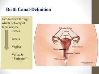
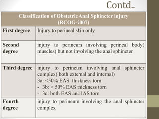
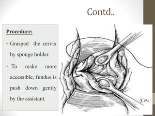
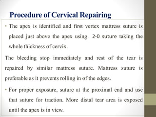
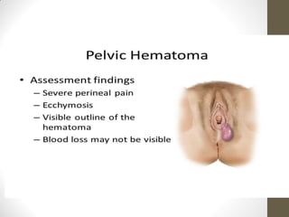
Injuries to Birth Canal
Prepared By:
Reena Bhagat
Senior Nursing Instructor
Maternal Health Nursing
BPKIHS
Objectives of the session
• Anatomy of birth canal
• Classification of injuries
• Causes, diagnosis and management of
injuries
Introduction
• Maternal birth canal injury following child birth
process are quite common and significant to maternal
morbidity and even to death.
• Second most frequent cause of PPH.
• PPH with a contracted uterus is usually due to a
cervical or vaginal tear.
• Prevention, early detection, prompt and effective
management are essential to minimize the morbidity
and gynecological problem in later life.
Meaning of birth canal
• The pathway through which the fetus is expelled during
parturition, leading from the uterus through cervix,
vagina and vulva is called birth canal.
• The birth canal is formed with the combination of
various bones and soft tissues.
• Injury to the canal occurs mainly in soft tissues and
rarely to the bony mass. Vagina, vulva and perineum
are often injured during expulsion stage of labor.
Birth Canal-Definition
Genital tract through
which delivery of
fetus occurs
uterus
cervix
Vagina
Vulva &
( Perineum)
Classification of Injury
Depending up on the anatomical structure
involved.
Depending up on type of injury
Example: Laceration, Hematoma, Rupture
Anatomical Classification
Injuries to Bony parts Injuries to Soft Tissues
Injury to Symphysis Pubis Injury to Vulva
Injury to Sacro-coccygeal
Joint
Perineal Tears
Injury to Sacro-iliac Joint Laceration of Vagina &
cervix
Rupture of Uterus
Causes of Injury
Incomplete dilation of cervix
Instrumentation (Wrong application)
Precipitate labor
Protracted labor due to borderline CPD
Obstructed labor
Iatrogenic
Injuries to soft tissues: Vulva
• Lacerations: Lacerations of the vulval skin
posteriorly
• Paraurethral tears: Paraurethral tear on the inner
aspect of the labia minora.
Paraurethral tear may be associated with brisk
hemorrhage and should be repaired by interrupted
catgut sutures, preferably after introduction of a
rubber catheter into the bladder to prevent injury of
the urethra.
Injuries to Soft Tissues: Perineal Tears
Incidence: Overall risk is 1% of all vaginal deliveries.
Causes: Perineal injury (mainly the third and fourth
degree) results from
(i) over stretching and/ or
(ii) rapid stretching of the perineum especially when the
perineum is inelastic (elderly primigravida, perineal
scar).
PREVENTION:
• Proper conduct in the second stage of labor
• Taking due care of the perineum
Contd..
Risks factors for Third Degree Perineal Tear
 Big baby (weight ≥ 3
kg)
 Nulliparity
 Outlet contraction
with narrow pubic
arch
 Shoulder dystocia
 Forceps delivery
 Scar in the perineum
(perineorrhaphy,
episiotomy)
 Face to pubis delivery
 Midline episiotomy
 Precipitate labor
Perineal Tears
Contd..
Classification of Obstetric Anal Sphincter injury
(RCOG-2007)
First degree Injury to perineal skin only
Second
degree
injury to perineum involving perineal body(
muscles) but not involving the anal sphincter
Third degree injury to perineum involving anal sphincter
complex( both external and internal)
3a: <50% EAS thickness torn
- 3b: > 50% EAS thickness torn
- 3c: both EAS and IAS torn
Fourth
degree
injury to perineum involving the anal sphincter
complex
Management
 Recent tear : Repaired immediately following the
delivery of the placenta.
- this reduces the chance of infection and minimizes
the blood loss
 Delay beyond 24 hours:
- repair should be withheld
- Antibiotics to be started to prevent infection.
The complete tear should be repaired after 3 months
if delayed beyond 24hours.
Contd….
REPAIR OF COMPLETE PERINEAL TEAR
Step 1:
Lithotomy position –repair done with local
infiltration of lignocaine hydrochloride
Step 2:
• Rectal and anal mucosa
• The rectal muscles including the pararectal facia
• The torn ends of sphincter ani externus(EAS)
• Overlapping or end to end approximation method
for repair
16
Contd..
Step3:
Repair of perineal muscle done by interrupted
sutures.
Step 4:
The Vaginal wall and the perineal skin are apposed
by interrupted sutures
Contd..
Aftercare:
• Similar to that following episiotomy.
• Special care following repair of complete tear.
• A low residual diet consisting of milk, bread, egg,
biscuits, fish, sweets, etc. is given from third day
onward.
• Lactulose 8 ml twice daily
• Broad-spectrum antibiotics( IV Cefuroxime
1.5gm)
Contd…
• Metronidazole 400 mg thrice daily, continued for
5-7 days
• Physiotherapy and pelvic floor exercises
• Review again 6-12 weeks postpartum
• In case of persistent incontinence of flatus and
feces, endoanal USG and anorectal manometry
should be considered to detect any residual
defects (20–30%). Consultation with a colorectal
surgeon may be needed
Contd….
Plan for Future Delivery:
• Need to have institutional delivery.
• Vaginal delivery may be allowed in a selected case
with or without episiotomy.
• Women having symptoms or with abnormal
endoanal USG and/or manometry should be
delivered by elective cesarean birth.
Vagina
• Isolated vaginal tears or lacerations without
involvement of the perineum or cervix are not so
common.
• Following instrumental or manipulative delivery.
• On such cases, tears are extensive and often
associated with brisk hemorrhage.
Causes of vaginal lacerations
• Can be traumatic or spontaneous.
• These are injuries those occur during forceps
delivery, fetal malpresentation, insufficient
distensibility of vaginal wall and large fetal head.
22
Treatment
• Tears with brisk hemorrhage require exploration
under general anesthesia with good light.
• Tears are repaired by interrupted or continuous
sutures.
• In addition to sutures, hemostasis may be
achieved by intravaginal plugging by roller
gauze.
• Plug should be removed after 24 hours.
• Selective arterial embolization may also be done
if bleeding persists.
Embolization
Colporrhexis
• Rupture of the vault of the vagina is called
colporrhexis,
1. Primary( vault only involved)
2. Secondary( associated with cervical tear); common
3. Complete( peritoneum is opened up)
• Posterior fornix usually rupture.
• Cervical tear is usually associated with lateral fornix.
Contd..
If tear is limited to the vault close to the cervix ,
repair is done from below.
If tear extends high up , laparotomy is to be done
simultaneously with resuscitative measures.
Evacuation of hematoma and arterial ligation may
be needed.
Treatment:
Cervix
• Commonest cause of traumatic postpartum
hemorrhage
Causes:
Iatrogenic: Attempted forceps or Breech
extraction through incompletely dilated cervix.
Rigid cervix: congenital or scar from previous
operations amputation, conization or presence of
lesion like carcinoma cervix
Strong uterine contractions
Cervical Tear
Contd..
Detachment:
 ANNULAR : Detachment of the cervix may be
annular which involved the entire circumference
of the cervix.
 Following prolonged labor in primary cervical
dystocia.
ANTERIOR LIP : May involve only anterior lip
when it is nipped between the head and
symphysis pubis.
Degrees of Cervical Lacerations
• First degree lacerations - Length of cervical rupture
not over 2 cm
• Second degree lacerations - Length of rupture >2
cm but does not extend to vaginal fornices
• Third degree lacerations - Ruptured area extends to
the vaginal fornices and is very dangerous
Contd..
Diagnosis:
Excessive vaginal bleeding immediately following
delivery in presence of a hard and contracted uterus ,
raises the suspicion of a traumatic bleeding
Dangers
Early Late
 Deep cervical tears-
Severe postpartum
haemorrhage
 Broad ligament
hematoma
 Pelvic cellulitis
 Thrombophlebitis
 Ectropion
 Cervical incompetence
Contd…
Treatment:
• Only deep cervical tear associated with bleeding
should be repaired soon after delivery of placenta.
• Repair should be done under general anesthesia , in
lithotomy position with a good light.
Prerequisites are :
 Speculum
 Retractors
 Two sponge holding forceps
 Assistant.
Contd..
Procedure:
• Grasped the cervix
by sponge holder.
• To make more
accessible, fundus is
push down gently
by the assistant.
Procedure of Cervical Repairing
• The apex is identified and first vertex mattress suture is
placed just above the apex using 2-0 suture taking the
whole thickness of cervix.
The bleeding stop immediately and rest of the tear is
repaired by similar mattress suture. Mattress suture is
preferable as it prevents rolling in of the edges.
• For proper exposure, suture at the proximal end and use
that suture for traction. More distal tear area is exposed
until the apex is in view.
Pelvic Hematoma
Definition
Collection of blood anywhere in the area between
the pelvic peritoneum and the peineal skin is called
pelvic hematoma.
Anatomical Types:
• Infralevator hematoma: Common
• Supralevator hematoma: Rare
• Commonest one is vulval hematoma
Contd….
Infralevator Hematoma:
Etiology:
1. Improper hemostasis during repair of vaginal or
perineal tears or episiotomy wounds.
(a) Failure to take precaution while suturing the apex
of the tear
(b) Failure to obliterate the dead space while suturing
the vaginal walls.
2. Rupture of paravaginal venous plexus either
spontaneously or following instrumental delivery
Contd..
Symptoms:
1. Persistent , severe pain on the perineal region
2. There may be rectal tenesmus or bearing down
efforts when extension occurs to the ischiorectal
fossa.
3. There may be even retention of urine.
Signs:
• Variable degrees of shock
• Local examination : tense swelling at the vulva
Contd,,,
Treatment:
• Small hematoma(<5cm) conservatively with cold
compress
• Larger hematomas explored in theater under general
anesthesia
• Blood clots are to be scooped out and the bleeding
points are to be secured
• The dead space is to be obliterated
• A foleys catheter is inserted
• Prophylactic antibiotic is to be administered
Contd,,,
 Supralevator hematoma
Causes:
• Extension of cervical laceration
• Lower uterine segment rupture
• Spontaneous rupture of paravaginal venous plexus
Contd…
Diagnosis:
• Unexplained shock with features of internal
hemorrhage following delivery raises the suspicion
• Swelling above the inguinal ligament pushing
uterus to the contra-lateral side
• Vaginal examination:
Occlusion of the vaginal canal by a bulge (or)
Boggy swelling felt through the fornix
• Rectal examination: Boggy mass
• Ultrasonography
Contd..
Management:
• Treatment of shock and Laparotomy
• Anterior leaf of the broad ligament peritoneum is
incised and the blood clot is scooped out.
• Random blind sutures should not be placed to
prevent ureteric damage
• Tie the anterior division of the internal iliac artery
Rupture of Uterus
Definition : Disruption in the continuity of the all
uterine layers (endometrium, myometrium and
serosa) any time beyond 28 weeks of pregnancy is
called rupture of the uterus
Perforation
Rupture of a rudimentary pregnant horn
Incidence : 1 in 2000 to 1 in 200 deliveries
Increased prevalence of scar rupture following
increased incidence of cesarean section over the
years.
Uterine Rupture
Contd…
Etiology:
The causes of rupture of the uterus are broadly
divided into:
• Spontaneous
• Scar Rupture
• Iatrogenic
Contd..
Spontaneous:
During pregnancy : Rare
Causes:
1. Previous damage to the uterine walls, dilatation
and curettage or manual removal of placenta
2. Grand multipara
3. Congenital malformation
4. Couvelaire uterus
oUsually complete
oInvolves upper segment
oLater months of pregnancy
Contd…
During Labor: Rupture of uterus may be due to
• Obstructive rupture: end result of an obstructed labor
• Non-obstructive rupture : grand multipara are usually in
early labor. Involves the fundal area and is complete.
Scar Rupture:
The incidence of lower segment scar rupture is about 1-
2%,while that following classical one is 5-10 times higher.
During Pregnancy:
Classical cesarean or hysterectomy scar is likely to give
way during later months of pregnancy
Contd..
During labour:
The classical or hysterectomy scar or cornual
resection for ectopic pregnancy is more
vulnerable to rupture during labor. Although rare,
lower segment scar predominantly ruptures
during labor.
Contd…
Iatrogenic or Traumatic:
During pregnancy:
Use of oxytocin
Prostaglandins
Forcible external version
Fall or blow on the abdomen
During labor:
Internal podalic version
Destructive operation
Manual removal of placenta
Forceps or breech extraction in incompletely dilated
cervix
Injudicious administration of oxytocin
Contd..
Pathology
• Types: Complete and Incomplete rupture depending
whether the peritoneal coat is involved or not.
 Incomplete Rupture :
• Rupture of lower segment scar
• Extension of a cervical tear into the lower segment
 Complete Rupture :
• disruption of the scar in upper segment
• spontaneous rupture of both obstructive and non-
obstructive type
Contd…
Sites:
Spontaneous non-obstructive rupture:
Involves the upper segment and often involves the fundus
Spontaneous Obstructive type :
• Involves the anterior lower segment transversely
• May involve the cervix and Vaginal wall(colporrhexis).
• The bladder may be involved, at times.
• Rupture over the previous scar
• Rent over the lower segment scar may extend to one or
both the sides to involve the major branches of uterine
vessels.
Traumatic rupture after destructive operations is similar to
that met in spontaneous obstructive variety.
Contd..
Scar Dehiscence:
• Disruption of part of scar
• Fetal membranes intact
• Bleeding is almost nil or minimal
Scar Rupture
• Disruption of the entire length
• Complete separation of all layers
• Rupture of the membranes
• Varying amount of bleeding from the margins or from
its extension.
• The uterine cavity and peritoneal cavity become
continuous.
Contd…
Fetus and Placenta :
• In incomplete rupture: In incomplete rupture,
both the fetus and placenta remain inside the
uterine cavity or part of the fetus may occupy in
between the layers of broad ligament
• In complete rupture: In complete rupture, the
fetus with or without the placenta usually escapes
out of the uterus. The uterus remains contracted.
Blood loss is not much unless major vessels are
affected.
Contd..
Prognosis : Depends on
• Manner in which labor is managed ,
• Type of rupture,
• Morbid pathological changes , effective management.
• Lower segment scar rupture: comparatively better
prognosis.
• But, rupture following obstructed labor either
spontaneous or due to instrumentation gives maternal
death rate of 20% or more.
The major causes of death:
Hemorrhage,
Shock and sepsis.
Late sequelae:
- Intestinal obstruction
- Scar rupture in subsequent pregnancies
Contd..
Diagnosis of Rupture Uterus
That one should be conscious of the entity for an early
diagnosis
During pregnancy :
Scar Rupture Spontaneous Iatrogenic
Scar Rupture: Classical or hysterectomy:
oDull abdominal pain over scar area with slight vaginal
bleeding
oTenderness on uterine palpation.
oFHS may be irregular or absent (silent phase).
oSooner or later, rupture becomes complete
oThere is a sense of something giving way accompanied
by acute abdominal pain and collapse.
Contd…
Spontaneous rupture in uninjured uterus :
• Rupture is usually confined to high parous women
• The onset is usually acute but sometimes insidious.
• In acute types, diagnosis is established by the
presence of features of shock, acute tenderness on
abdominal examination, palpation of superficial
fetal parts if rupture is complete and absence of
fetal heart rate.
• With insidious onset, diagnosis is confused with
concealed accidental hemorrhage or rectus sheath
hematoma.
Contd..
Rupture following fall, blow or external version or
use of oxytocics
• There is history of such an accident followed by
acute pain abdomen and slight vaginal bleeding
• The confirmation is done by laparotomy
• Too often confused with accidental hemorrhage
During Labor:
Scar Rupture
Spontaneous Obstructive
Spontaneous Non-obstructive
Iatrogenic
Contd..
Scar Rupture:
Classical or hysterectomy scar rupture: The features are
the same as those occur during pregnancy. The onset is
usually acute.
Lower segment scar rupture:
 The onset is insidious.
 There is no classical feature of lower segment scar
rupture.
 The confirmation is by laparotomy.
 The features of scar rupture are not as dramatic as those
following obstructed labor and hence called “silent
rupture”.
Contd..
Spontaneous obstructive rupture :
This type has got distinct premonitory phase prior to
rupture
Premonitory phase
 Patient is usually a multipara who is in labor with
features of obstruction
 On examination, dehydrated exhausted pulse rate and
temperature rise
 Abdominal examination reveals tender lower segment
bandl’s ring may be visible evidence of fetal distress
FHS may be absent.
 On vaginal examination, the presenting part is found
jammed in the pelvis and vagina becomes dry and
edematous.
Contd..
Phase of rupture
1. Sense of something giving way at the height of
uterine contraction.
2. The constant pain changes to dull aching pain
with cessation of uterine contractions.
3. General examination : Features of exhaustion
and shock
4. Abdominal examination reveals
• Superficial fetal parts
• Absence of FHS
• Absence uterine contour
• Two separate swellings one- contracted uterus and
other- fetal ovoid.
Contd..
5. Vaginal examination reveals
• Recession of the presenting part
• Varying degrees of bleeding.
Spontaneous non obstructive rupture
This is rare and solely confined to high parous women
The height of uterine contraction is suddenly seized with an
agonizing bursting pain followed by a relief, with cessation of
contractions. Diagnostic features of the catastrophe :
 Presence of shock
 Internal hemorrhage
 Tenderness the uterus
 Varying amount of vaginal bleeding.
Contd…
 Rupture following manipulative or instrumental
delivery :
• Sudden deterioration of the general condition with
varying amount of vaginal bleeding following
manipulative or instrumental delivery.
• Exploration of uterus to feel the rent confirms the
diagnosis
• Shortening of the cord immediately following a
difficult vaginal delivery is pathognomonic of uterine
rupture, the placenta being extruded out into the
abdominal cavity, through the rent in the uterus.
Contd…
Management of Rupture Uterus
Prophylaxis:
At risk mothers should have mandatory hospital delivery
these are
a) Contracted pelvis
b) Previous history of caesarean section
c) Uncorrected transverse lie
d) Grand multiparity
e) Known case of hydrocephalus
 General anesthesia should not be used give undue force
in external version
 Undue delay in the progress of labor
Contd..
• Judicious selection of cases with previous history
of cesarean sections for vaginal delivery.
• Judicious selection of cases and careful watch are
mandatory during oxytocin infusion either for
induction or augmentation of labor.
• There is hardly any place of internal podalic
version in singleton fetus in present day obstetrics.
It should never be done in obstructed labor as an
alternative to destructive operation or cesarean
delivery.
Contd…
• Attempted forceps delivery or breech extraction
through incompletely dilated cervix should be
avoided.
• Destructive vaginal operations should be
performed by skilled personnel and exploration
of the uterus should be done as a routine
following delivery.
• Manual removal in morbid adherent placenta
should be done by a senior person.
Contd..
Treatment:
- Resuscitation - Laparotomy
Depending upon the state of the clinical condition,
either resuscitation is to be done followed by
laparotomy or in acute conditions, resuscitation
and laparotomy are to be done simultaneously.
Contd..
Laparotomy: Any of the three procedures may be
adopted following laparotomy.
Hysterectomy:
• It is the surgery for rupture uterus
• This is specially indicated in spontaneous obstructive
rupture.
• Preferable to perform a quick subtotal hysterectomy
rather than total hysterectomy. Chance of injury to the
ureters or bladder is thereby minimized.
Contd..
Repair :
• This is mostly applicable to a scar rupture where the
margins are clean.
• Repair is done by excision of the fibrous tissue at the
margins.
• Remote prognosis during future pregnancy is very
much unfavorable because of high risk of scar
rupture.
Repair and sterilization: This is mostly done in
patients with a clean cut scar rupture having desired
number of children.
Visceral Injuries: Bladder
Causes: Obstetrical injury to the bladder may be due to:
a) Traumatic:
1. Instrumental vaginal delivery such as destructive
operations or forceps delivery specially with
Kielland.
2. Abdominal operation such as hysterectomy for
rupture uterus or caesarean section.
b) Sloughing fistula
It results, from prolonged compression effect on the
bladder between the head and symphysis pubis in
obstructed labor.
Contd..
Diagnosis:
a) Traumatic:
1. Urine dribbles out soon following the operative
delivery. Blood stained urine following cesarean
section or hysterectomy is suggestive of bladder
injury.
2. Margins are clean cut with oozing surfaces.
b) Sloughing fistula
1. History of prolonged labor
2. Dribbling of urine occurs after varying interval
following delivery (5–7 days)
3. Margins devitalized and necrosed
4. Missing of a chunk of tissue.
Contd…
Management
Traumatic fistula:
Immediate local repair is preferable, if the local tissues
are healthy.
In unfavorable condition, a self-retaining catheter is
introduced and to be kept for 10–14 days or even
longer. Urinary antiseptics are prescribed.
In favorable condition, there may be spontaneous
closure of the fistula. If it fails, repair is to be done
after 3 months.
Contd..
Sloughing fistula:
• Repair should not be attempted as the conditions are
not ideal, instead, a self retaining catheter is placed
as outlined above.
• Repair is to be done after 3 months.
Contd..
Rectum:
Rectal injury is rare in obstetrics.
Prolonged compression of the rectum by the head in
mid pelvic contraction with a flat sacrum predisposes to
ischemic necrosis of the anterior rectal wall and results
in recto-vaginal fistula.
The repair in such cases should be postponed for at least
3 months.
Contd..
Urethra:
 Urethral injury may be traumatic resulting from
instrumental delivery.
 Principles in management are similar to those of
bladder injury.
Injuries to Symphysis Pubis
oDuring forcible extraction of the head by forceps
or in breech delivery.
oNot so serious
oUrethra & bladder may be involved – complicate
the case
Diagnosis: Pain at pubic region or movement gap
may be felt tender pubic symphysis.
Treatment: Bed rest for 2-3 week, analgesics, firm
binder around the pelvis; bladder care.
Dislocation of Coccyx
During extraction where sub-pubic angle is narrow
Diagnosis
oPain at the region of coccyx while sitting
oMobile or displaced coccyx
Treatment
Excise the coccyx
Injury to Sacro-iliac Joint:
Results after injury to Symphysis Pubis,
Symphysiotomy or Pubiotomy.
Diagnosis:
Ligaments are torn & Flaring out of the iliac bones
Treatment:
Bed Rest; Strapping of pelvis for 2-3 weeks
References:
• Dutta D.C; Textbook of Obstetrics,9th edition,
New Delhi, India, Jaypee Brothers Medical
Publishers(P) Ltd., 2018, page no. 421- 431.
• Myles Textbook of Midwives, 16th edition,
United Kingdom, Churchill Livingstone Elsevier
ltd., 2014, Page No. 629-634
THANK YOU

injuries to birth canal.pdf

  • 1.
    Injuries to BirthCanal Prepared By: Reena Bhagat Senior Nursing Instructor Maternal Health Nursing BPKIHS
  • 2.
    Objectives of thesession • Anatomy of birth canal • Classification of injuries • Causes, diagnosis and management of injuries
  • 3.
    Introduction • Maternal birthcanal injury following child birth process are quite common and significant to maternal morbidity and even to death. • Second most frequent cause of PPH. • PPH with a contracted uterus is usually due to a cervical or vaginal tear. • Prevention, early detection, prompt and effective management are essential to minimize the morbidity and gynecological problem in later life.
  • 4.
    Meaning of birthcanal • The pathway through which the fetus is expelled during parturition, leading from the uterus through cervix, vagina and vulva is called birth canal. • The birth canal is formed with the combination of various bones and soft tissues. • Injury to the canal occurs mainly in soft tissues and rarely to the bony mass. Vagina, vulva and perineum are often injured during expulsion stage of labor.
  • 5.
    Birth Canal-Definition Genital tractthrough which delivery of fetus occurs uterus cervix Vagina Vulva & ( Perineum)
  • 6.
    Classification of Injury Dependingup on the anatomical structure involved. Depending up on type of injury Example: Laceration, Hematoma, Rupture
  • 7.
    Anatomical Classification Injuries toBony parts Injuries to Soft Tissues Injury to Symphysis Pubis Injury to Vulva Injury to Sacro-coccygeal Joint Perineal Tears Injury to Sacro-iliac Joint Laceration of Vagina & cervix Rupture of Uterus
  • 8.
    Causes of Injury Incompletedilation of cervix Instrumentation (Wrong application) Precipitate labor Protracted labor due to borderline CPD Obstructed labor Iatrogenic
  • 9.
    Injuries to softtissues: Vulva • Lacerations: Lacerations of the vulval skin posteriorly • Paraurethral tears: Paraurethral tear on the inner aspect of the labia minora. Paraurethral tear may be associated with brisk hemorrhage and should be repaired by interrupted catgut sutures, preferably after introduction of a rubber catheter into the bladder to prevent injury of the urethra.
  • 10.
    Injuries to SoftTissues: Perineal Tears Incidence: Overall risk is 1% of all vaginal deliveries. Causes: Perineal injury (mainly the third and fourth degree) results from (i) over stretching and/ or (ii) rapid stretching of the perineum especially when the perineum is inelastic (elderly primigravida, perineal scar). PREVENTION: • Proper conduct in the second stage of labor • Taking due care of the perineum
  • 11.
    Contd.. Risks factors forThird Degree Perineal Tear  Big baby (weight ≥ 3 kg)  Nulliparity  Outlet contraction with narrow pubic arch  Shoulder dystocia  Forceps delivery  Scar in the perineum (perineorrhaphy, episiotomy)  Face to pubis delivery  Midline episiotomy  Precipitate labor
  • 12.
  • 13.
    Contd.. Classification of ObstetricAnal Sphincter injury (RCOG-2007) First degree Injury to perineal skin only Second degree injury to perineum involving perineal body( muscles) but not involving the anal sphincter Third degree injury to perineum involving anal sphincter complex( both external and internal) 3a: <50% EAS thickness torn - 3b: > 50% EAS thickness torn - 3c: both EAS and IAS torn Fourth degree injury to perineum involving the anal sphincter complex
  • 14.
    Management  Recent tear: Repaired immediately following the delivery of the placenta. - this reduces the chance of infection and minimizes the blood loss  Delay beyond 24 hours: - repair should be withheld - Antibiotics to be started to prevent infection. The complete tear should be repaired after 3 months if delayed beyond 24hours.
  • 15.
    Contd…. REPAIR OF COMPLETEPERINEAL TEAR Step 1: Lithotomy position –repair done with local infiltration of lignocaine hydrochloride Step 2: • Rectal and anal mucosa • The rectal muscles including the pararectal facia • The torn ends of sphincter ani externus(EAS) • Overlapping or end to end approximation method for repair
  • 16.
  • 17.
    Contd.. Step3: Repair of perinealmuscle done by interrupted sutures. Step 4: The Vaginal wall and the perineal skin are apposed by interrupted sutures
  • 18.
    Contd.. Aftercare: • Similar tothat following episiotomy. • Special care following repair of complete tear. • A low residual diet consisting of milk, bread, egg, biscuits, fish, sweets, etc. is given from third day onward. • Lactulose 8 ml twice daily • Broad-spectrum antibiotics( IV Cefuroxime 1.5gm)
  • 19.
    Contd… • Metronidazole 400mg thrice daily, continued for 5-7 days • Physiotherapy and pelvic floor exercises • Review again 6-12 weeks postpartum • In case of persistent incontinence of flatus and feces, endoanal USG and anorectal manometry should be considered to detect any residual defects (20–30%). Consultation with a colorectal surgeon may be needed
  • 20.
    Contd…. Plan for FutureDelivery: • Need to have institutional delivery. • Vaginal delivery may be allowed in a selected case with or without episiotomy. • Women having symptoms or with abnormal endoanal USG and/or manometry should be delivered by elective cesarean birth.
  • 21.
    Vagina • Isolated vaginaltears or lacerations without involvement of the perineum or cervix are not so common. • Following instrumental or manipulative delivery. • On such cases, tears are extensive and often associated with brisk hemorrhage.
  • 22.
    Causes of vaginallacerations • Can be traumatic or spontaneous. • These are injuries those occur during forceps delivery, fetal malpresentation, insufficient distensibility of vaginal wall and large fetal head. 22
  • 23.
    Treatment • Tears withbrisk hemorrhage require exploration under general anesthesia with good light. • Tears are repaired by interrupted or continuous sutures. • In addition to sutures, hemostasis may be achieved by intravaginal plugging by roller gauze. • Plug should be removed after 24 hours. • Selective arterial embolization may also be done if bleeding persists.
  • 24.
  • 25.
    Colporrhexis • Rupture ofthe vault of the vagina is called colporrhexis, 1. Primary( vault only involved) 2. Secondary( associated with cervical tear); common 3. Complete( peritoneum is opened up) • Posterior fornix usually rupture. • Cervical tear is usually associated with lateral fornix.
  • 26.
    Contd.. If tear islimited to the vault close to the cervix , repair is done from below. If tear extends high up , laparotomy is to be done simultaneously with resuscitative measures. Evacuation of hematoma and arterial ligation may be needed. Treatment:
  • 27.
    Cervix • Commonest causeof traumatic postpartum hemorrhage Causes: Iatrogenic: Attempted forceps or Breech extraction through incompletely dilated cervix. Rigid cervix: congenital or scar from previous operations amputation, conization or presence of lesion like carcinoma cervix Strong uterine contractions
  • 28.
  • 29.
    Contd.. Detachment:  ANNULAR :Detachment of the cervix may be annular which involved the entire circumference of the cervix.  Following prolonged labor in primary cervical dystocia. ANTERIOR LIP : May involve only anterior lip when it is nipped between the head and symphysis pubis.
  • 30.
    Degrees of CervicalLacerations • First degree lacerations - Length of cervical rupture not over 2 cm • Second degree lacerations - Length of rupture >2 cm but does not extend to vaginal fornices • Third degree lacerations - Ruptured area extends to the vaginal fornices and is very dangerous
  • 31.
    Contd.. Diagnosis: Excessive vaginal bleedingimmediately following delivery in presence of a hard and contracted uterus , raises the suspicion of a traumatic bleeding Dangers Early Late  Deep cervical tears- Severe postpartum haemorrhage  Broad ligament hematoma  Pelvic cellulitis  Thrombophlebitis  Ectropion  Cervical incompetence
  • 32.
    Contd… Treatment: • Only deepcervical tear associated with bleeding should be repaired soon after delivery of placenta. • Repair should be done under general anesthesia , in lithotomy position with a good light. Prerequisites are :  Speculum  Retractors  Two sponge holding forceps  Assistant.
  • 33.
    Contd.. Procedure: • Grasped thecervix by sponge holder. • To make more accessible, fundus is push down gently by the assistant.
  • 34.
    Procedure of CervicalRepairing • The apex is identified and first vertex mattress suture is placed just above the apex using 2-0 suture taking the whole thickness of cervix. The bleeding stop immediately and rest of the tear is repaired by similar mattress suture. Mattress suture is preferable as it prevents rolling in of the edges. • For proper exposure, suture at the proximal end and use that suture for traction. More distal tear area is exposed until the apex is in view.
  • 35.
    Pelvic Hematoma Definition Collection ofblood anywhere in the area between the pelvic peritoneum and the peineal skin is called pelvic hematoma. Anatomical Types: • Infralevator hematoma: Common • Supralevator hematoma: Rare • Commonest one is vulval hematoma
  • 38.
    Contd…. Infralevator Hematoma: Etiology: 1. Improperhemostasis during repair of vaginal or perineal tears or episiotomy wounds. (a) Failure to take precaution while suturing the apex of the tear (b) Failure to obliterate the dead space while suturing the vaginal walls. 2. Rupture of paravaginal venous plexus either spontaneously or following instrumental delivery
  • 39.
    Contd.. Symptoms: 1. Persistent ,severe pain on the perineal region 2. There may be rectal tenesmus or bearing down efforts when extension occurs to the ischiorectal fossa. 3. There may be even retention of urine. Signs: • Variable degrees of shock • Local examination : tense swelling at the vulva
  • 40.
    Contd,,, Treatment: • Small hematoma(<5cm)conservatively with cold compress • Larger hematomas explored in theater under general anesthesia • Blood clots are to be scooped out and the bleeding points are to be secured • The dead space is to be obliterated • A foleys catheter is inserted • Prophylactic antibiotic is to be administered
  • 41.
    Contd,,,  Supralevator hematoma Causes: •Extension of cervical laceration • Lower uterine segment rupture • Spontaneous rupture of paravaginal venous plexus
  • 42.
    Contd… Diagnosis: • Unexplained shockwith features of internal hemorrhage following delivery raises the suspicion • Swelling above the inguinal ligament pushing uterus to the contra-lateral side • Vaginal examination: Occlusion of the vaginal canal by a bulge (or) Boggy swelling felt through the fornix • Rectal examination: Boggy mass • Ultrasonography
  • 43.
    Contd.. Management: • Treatment ofshock and Laparotomy • Anterior leaf of the broad ligament peritoneum is incised and the blood clot is scooped out. • Random blind sutures should not be placed to prevent ureteric damage • Tie the anterior division of the internal iliac artery
  • 44.
    Rupture of Uterus Definition: Disruption in the continuity of the all uterine layers (endometrium, myometrium and serosa) any time beyond 28 weeks of pregnancy is called rupture of the uterus Perforation Rupture of a rudimentary pregnant horn Incidence : 1 in 2000 to 1 in 200 deliveries Increased prevalence of scar rupture following increased incidence of cesarean section over the years.
  • 45.
  • 46.
    Contd… Etiology: The causes ofrupture of the uterus are broadly divided into: • Spontaneous • Scar Rupture • Iatrogenic
  • 47.
    Contd.. Spontaneous: During pregnancy :Rare Causes: 1. Previous damage to the uterine walls, dilatation and curettage or manual removal of placenta 2. Grand multipara 3. Congenital malformation 4. Couvelaire uterus oUsually complete oInvolves upper segment oLater months of pregnancy
  • 48.
    Contd… During Labor: Ruptureof uterus may be due to • Obstructive rupture: end result of an obstructed labor • Non-obstructive rupture : grand multipara are usually in early labor. Involves the fundal area and is complete. Scar Rupture: The incidence of lower segment scar rupture is about 1- 2%,while that following classical one is 5-10 times higher. During Pregnancy: Classical cesarean or hysterectomy scar is likely to give way during later months of pregnancy
  • 49.
    Contd.. During labour: The classicalor hysterectomy scar or cornual resection for ectopic pregnancy is more vulnerable to rupture during labor. Although rare, lower segment scar predominantly ruptures during labor.
  • 50.
    Contd… Iatrogenic or Traumatic: Duringpregnancy: Use of oxytocin Prostaglandins Forcible external version Fall or blow on the abdomen During labor: Internal podalic version Destructive operation Manual removal of placenta Forceps or breech extraction in incompletely dilated cervix Injudicious administration of oxytocin
  • 52.
    Contd.. Pathology • Types: Completeand Incomplete rupture depending whether the peritoneal coat is involved or not.  Incomplete Rupture : • Rupture of lower segment scar • Extension of a cervical tear into the lower segment  Complete Rupture : • disruption of the scar in upper segment • spontaneous rupture of both obstructive and non- obstructive type
  • 53.
    Contd… Sites: Spontaneous non-obstructive rupture: Involvesthe upper segment and often involves the fundus Spontaneous Obstructive type : • Involves the anterior lower segment transversely • May involve the cervix and Vaginal wall(colporrhexis). • The bladder may be involved, at times. • Rupture over the previous scar • Rent over the lower segment scar may extend to one or both the sides to involve the major branches of uterine vessels. Traumatic rupture after destructive operations is similar to that met in spontaneous obstructive variety.
  • 54.
    Contd.. Scar Dehiscence: • Disruptionof part of scar • Fetal membranes intact • Bleeding is almost nil or minimal Scar Rupture • Disruption of the entire length • Complete separation of all layers • Rupture of the membranes • Varying amount of bleeding from the margins or from its extension. • The uterine cavity and peritoneal cavity become continuous.
  • 56.
    Contd… Fetus and Placenta: • In incomplete rupture: In incomplete rupture, both the fetus and placenta remain inside the uterine cavity or part of the fetus may occupy in between the layers of broad ligament • In complete rupture: In complete rupture, the fetus with or without the placenta usually escapes out of the uterus. The uterus remains contracted. Blood loss is not much unless major vessels are affected.
  • 57.
    Contd.. Prognosis : Dependson • Manner in which labor is managed , • Type of rupture, • Morbid pathological changes , effective management. • Lower segment scar rupture: comparatively better prognosis. • But, rupture following obstructed labor either spontaneous or due to instrumentation gives maternal death rate of 20% or more. The major causes of death: Hemorrhage, Shock and sepsis. Late sequelae: - Intestinal obstruction - Scar rupture in subsequent pregnancies
  • 58.
    Contd.. Diagnosis of RuptureUterus That one should be conscious of the entity for an early diagnosis During pregnancy : Scar Rupture Spontaneous Iatrogenic Scar Rupture: Classical or hysterectomy: oDull abdominal pain over scar area with slight vaginal bleeding oTenderness on uterine palpation. oFHS may be irregular or absent (silent phase). oSooner or later, rupture becomes complete oThere is a sense of something giving way accompanied by acute abdominal pain and collapse.
  • 59.
    Contd… Spontaneous rupture inuninjured uterus : • Rupture is usually confined to high parous women • The onset is usually acute but sometimes insidious. • In acute types, diagnosis is established by the presence of features of shock, acute tenderness on abdominal examination, palpation of superficial fetal parts if rupture is complete and absence of fetal heart rate. • With insidious onset, diagnosis is confused with concealed accidental hemorrhage or rectus sheath hematoma.
  • 60.
    Contd.. Rupture following fall,blow or external version or use of oxytocics • There is history of such an accident followed by acute pain abdomen and slight vaginal bleeding • The confirmation is done by laparotomy • Too often confused with accidental hemorrhage During Labor: Scar Rupture Spontaneous Obstructive Spontaneous Non-obstructive Iatrogenic
  • 61.
    Contd.. Scar Rupture: Classical orhysterectomy scar rupture: The features are the same as those occur during pregnancy. The onset is usually acute. Lower segment scar rupture:  The onset is insidious.  There is no classical feature of lower segment scar rupture.  The confirmation is by laparotomy.  The features of scar rupture are not as dramatic as those following obstructed labor and hence called “silent rupture”.
  • 62.
    Contd.. Spontaneous obstructive rupture: This type has got distinct premonitory phase prior to rupture Premonitory phase  Patient is usually a multipara who is in labor with features of obstruction  On examination, dehydrated exhausted pulse rate and temperature rise  Abdominal examination reveals tender lower segment bandl’s ring may be visible evidence of fetal distress FHS may be absent.  On vaginal examination, the presenting part is found jammed in the pelvis and vagina becomes dry and edematous.
  • 63.
    Contd.. Phase of rupture 1.Sense of something giving way at the height of uterine contraction. 2. The constant pain changes to dull aching pain with cessation of uterine contractions. 3. General examination : Features of exhaustion and shock 4. Abdominal examination reveals • Superficial fetal parts • Absence of FHS • Absence uterine contour • Two separate swellings one- contracted uterus and other- fetal ovoid.
  • 64.
    Contd.. 5. Vaginal examinationreveals • Recession of the presenting part • Varying degrees of bleeding. Spontaneous non obstructive rupture This is rare and solely confined to high parous women The height of uterine contraction is suddenly seized with an agonizing bursting pain followed by a relief, with cessation of contractions. Diagnostic features of the catastrophe :  Presence of shock  Internal hemorrhage  Tenderness the uterus  Varying amount of vaginal bleeding.
  • 65.
    Contd…  Rupture followingmanipulative or instrumental delivery : • Sudden deterioration of the general condition with varying amount of vaginal bleeding following manipulative or instrumental delivery. • Exploration of uterus to feel the rent confirms the diagnosis • Shortening of the cord immediately following a difficult vaginal delivery is pathognomonic of uterine rupture, the placenta being extruded out into the abdominal cavity, through the rent in the uterus.
  • 66.
    Contd… Management of RuptureUterus Prophylaxis: At risk mothers should have mandatory hospital delivery these are a) Contracted pelvis b) Previous history of caesarean section c) Uncorrected transverse lie d) Grand multiparity e) Known case of hydrocephalus  General anesthesia should not be used give undue force in external version  Undue delay in the progress of labor
  • 67.
    Contd.. • Judicious selectionof cases with previous history of cesarean sections for vaginal delivery. • Judicious selection of cases and careful watch are mandatory during oxytocin infusion either for induction or augmentation of labor. • There is hardly any place of internal podalic version in singleton fetus in present day obstetrics. It should never be done in obstructed labor as an alternative to destructive operation or cesarean delivery.
  • 68.
    Contd… • Attempted forcepsdelivery or breech extraction through incompletely dilated cervix should be avoided. • Destructive vaginal operations should be performed by skilled personnel and exploration of the uterus should be done as a routine following delivery. • Manual removal in morbid adherent placenta should be done by a senior person.
  • 69.
    Contd.. Treatment: - Resuscitation -Laparotomy Depending upon the state of the clinical condition, either resuscitation is to be done followed by laparotomy or in acute conditions, resuscitation and laparotomy are to be done simultaneously.
  • 70.
    Contd.. Laparotomy: Any ofthe three procedures may be adopted following laparotomy. Hysterectomy: • It is the surgery for rupture uterus • This is specially indicated in spontaneous obstructive rupture. • Preferable to perform a quick subtotal hysterectomy rather than total hysterectomy. Chance of injury to the ureters or bladder is thereby minimized.
  • 71.
    Contd.. Repair : • Thisis mostly applicable to a scar rupture where the margins are clean. • Repair is done by excision of the fibrous tissue at the margins. • Remote prognosis during future pregnancy is very much unfavorable because of high risk of scar rupture. Repair and sterilization: This is mostly done in patients with a clean cut scar rupture having desired number of children.
  • 72.
    Visceral Injuries: Bladder Causes:Obstetrical injury to the bladder may be due to: a) Traumatic: 1. Instrumental vaginal delivery such as destructive operations or forceps delivery specially with Kielland. 2. Abdominal operation such as hysterectomy for rupture uterus or caesarean section. b) Sloughing fistula It results, from prolonged compression effect on the bladder between the head and symphysis pubis in obstructed labor.
  • 73.
    Contd.. Diagnosis: a) Traumatic: 1. Urinedribbles out soon following the operative delivery. Blood stained urine following cesarean section or hysterectomy is suggestive of bladder injury. 2. Margins are clean cut with oozing surfaces. b) Sloughing fistula 1. History of prolonged labor 2. Dribbling of urine occurs after varying interval following delivery (5–7 days) 3. Margins devitalized and necrosed 4. Missing of a chunk of tissue.
  • 74.
    Contd… Management Traumatic fistula: Immediate localrepair is preferable, if the local tissues are healthy. In unfavorable condition, a self-retaining catheter is introduced and to be kept for 10–14 days or even longer. Urinary antiseptics are prescribed. In favorable condition, there may be spontaneous closure of the fistula. If it fails, repair is to be done after 3 months.
  • 75.
    Contd.. Sloughing fistula: • Repairshould not be attempted as the conditions are not ideal, instead, a self retaining catheter is placed as outlined above. • Repair is to be done after 3 months.
  • 76.
    Contd.. Rectum: Rectal injury israre in obstetrics. Prolonged compression of the rectum by the head in mid pelvic contraction with a flat sacrum predisposes to ischemic necrosis of the anterior rectal wall and results in recto-vaginal fistula. The repair in such cases should be postponed for at least 3 months.
  • 77.
    Contd.. Urethra:  Urethral injurymay be traumatic resulting from instrumental delivery.  Principles in management are similar to those of bladder injury.
  • 78.
    Injuries to SymphysisPubis oDuring forcible extraction of the head by forceps or in breech delivery. oNot so serious oUrethra & bladder may be involved – complicate the case Diagnosis: Pain at pubic region or movement gap may be felt tender pubic symphysis. Treatment: Bed rest for 2-3 week, analgesics, firm binder around the pelvis; bladder care.
  • 79.
    Dislocation of Coccyx Duringextraction where sub-pubic angle is narrow Diagnosis oPain at the region of coccyx while sitting oMobile or displaced coccyx Treatment Excise the coccyx
  • 80.
    Injury to Sacro-iliacJoint: Results after injury to Symphysis Pubis, Symphysiotomy or Pubiotomy. Diagnosis: Ligaments are torn & Flaring out of the iliac bones Treatment: Bed Rest; Strapping of pelvis for 2-3 weeks
  • 81.
    References: • Dutta D.C;Textbook of Obstetrics,9th edition, New Delhi, India, Jaypee Brothers Medical Publishers(P) Ltd., 2018, page no. 421- 431. • Myles Textbook of Midwives, 16th edition, United Kingdom, Churchill Livingstone Elsevier ltd., 2014, Page No. 629-634
  • 82.