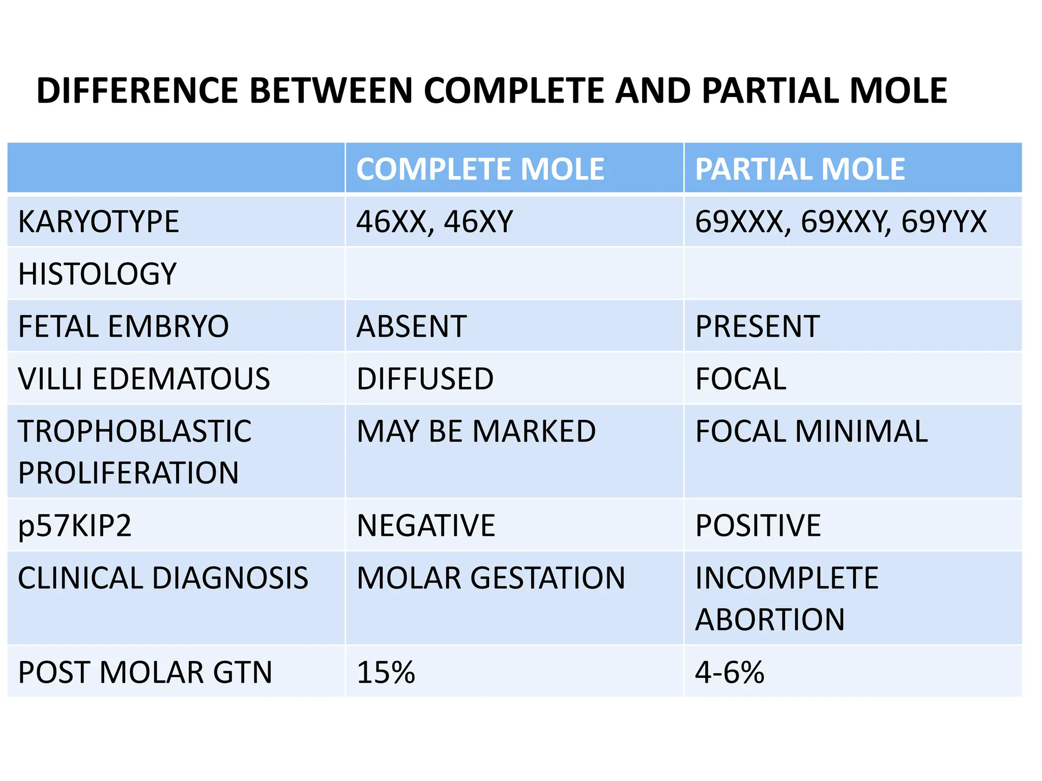
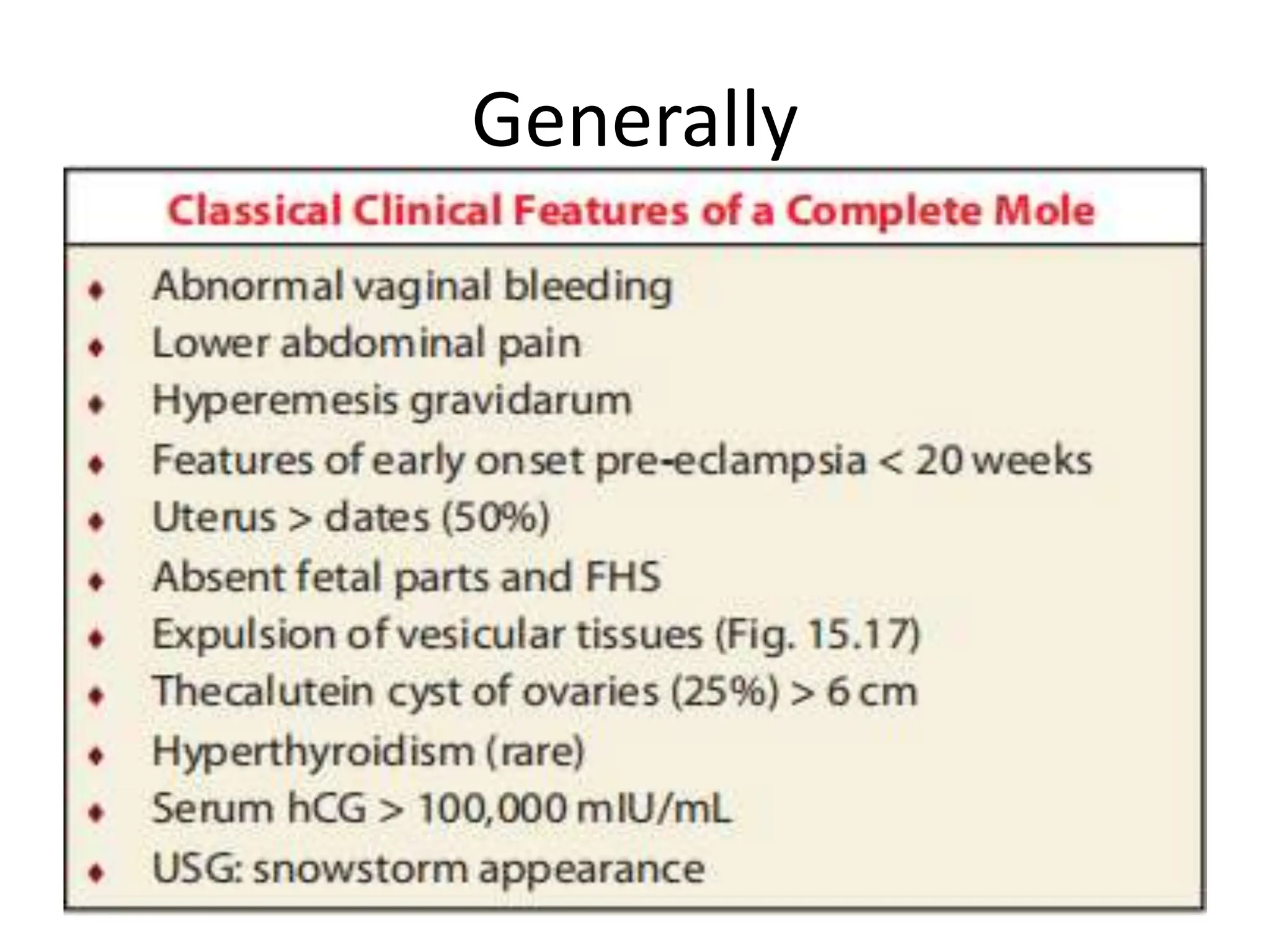
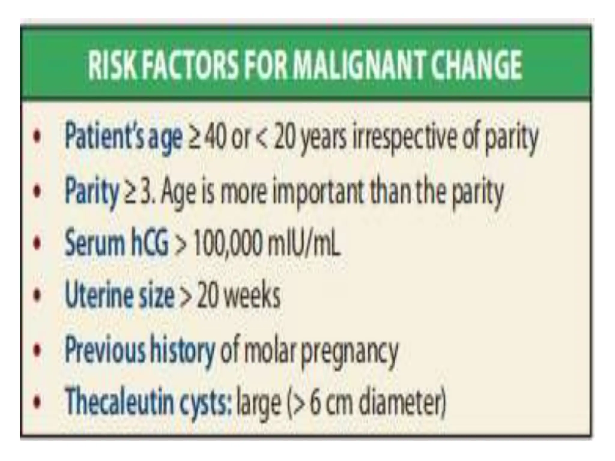
Dr.Massam
MOLAR PREGNANCY
Objectives
• Definitions
• Risk factors
• Clinical features
• Complications
• Investigations
• Treatments
• Follow up plans
Definitions
• Molar pregnancy is one of the disease conditions
originating from the trophoblast (placenta).
• These disease conditions are collectively called the
Gestational trophoblastic disease (GTD)
• Apart from molar pregnancy, other GTD are:-
– Placental site trophoblastic tumours
– Choriocarcinomas
– Invasive mole
Definitions
• Molar pregnancy- abnormal form of pregnancy in
which a non viable fertilized egg implants in the
uterus and will fail to come to term.
• Is a gestational trophoblastic disease (GTD)
• Occurs when a fertilized egg does not contain an
original maternal nucleus i.e. an empty egg + sperm
Modified WHO classification of
GTD
Molar pregnancies:
1. Hydatidiform mole
-Complete mole
-Partial mole
2. Invasive mole
Trophoblastic tumors:
1. Choriocarcinoma
2. Placenta site
trophoblastic tumor
(PSTT)
3. Epithelioid trophoblastic
tumor (ETT)
MOLAR PREGNANCY/HYDATIDIFORM MOLE
• Molar pregnancy as hydatidiform mole is divided into
two types:-
1. Complete moles
2. Partial moles
Complete moles
• A single sperm (abt 90%) or two sperms
(10%)..combining with an egg which has lost its DNA.
• No fetal tissue present
• 46,XX (Diploid), 46,XY (Diploid).
• 46 ,YY (diploid) is not observed
Complete Mole
• Empty ovum +23X sperm →23X
→duplicate→ 46XX (90%)
• Empty ovum + 23X →46XX
23X
• Empty ovum + 23X →46XY
23Y
Complete mole
Partial moles
• Partial moles occurs when a haploid egg is fertilized
by two sperms or by one sperm which reduplicates
itself yielding the genotype of 69,XXY (triploid) OR
92,XXXY (tetraploid)
• Fetal tissue is present (fetal erythrocytes +blood
vessels)
Partial Mole
• 23X ovum +23X sperm = 69XXX
23X
• 23X ovum +23Y sperm =69XYY
23Y
• 23X ovum +23X sperm =69XXY
23Y
Partial mole
Complete Partial
COMPLETE MOLE PARTIAL MOLE
KARYOTYPE 46,XX
46,XY
69,XXY
hCG
Uterine Size __
Fetal parts No YES
Components 2 sperms + empty egg 2 sperms + 1 egg
Risk of complications 15-20% malignant
trophoblatic disease
Low risk of malignancy
(<5%)
COMPLETE MOLE PARTIAL MOLE
KARYOTYPE 46XX, 46XY 69XXX, 69XXY, 69YYX
HISTOLOGY
FETAL EMBRYO ABSENT PRESENT
VILLI EDEMATOUS DIFFUSED FOCAL
TROPHOBLASTIC
PROLIFERATION
MAY BE MARKED FOCAL MINIMAL
p57KIP2 NEGATIVE POSITIVE
CLINICAL DIAGNOSIS MOLAR GESTATION INCOMPLETE
ABORTION
POST MOLAR GTN 15% 4-6%
DIFFERENCE BETWEEN COMPLETE AND PARTIAL MOLE
ETIOLOGY AND RISKS FACTORS
• The exact etiology is not well understood.
• Risk factors:
– Maternal age (extremes <15 & >45)
– Older paternal age
– Previous hx of GTD & Hx of spontaneous abortion
– Nutritional deficiency of carotene, folic acid, proteins and
vitamin A and high carbohydrates.
– AB blood group of the parents
– Smoking
CLINICAL FEATURES - Complete moles
• Vaginal bleeding-uterus may become distended by
large amount of blood + dark fluid may leak into the
vagina-50%
• Hyperemesis –severe nausea +vomiting
• Thyrotoxic features of tremors or tachycardia (2%).
It is probably due to increased chorionic thyrotropin
• Varying degree of lower abdominal pain
Clinical features - Complete mole
• Breathlessness due to pulmonary embolization of
the trophoblastic cells (2%).
• Expulsion of grape like vesicles per vagina is
diagnostic of vesicular mole. Actually, in
approximately 50% of cases the mole is not
suspected until it is expelled in part or whole.
• History of quickening is absent.
Generally
Clinical features - Partial moles
• Usually mimic the symptoms of incomplete /missed
abortion
• Vaginal bleeding + absence of fetal heart beats
Physical examination
Complete moles
• Large uterus than expected GA - excessive
trophoblastic growth + retained blood
• Pre eclampsia _2%
• Theca lutein cyst – ovarian cyst > 6 cm in diameters,
increased hCG , GA > 10 Weeks.Presents with
pressure or pelvic pain, regress after evacuation – up
to 12 weeks.
Physical examination con't...
• Features suggestive of early months of
pregnancy are evident.
• The patient looks more ill than can be
accounted for.
• Pallor
• The feel of the uterus is firm elastic (doughy).
• Fetal parts are not felt, nor any fetal
movements.
• Absence of fetal heart sound
Physical examination – PARTIAL MOLES
• Uterine enlargement than GA
• PRE ECLAMPSIA
COMPLICATIONS
• Perforation of the uterus
• Hemorrhage
• Disseminated intravascular coagulation
• Acute respiratory distress- trophoblastic embolism
esp with a large uterus than gestation age.
Complications con't...
• Pre-eclampsia
• Sepsis as no protective layer the vaginal organisms
can invade the uterus
• The development of choriocarcinoma in 2 -10%
DIFFERENTIAL DIAGNOSIS
• Hyperemesis gravidarum
• Hypertension
• Hyperthyroidism/Thyrotoxicosis
• Abortion
INVESTIGATIONS
NB: Most of the investigations are done at hospital level,
consider referral
• Quantitative beta –Hcg levels , hCG >100,000 mIU/mL, may
indicates trophoblastic growth.
• Full blood picture- Hb- anemia??, platelets –
coagulopathy??
• Ultrasound – bunch of grapes or honeycombed uterus or
snow storm appearance, (intrauterine mass containing
many small cysts)
• Chest X ray- lungs primary site of metastatis
• Histology
 Complete moles - oedematous placenta villi,
hyperplasia of trophoblast, no fetal tissue
 Incomplete moles - oedematous villi, trophoblastic
proliferation.
TREATMENT
• Also consider referral when at dispensary or health
center level
• Blood transfusion, if anaemic
• Fresh frozen plasma - DIC
• Contraception for 6 month
TREATMENT
• SURGICAL CARE
 Evacuation of the uterus by dilation and curettage
 IV Oxytocin is used after the dilation of the cervix at
the initiation of suctioning and postoperatively to
reduce likelyhood of hemorrhage
PROGNOSIS
• Complete hydatidiform mole - 15-20% risk of
developing into choriocarcinoma and 15% to invasive
mole. Accounts for 50% of all cases of
choriocarcinoma.
• Incomplete moles can become invasive (<5%) but not
choriocarcinoma
• One percent of women with molar pregnacy may
have recurrence
MONITORING
• Serial quantitative beta hCG
 Weekly until levels are within reference range for 3-4
weeks
 hCG levels should consistently drop,never increase,
and should get back to normal (8-12 weeks) after
evacuation
 Once reference range reached (3-4 weeks) , continue
checking monthly for 6 month
MONITORING
 If the level of hCG plateau or rise ,think of malignant
transformation.
 Effective contraception- if IUD was chosen , to avoid
perforation and bleeding , insertion should wait for
uterus involution to occur and normalization of
serum hCG levels
Key points
• Molar pregnancy is one among the diseases called
the Gestational Trophoblastic Diseases (GTD)
• A doctor has to diagnose proparly and provide
appropriate management.
Evaluation
• How a molar pregnancy is formed?
• What are the clinical features of a complete molar?
• What are the risk factors for a molar pregnancy to
turn into malignancy?
Reference
• DC Dutta's Textbook of Obstetrics 7th Edition

Molar Pregnancy Complete Vs Incomplete Hydatidiform Mole.pptxpptx

  • 1.
  • 2.
    Objectives • Definitions • Riskfactors • Clinical features • Complications • Investigations • Treatments • Follow up plans
  • 3.
    Definitions • Molar pregnancyis one of the disease conditions originating from the trophoblast (placenta). • These disease conditions are collectively called the Gestational trophoblastic disease (GTD) • Apart from molar pregnancy, other GTD are:- – Placental site trophoblastic tumours – Choriocarcinomas – Invasive mole
  • 4.
    Definitions • Molar pregnancy-abnormal form of pregnancy in which a non viable fertilized egg implants in the uterus and will fail to come to term. • Is a gestational trophoblastic disease (GTD) • Occurs when a fertilized egg does not contain an original maternal nucleus i.e. an empty egg + sperm
  • 5.
    Modified WHO classificationof GTD Molar pregnancies: 1. Hydatidiform mole -Complete mole -Partial mole 2. Invasive mole Trophoblastic tumors: 1. Choriocarcinoma 2. Placenta site trophoblastic tumor (PSTT) 3. Epithelioid trophoblastic tumor (ETT)
  • 6.
    MOLAR PREGNANCY/HYDATIDIFORM MOLE •Molar pregnancy as hydatidiform mole is divided into two types:- 1. Complete moles 2. Partial moles
  • 7.
    Complete moles • Asingle sperm (abt 90%) or two sperms (10%)..combining with an egg which has lost its DNA. • No fetal tissue present • 46,XX (Diploid), 46,XY (Diploid). • 46 ,YY (diploid) is not observed
  • 8.
    Complete Mole • Emptyovum +23X sperm →23X →duplicate→ 46XX (90%) • Empty ovum + 23X →46XX 23X • Empty ovum + 23X →46XY 23Y
  • 9.
  • 10.
    Partial moles • Partialmoles occurs when a haploid egg is fertilized by two sperms or by one sperm which reduplicates itself yielding the genotype of 69,XXY (triploid) OR 92,XXXY (tetraploid) • Fetal tissue is present (fetal erythrocytes +blood vessels)
  • 11.
    Partial Mole • 23Xovum +23X sperm = 69XXX 23X • 23X ovum +23Y sperm =69XYY 23Y • 23X ovum +23X sperm =69XXY 23Y
  • 12.
  • 13.
    Complete Partial COMPLETE MOLEPARTIAL MOLE KARYOTYPE 46,XX 46,XY 69,XXY hCG Uterine Size __ Fetal parts No YES Components 2 sperms + empty egg 2 sperms + 1 egg Risk of complications 15-20% malignant trophoblatic disease Low risk of malignancy (<5%)
  • 14.
    COMPLETE MOLE PARTIALMOLE KARYOTYPE 46XX, 46XY 69XXX, 69XXY, 69YYX HISTOLOGY FETAL EMBRYO ABSENT PRESENT VILLI EDEMATOUS DIFFUSED FOCAL TROPHOBLASTIC PROLIFERATION MAY BE MARKED FOCAL MINIMAL p57KIP2 NEGATIVE POSITIVE CLINICAL DIAGNOSIS MOLAR GESTATION INCOMPLETE ABORTION POST MOLAR GTN 15% 4-6% DIFFERENCE BETWEEN COMPLETE AND PARTIAL MOLE
  • 15.
    ETIOLOGY AND RISKSFACTORS • The exact etiology is not well understood. • Risk factors: – Maternal age (extremes <15 & >45) – Older paternal age – Previous hx of GTD & Hx of spontaneous abortion – Nutritional deficiency of carotene, folic acid, proteins and vitamin A and high carbohydrates. – AB blood group of the parents – Smoking
  • 16.
    CLINICAL FEATURES -Complete moles • Vaginal bleeding-uterus may become distended by large amount of blood + dark fluid may leak into the vagina-50% • Hyperemesis –severe nausea +vomiting • Thyrotoxic features of tremors or tachycardia (2%). It is probably due to increased chorionic thyrotropin • Varying degree of lower abdominal pain
  • 17.
    Clinical features -Complete mole • Breathlessness due to pulmonary embolization of the trophoblastic cells (2%). • Expulsion of grape like vesicles per vagina is diagnostic of vesicular mole. Actually, in approximately 50% of cases the mole is not suspected until it is expelled in part or whole. • History of quickening is absent.
  • 18.
  • 19.
    Clinical features -Partial moles • Usually mimic the symptoms of incomplete /missed abortion • Vaginal bleeding + absence of fetal heart beats
  • 20.
    Physical examination Complete moles •Large uterus than expected GA - excessive trophoblastic growth + retained blood • Pre eclampsia _2% • Theca lutein cyst – ovarian cyst > 6 cm in diameters, increased hCG , GA > 10 Weeks.Presents with pressure or pelvic pain, regress after evacuation – up to 12 weeks.
  • 21.
    Physical examination con't... •Features suggestive of early months of pregnancy are evident. • The patient looks more ill than can be accounted for. • Pallor • The feel of the uterus is firm elastic (doughy). • Fetal parts are not felt, nor any fetal movements. • Absence of fetal heart sound
  • 22.
    Physical examination –PARTIAL MOLES • Uterine enlargement than GA • PRE ECLAMPSIA
  • 23.
    COMPLICATIONS • Perforation ofthe uterus • Hemorrhage • Disseminated intravascular coagulation • Acute respiratory distress- trophoblastic embolism esp with a large uterus than gestation age.
  • 24.
    Complications con't... • Pre-eclampsia •Sepsis as no protective layer the vaginal organisms can invade the uterus • The development of choriocarcinoma in 2 -10%
  • 25.
    DIFFERENTIAL DIAGNOSIS • Hyperemesisgravidarum • Hypertension • Hyperthyroidism/Thyrotoxicosis • Abortion
  • 26.
    INVESTIGATIONS NB: Most ofthe investigations are done at hospital level, consider referral • Quantitative beta –Hcg levels , hCG >100,000 mIU/mL, may indicates trophoblastic growth. • Full blood picture- Hb- anemia??, platelets – coagulopathy?? • Ultrasound – bunch of grapes or honeycombed uterus or snow storm appearance, (intrauterine mass containing many small cysts)
  • 27.
    • Chest Xray- lungs primary site of metastatis • Histology  Complete moles - oedematous placenta villi, hyperplasia of trophoblast, no fetal tissue  Incomplete moles - oedematous villi, trophoblastic proliferation.
  • 28.
    TREATMENT • Also considerreferral when at dispensary or health center level • Blood transfusion, if anaemic • Fresh frozen plasma - DIC • Contraception for 6 month
  • 29.
    TREATMENT • SURGICAL CARE Evacuation of the uterus by dilation and curettage  IV Oxytocin is used after the dilation of the cervix at the initiation of suctioning and postoperatively to reduce likelyhood of hemorrhage
  • 30.
    PROGNOSIS • Complete hydatidiformmole - 15-20% risk of developing into choriocarcinoma and 15% to invasive mole. Accounts for 50% of all cases of choriocarcinoma. • Incomplete moles can become invasive (<5%) but not choriocarcinoma • One percent of women with molar pregnacy may have recurrence
  • 32.
    MONITORING • Serial quantitativebeta hCG  Weekly until levels are within reference range for 3-4 weeks  hCG levels should consistently drop,never increase, and should get back to normal (8-12 weeks) after evacuation  Once reference range reached (3-4 weeks) , continue checking monthly for 6 month
  • 33.
    MONITORING  If thelevel of hCG plateau or rise ,think of malignant transformation.  Effective contraception- if IUD was chosen , to avoid perforation and bleeding , insertion should wait for uterus involution to occur and normalization of serum hCG levels
  • 34.
    Key points • Molarpregnancy is one among the diseases called the Gestational Trophoblastic Diseases (GTD) • A doctor has to diagnose proparly and provide appropriate management.
  • 35.
    Evaluation • How amolar pregnancy is formed? • What are the clinical features of a complete molar? • What are the risk factors for a molar pregnancy to turn into malignancy?
  • 36.
    Reference • DC Dutta'sTextbook of Obstetrics 7th Edition

Editor's Notes

  • #6 Extreme ages- commonly for complete mole Older maternal or parternal age – partial moles … it is thought that its due to increased rate of abnormal fertilization of the ova Hx of GTD – increases risk by 10 fold Hx of abortion 2 fold Cocs doubles the risk Lack of carotene and vit a common for complete moles Smokin/irregular menses – for partial moles
  • #10 Differ from partial in regard to their karyotype and histological appearance and clinical ppt Diploid karyotype 46xx/46xy and have paternal chromosomes in origin The ovum is fertilized by haploid sperm and thru meosis it duplicates Nuclear dna is entirely paternal and mitochondrial dna remains maternal origin
  • #12 Microscopically – enlarged edematous villi+ abnormal troph proliferation involving the entire placenta Macroscopically – cluster of vesicles – bunch of grape appearance No fetal tissue or amnion is produced in this case *serum free thyroxine levels are elevated as a consequence of thyrotropin like effect on bhcg
  • #13 Have triploid karyotype 69xxx,69xxy, 69xyy One maternal and 2 parternal haploid sets of chromosomes, the coexisting fetus is usually nonviable with multiple malformation
  • #15 Most contain fetal tissue and amnion in addition to placental tissue. Patient present with sign and symptoms of missed abortion or incomplete Trophoblastic proliferation is focal and bhcg doesn’t exceed 100,000miu/ml. usual not identified until post histological findings after curratings