Downloaded 114 times

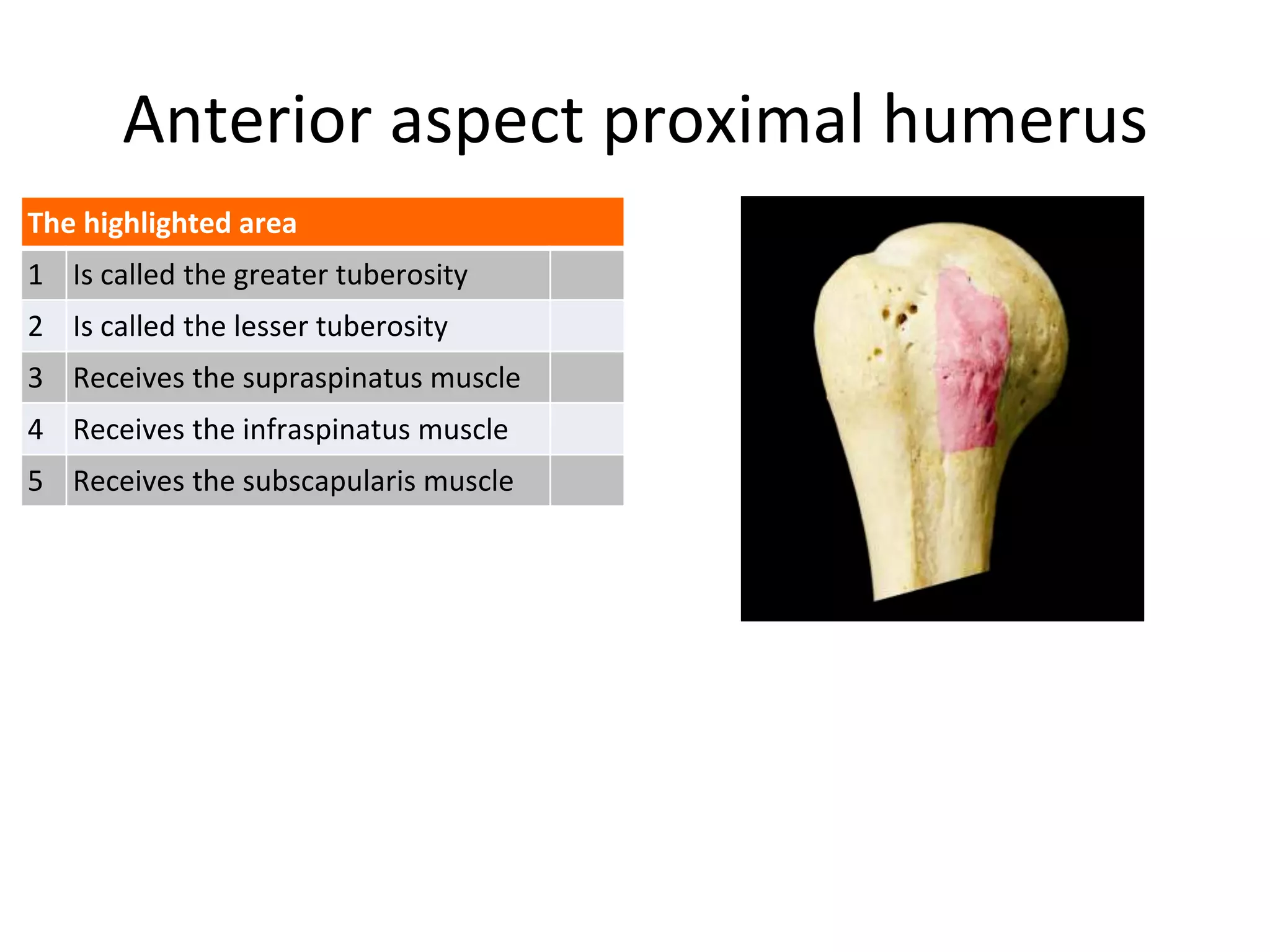
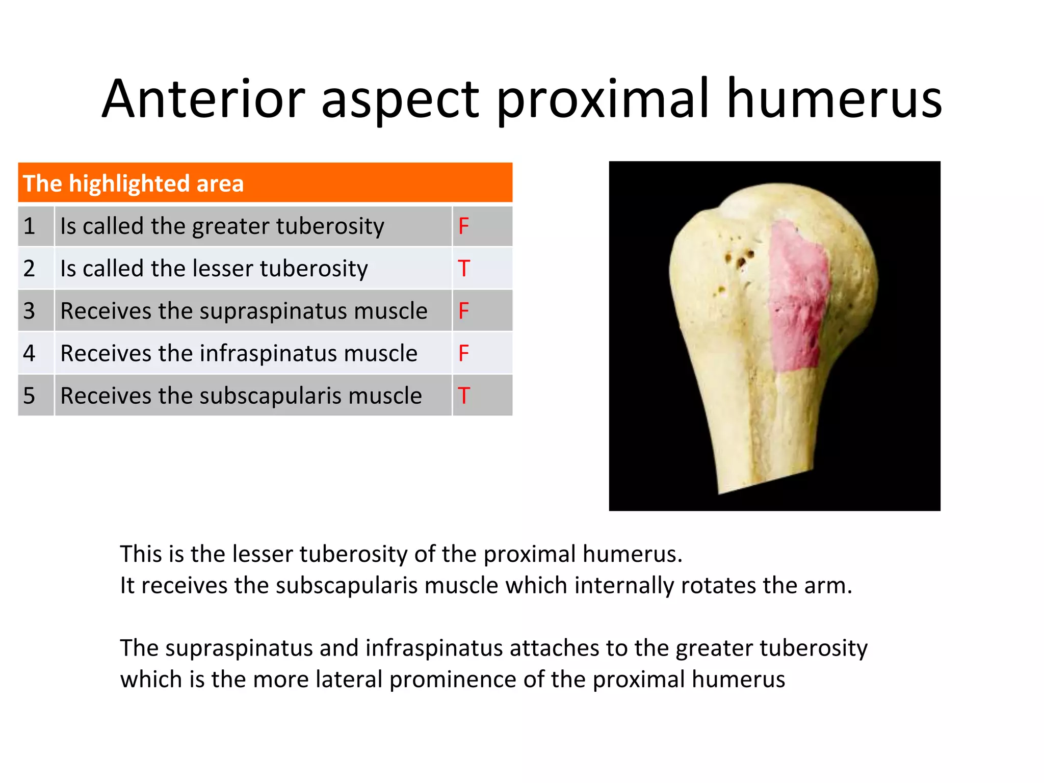
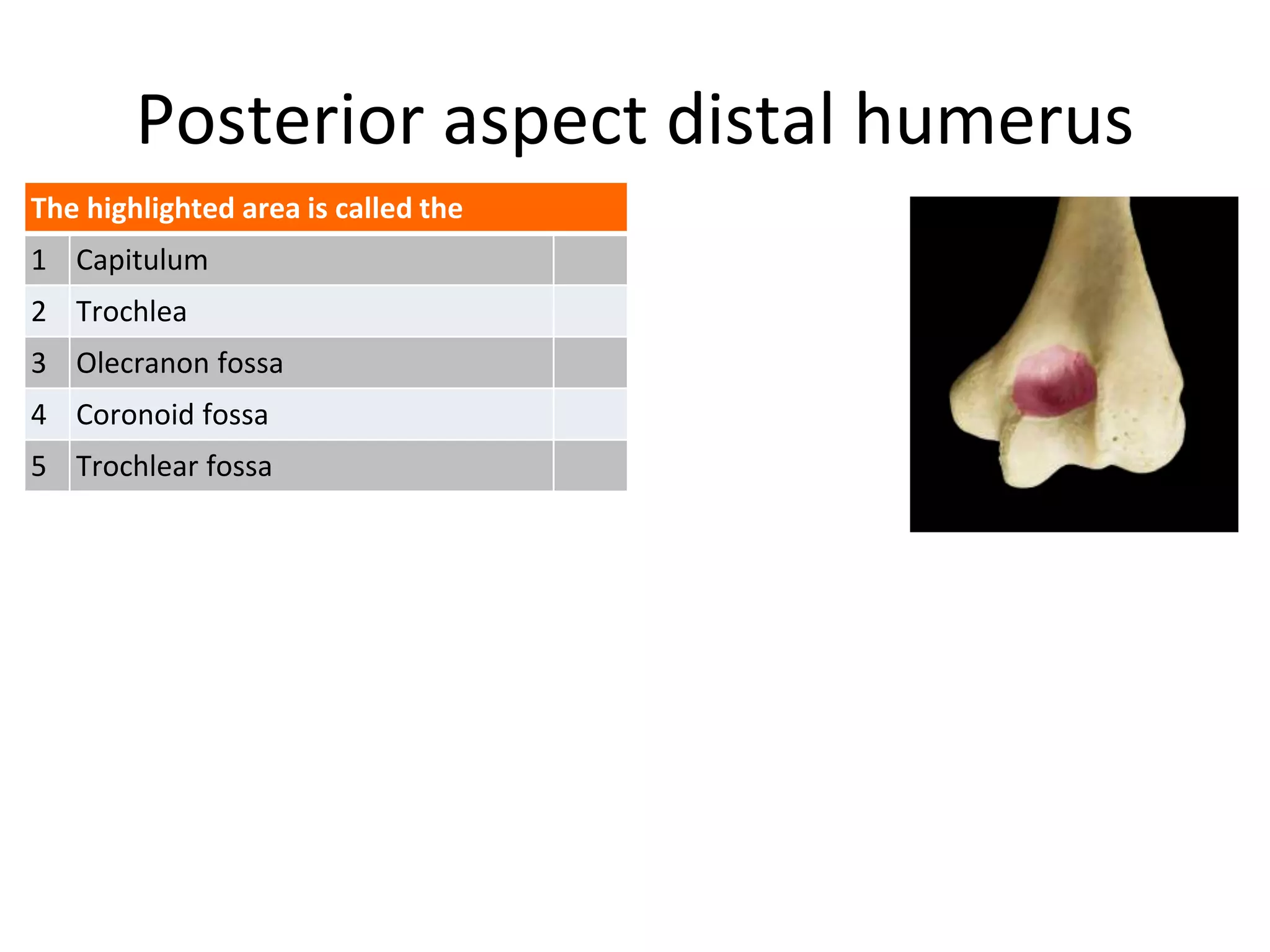
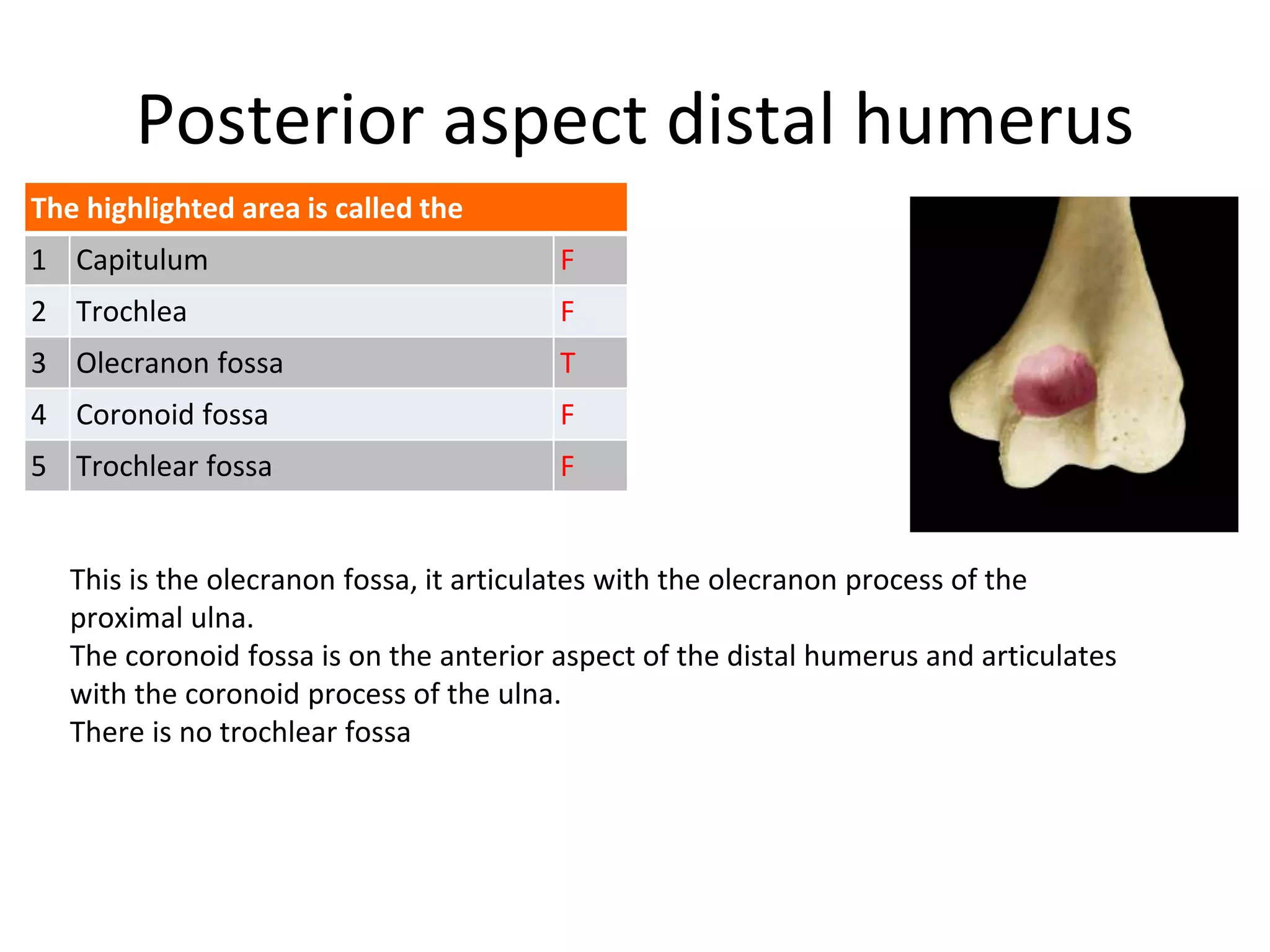
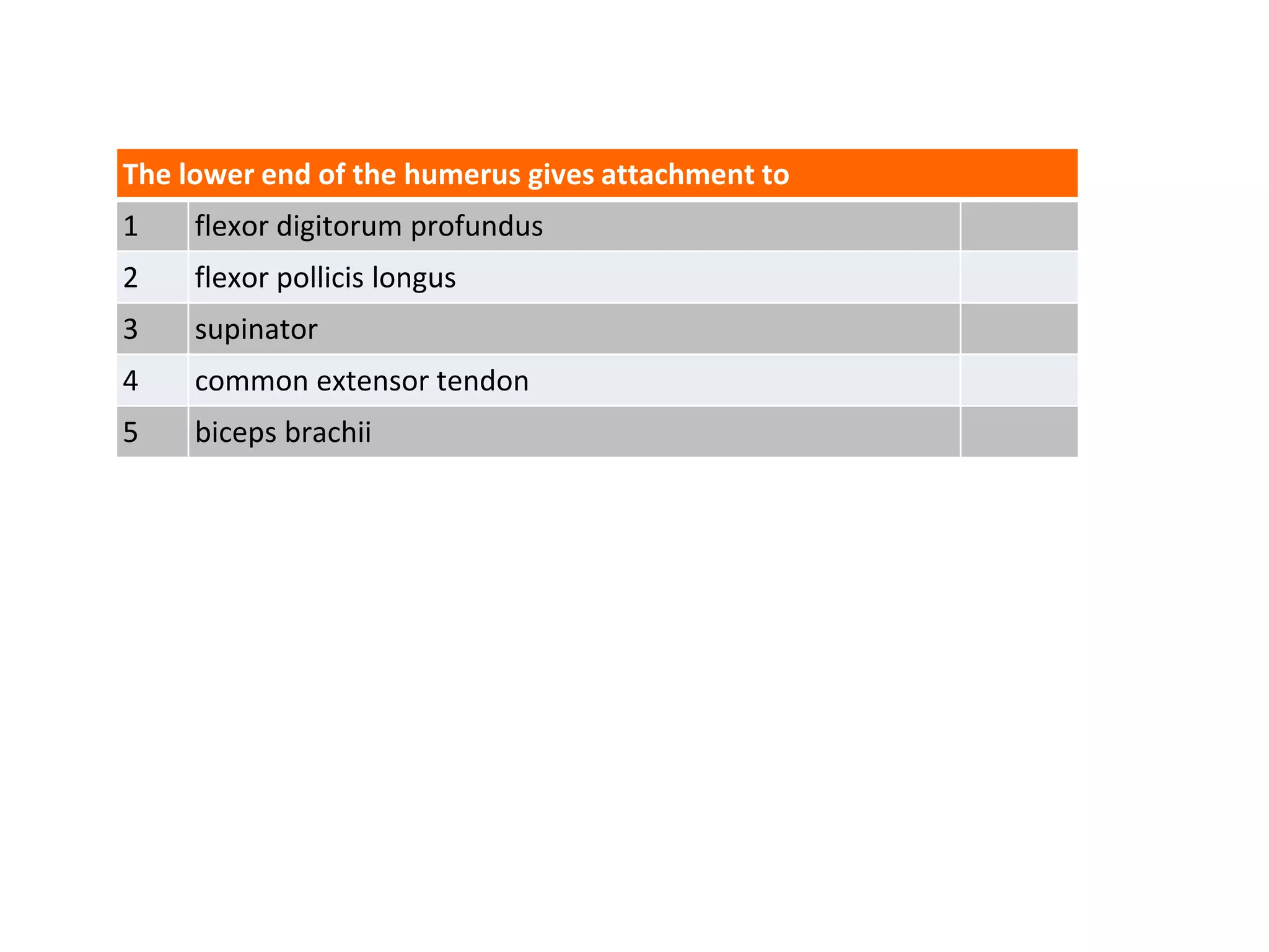
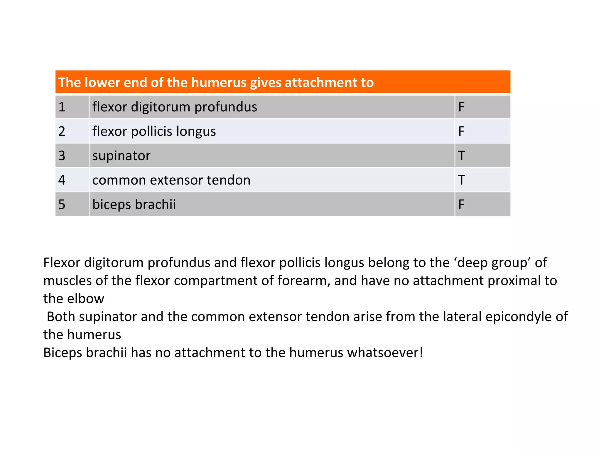
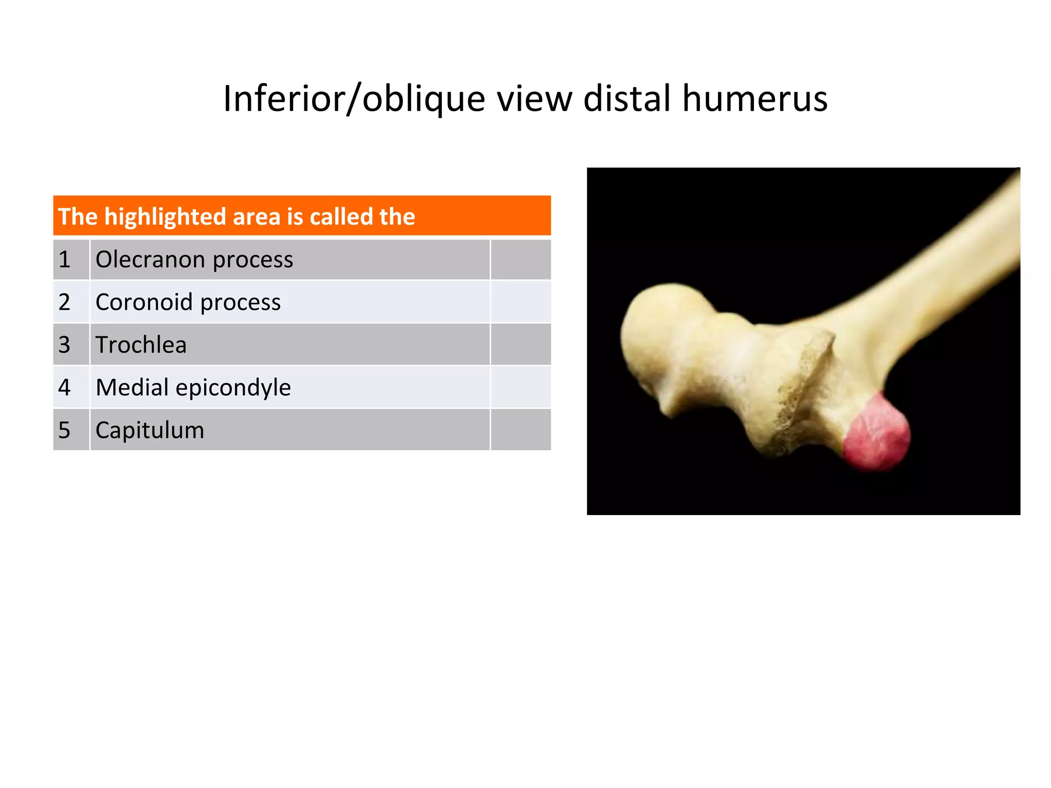
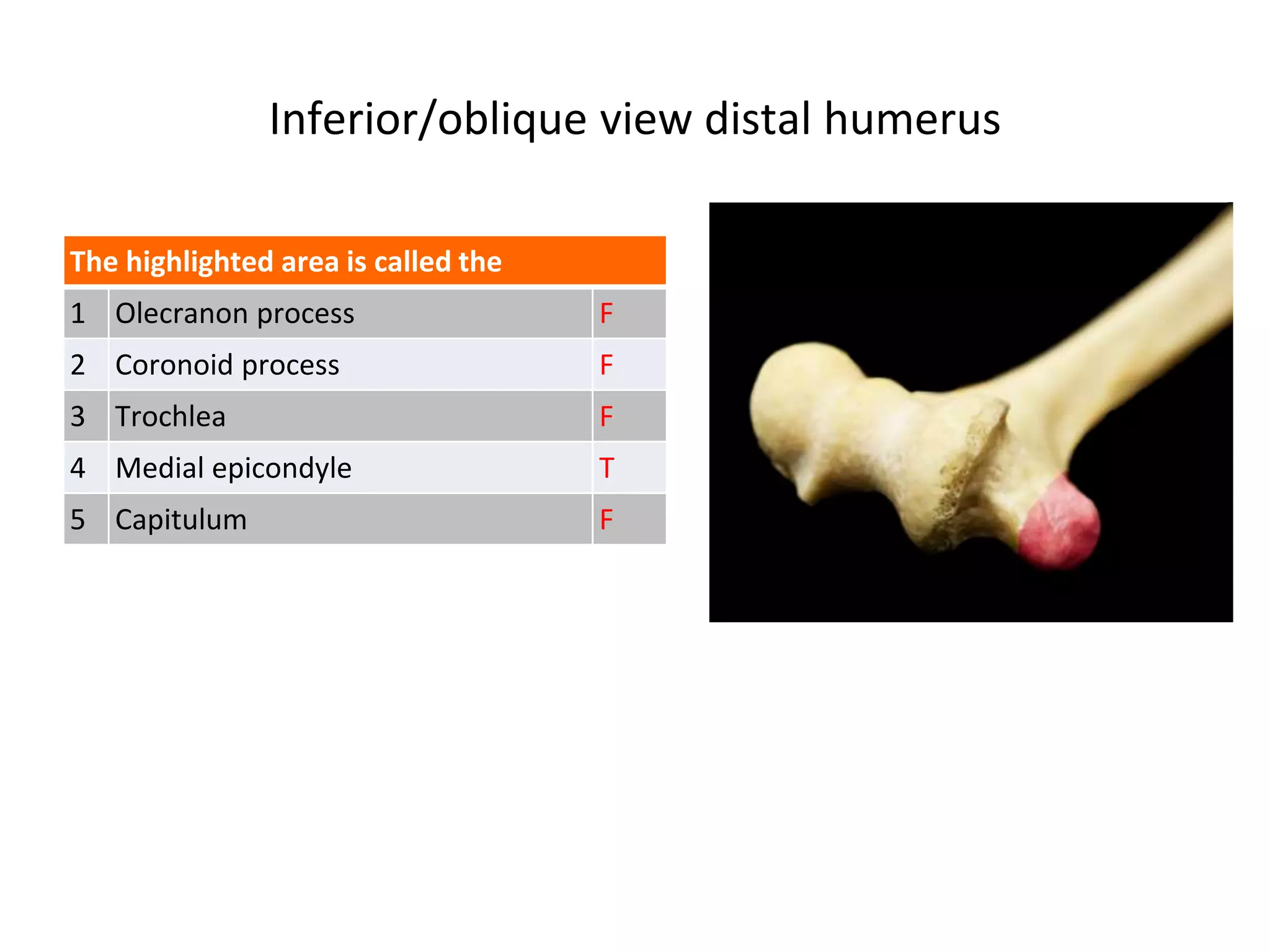
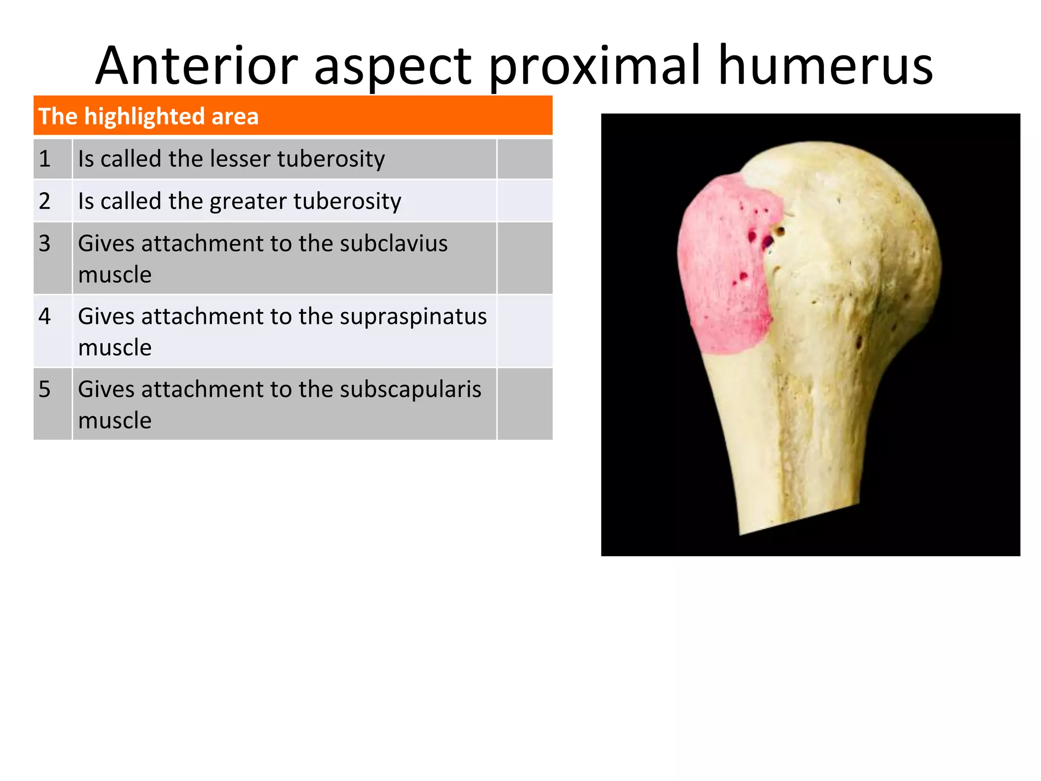
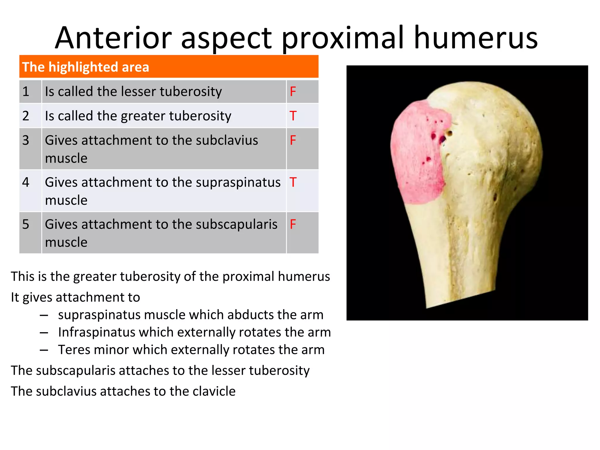
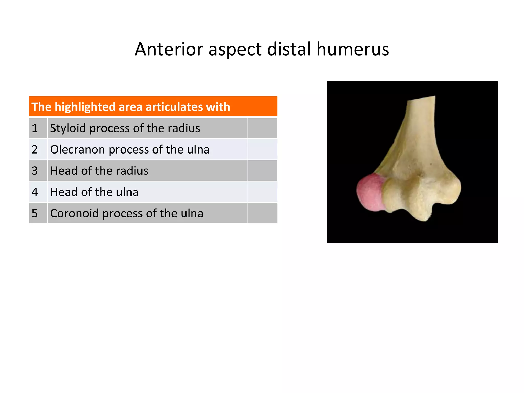
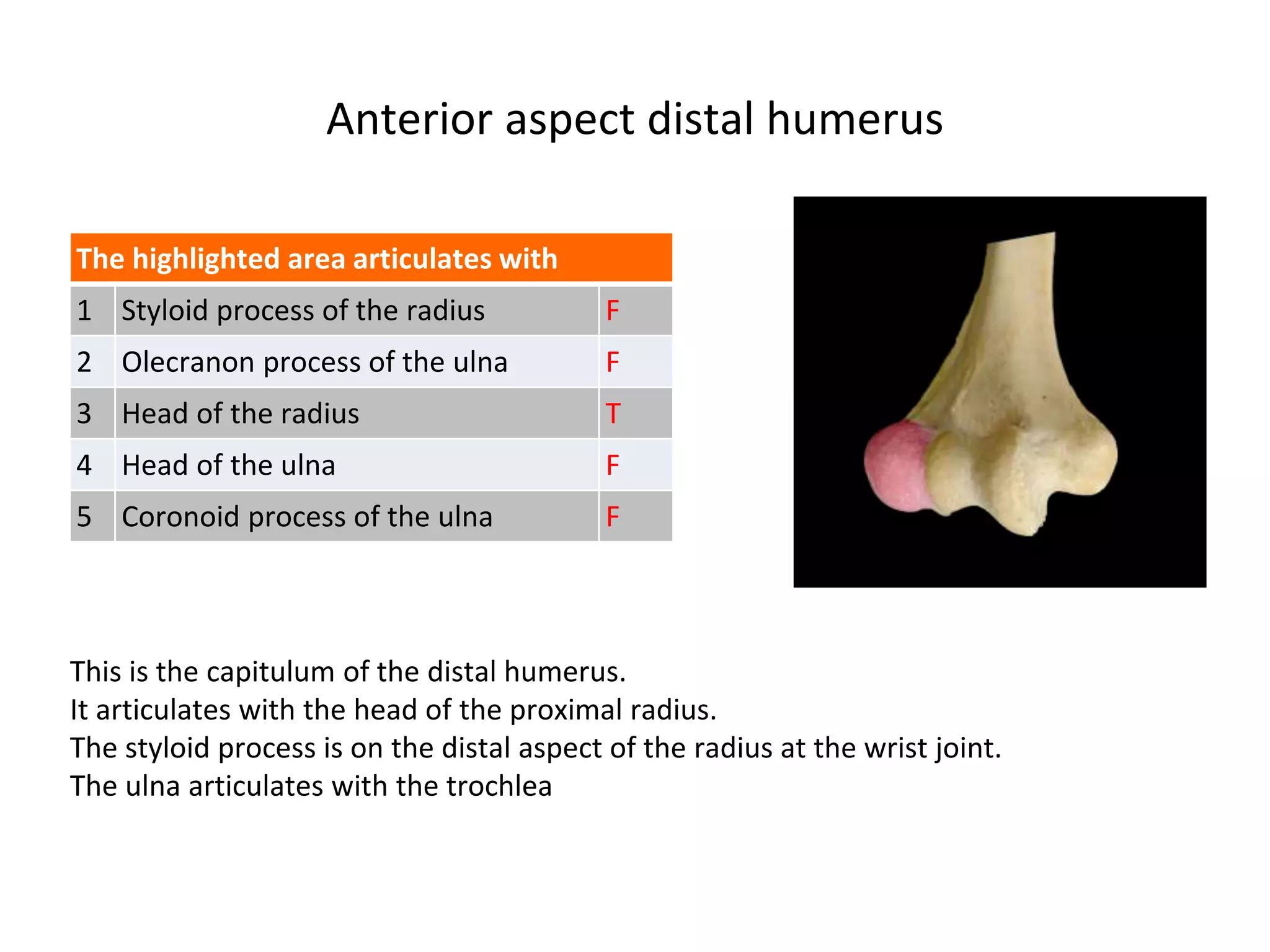
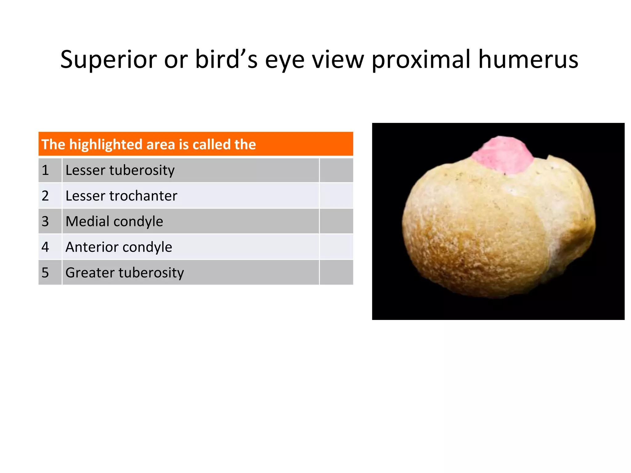

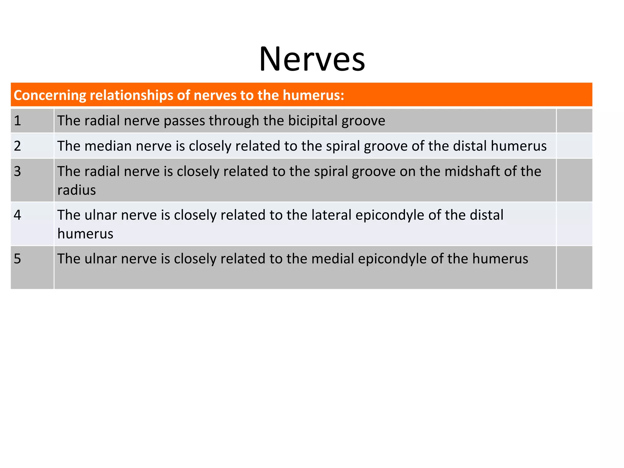
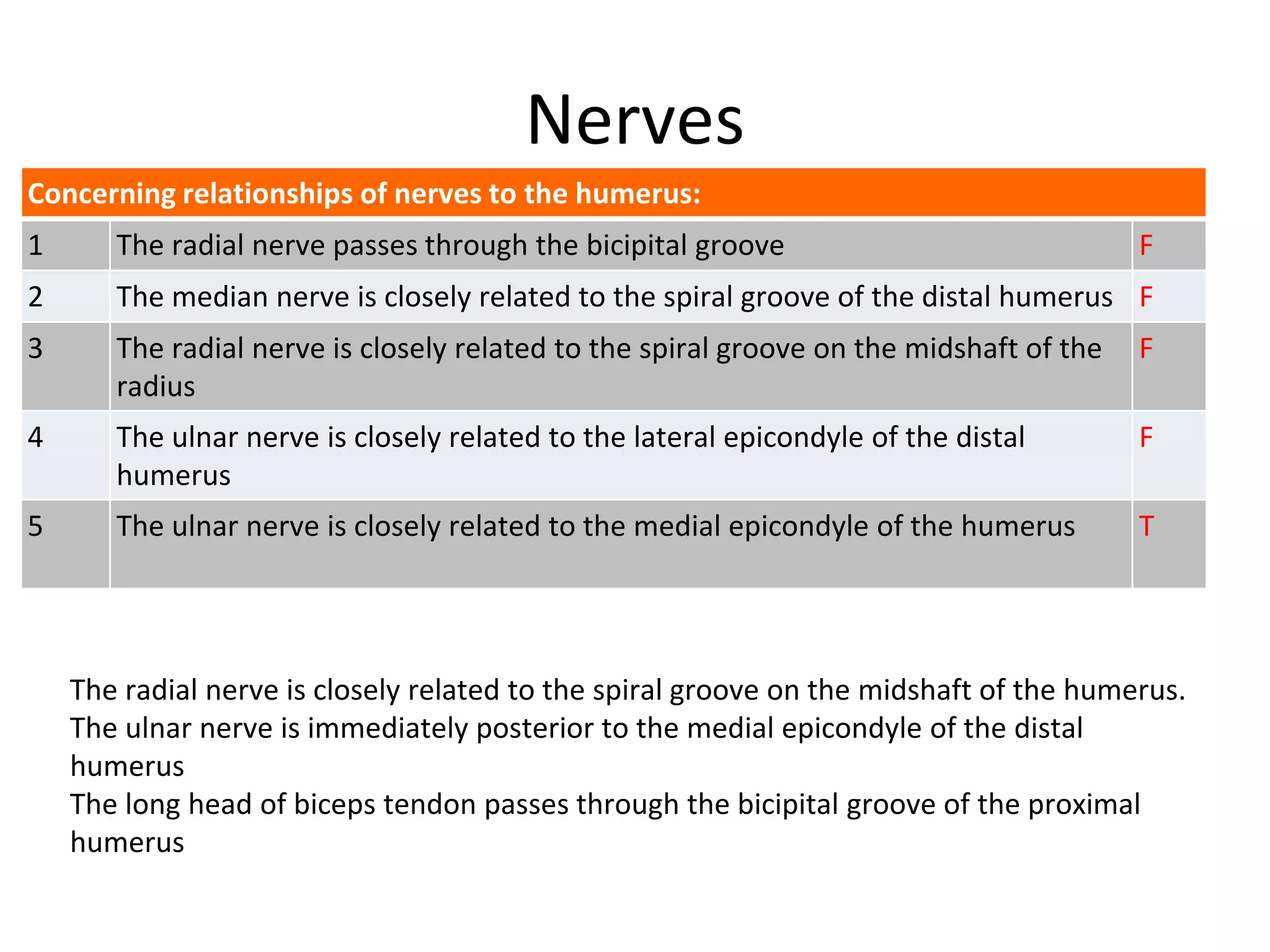
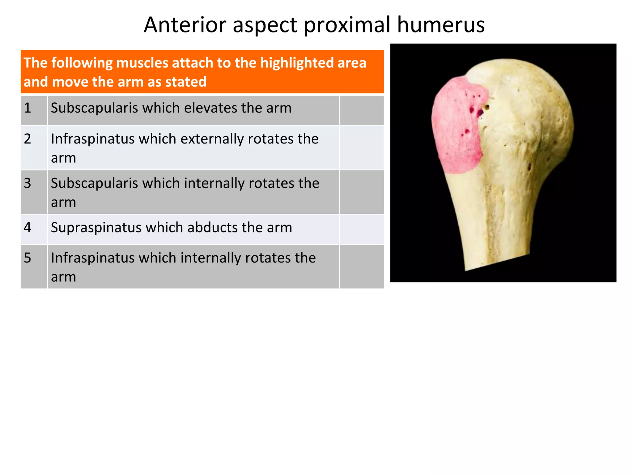
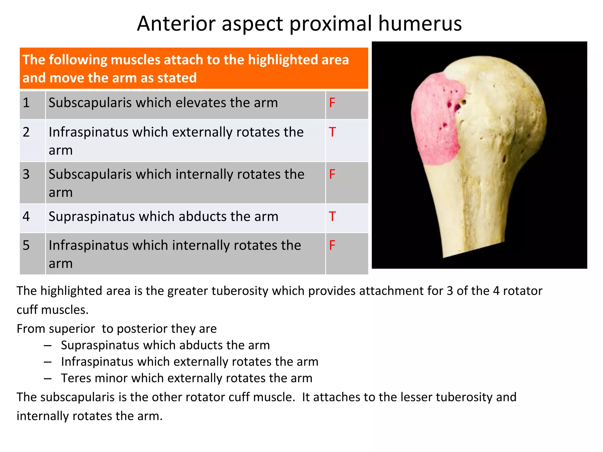

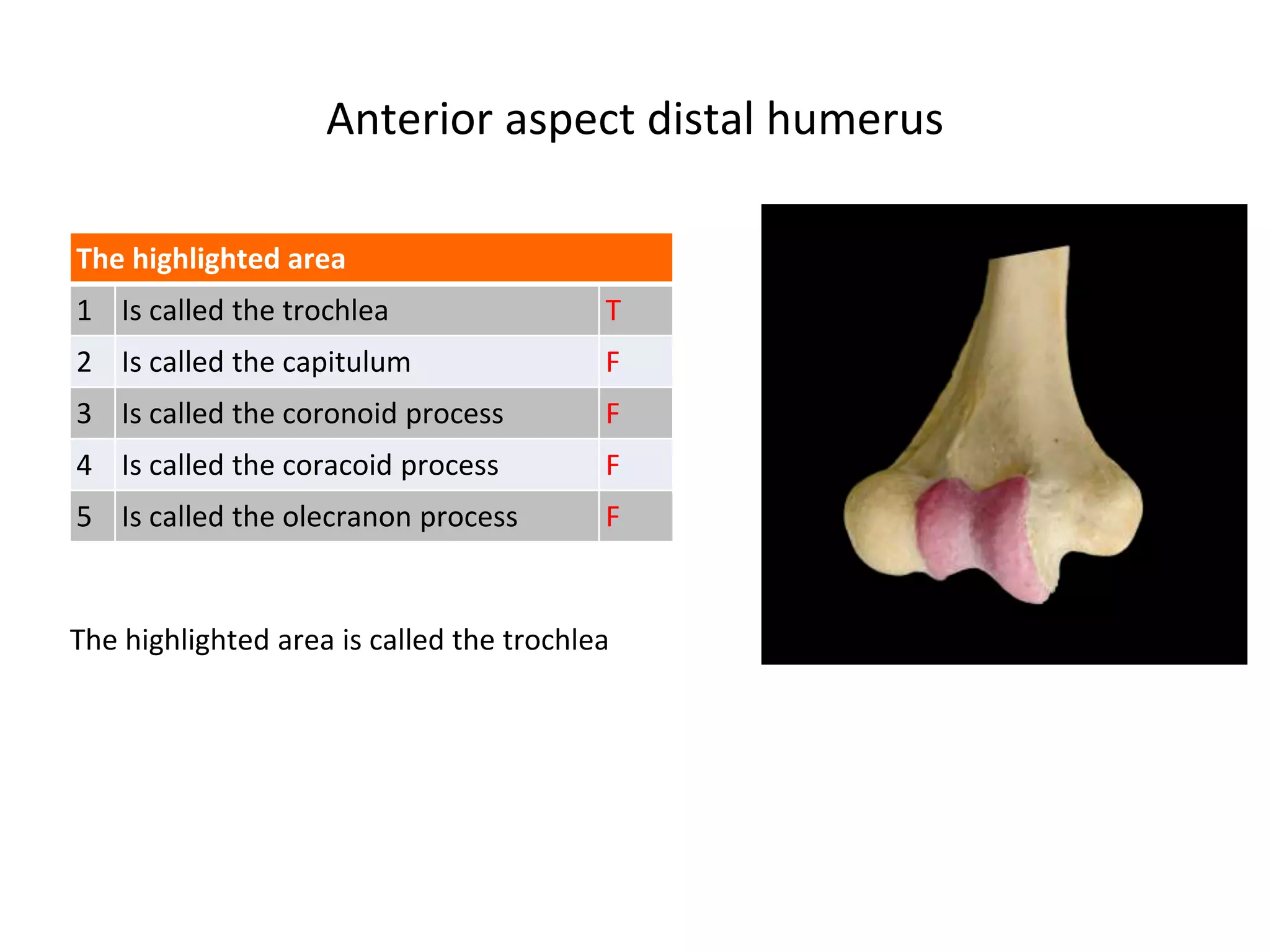
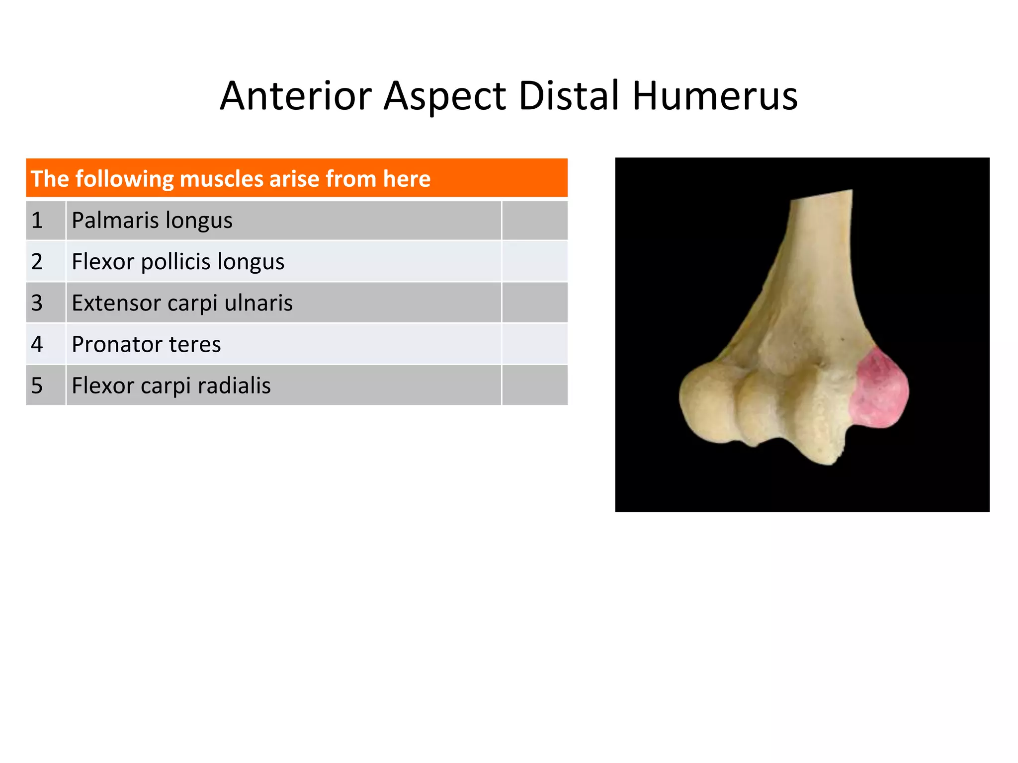

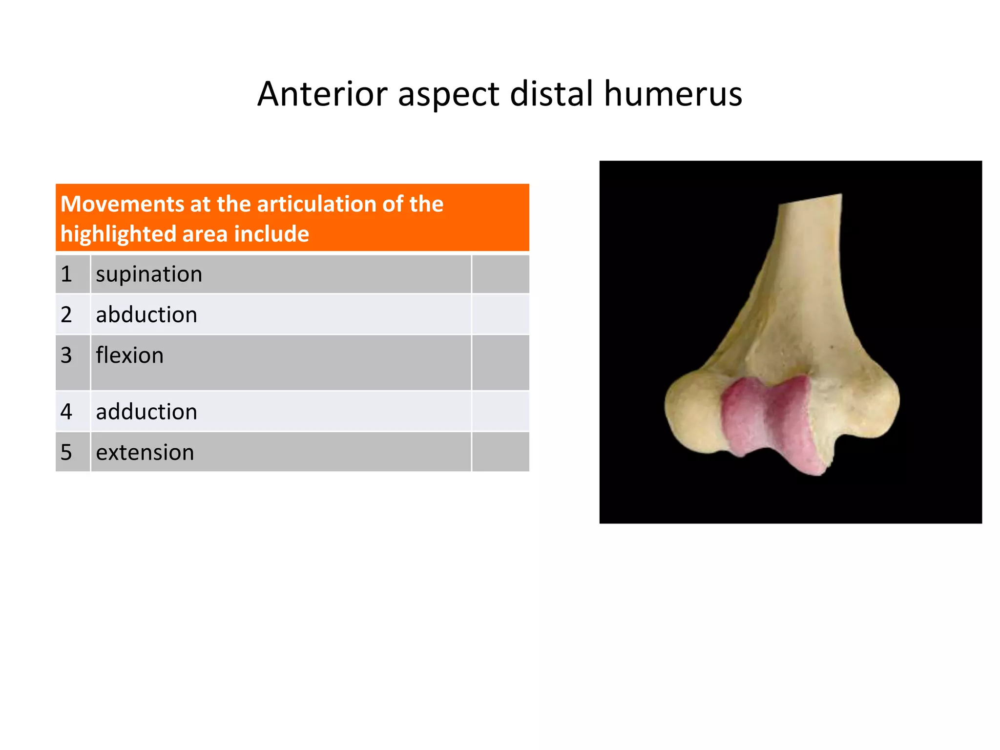
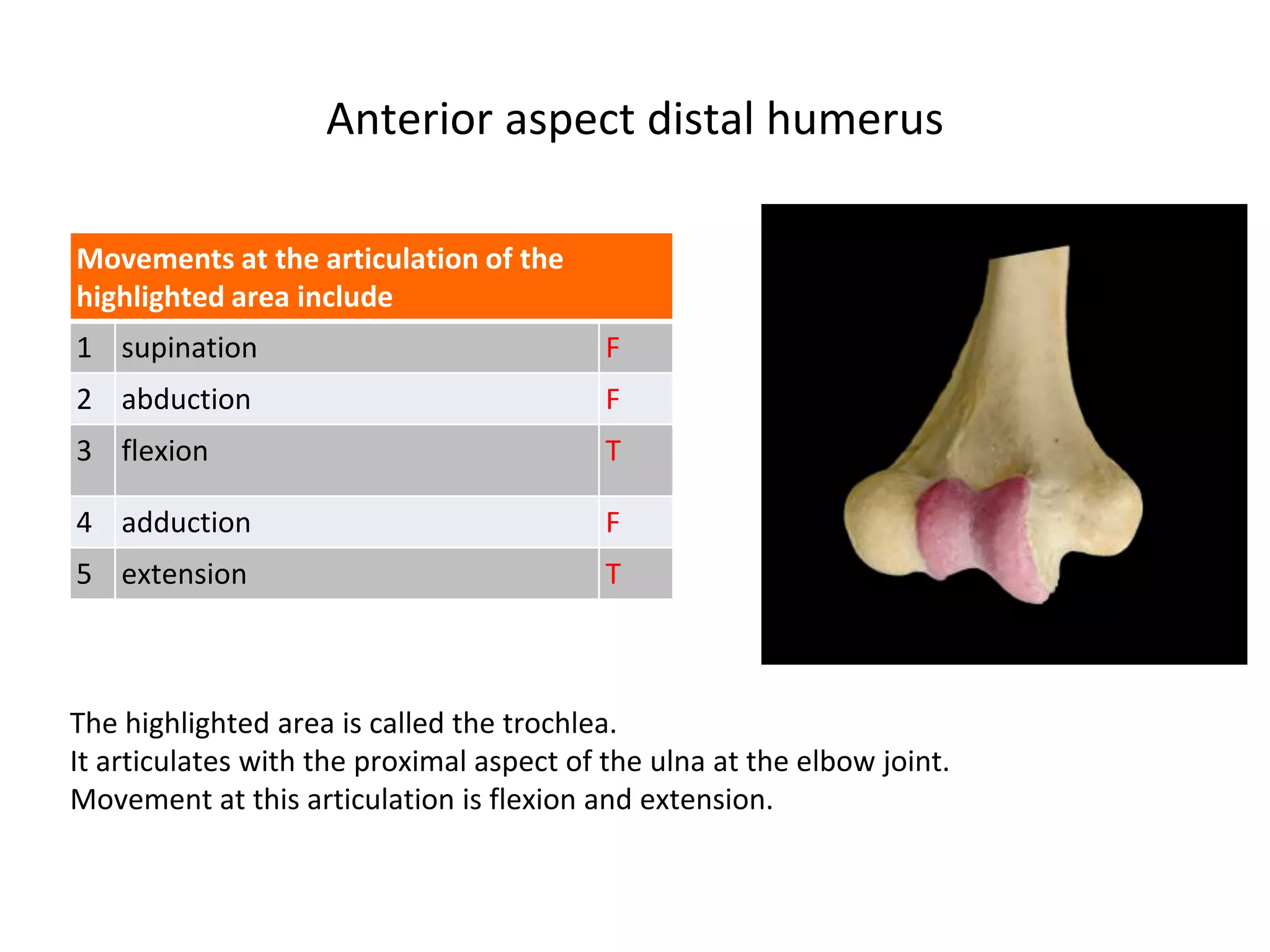

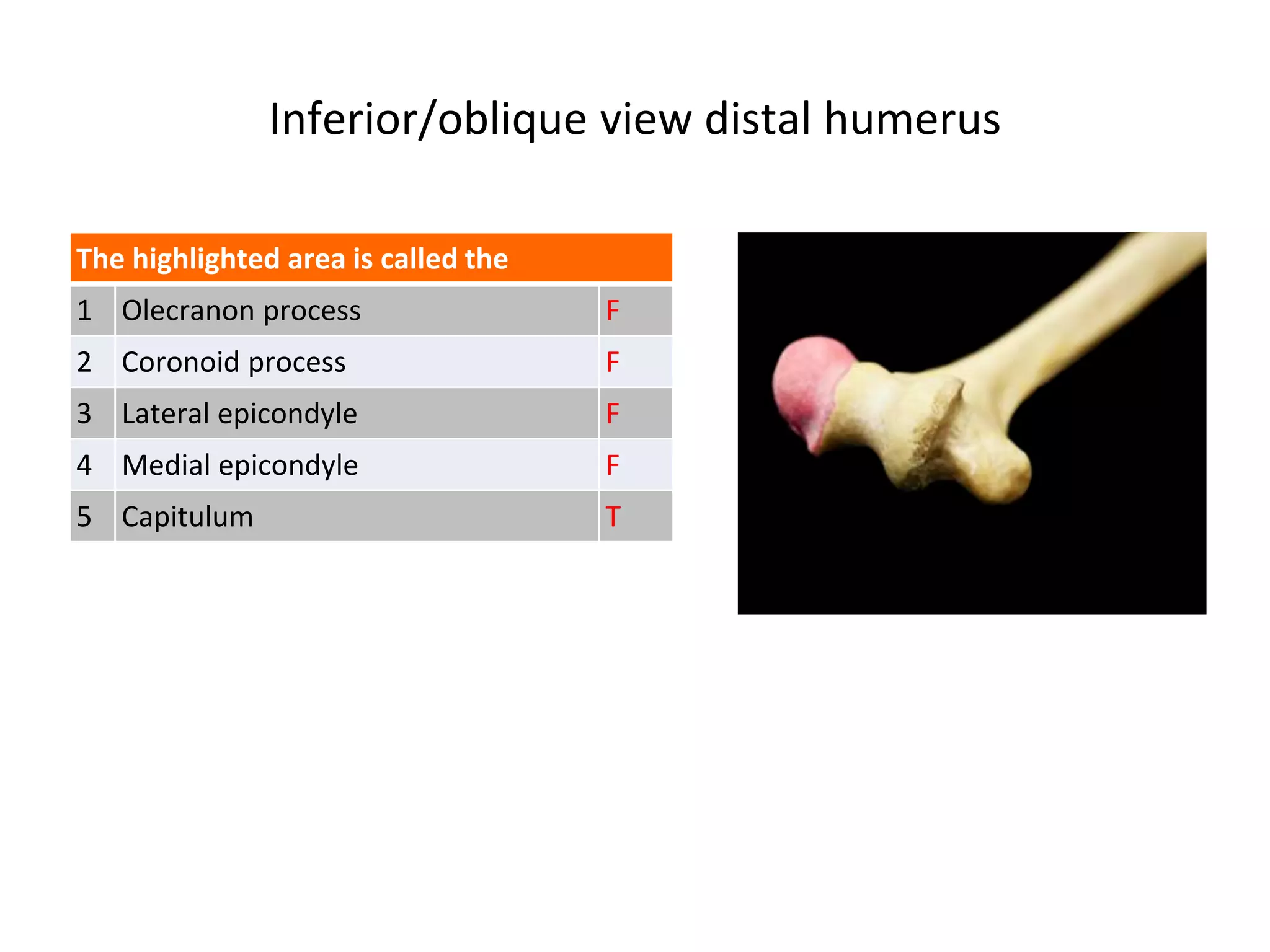


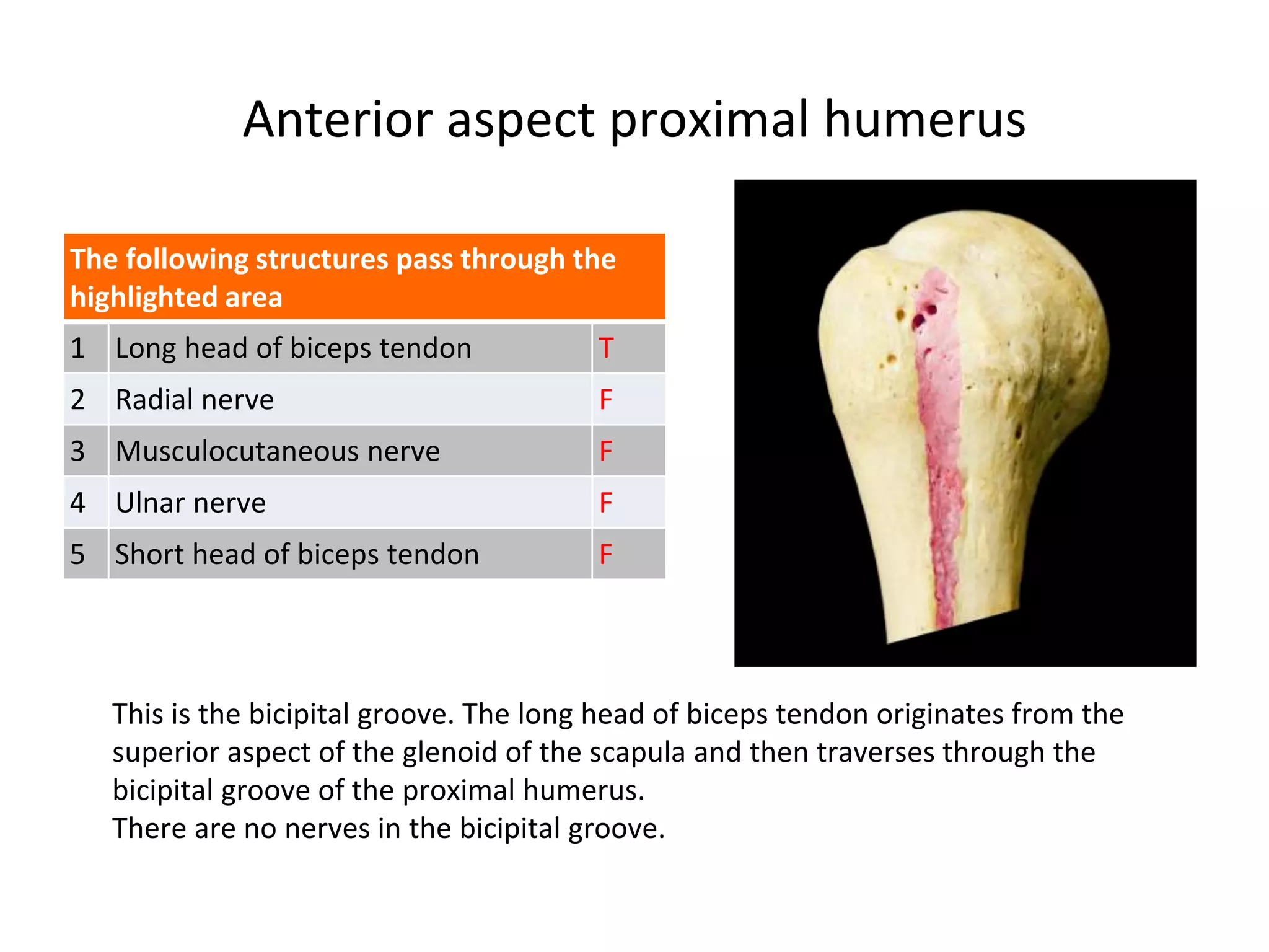
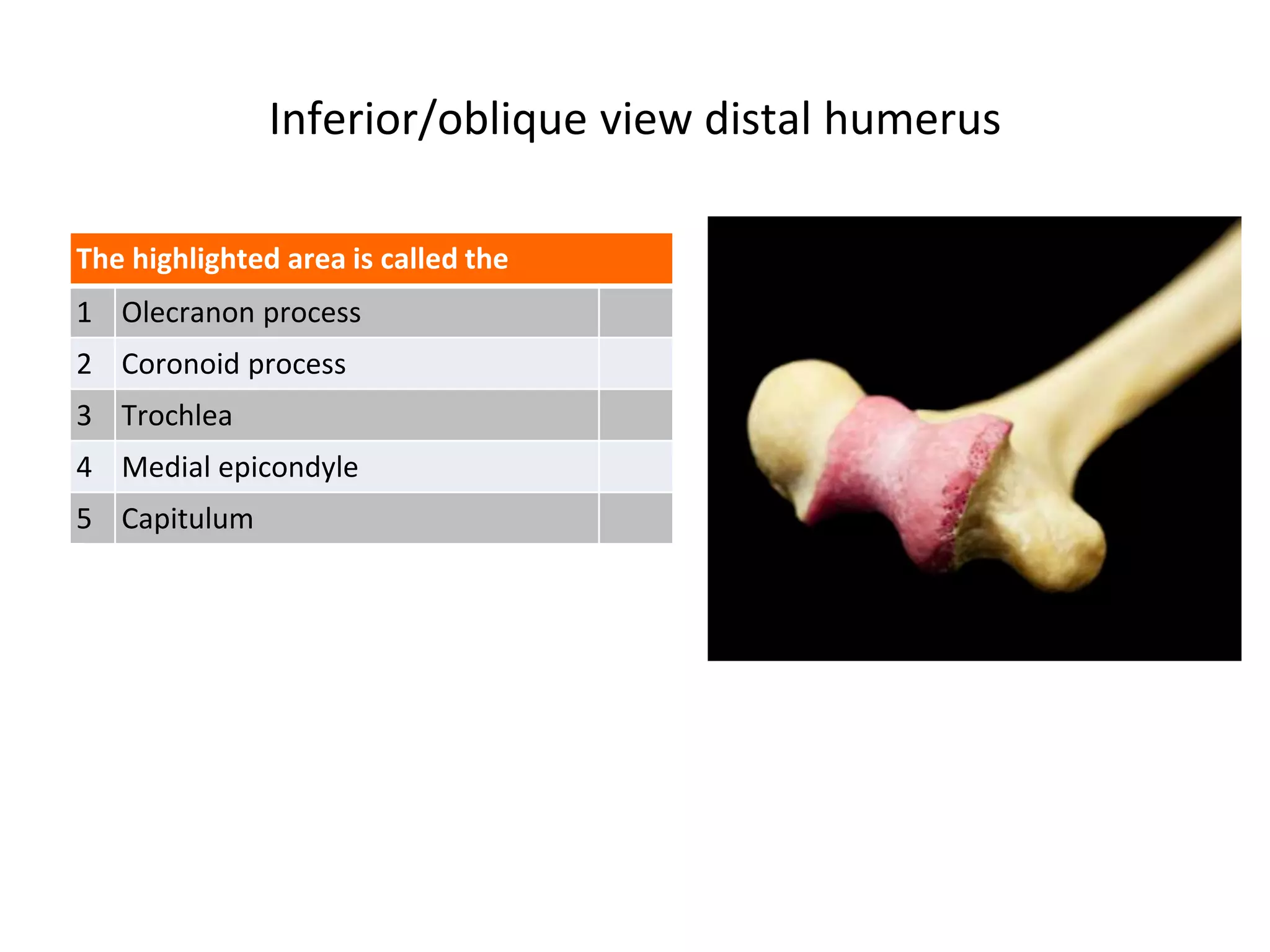


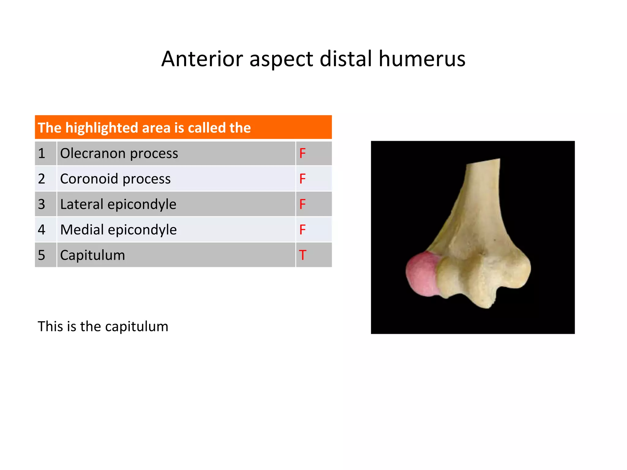

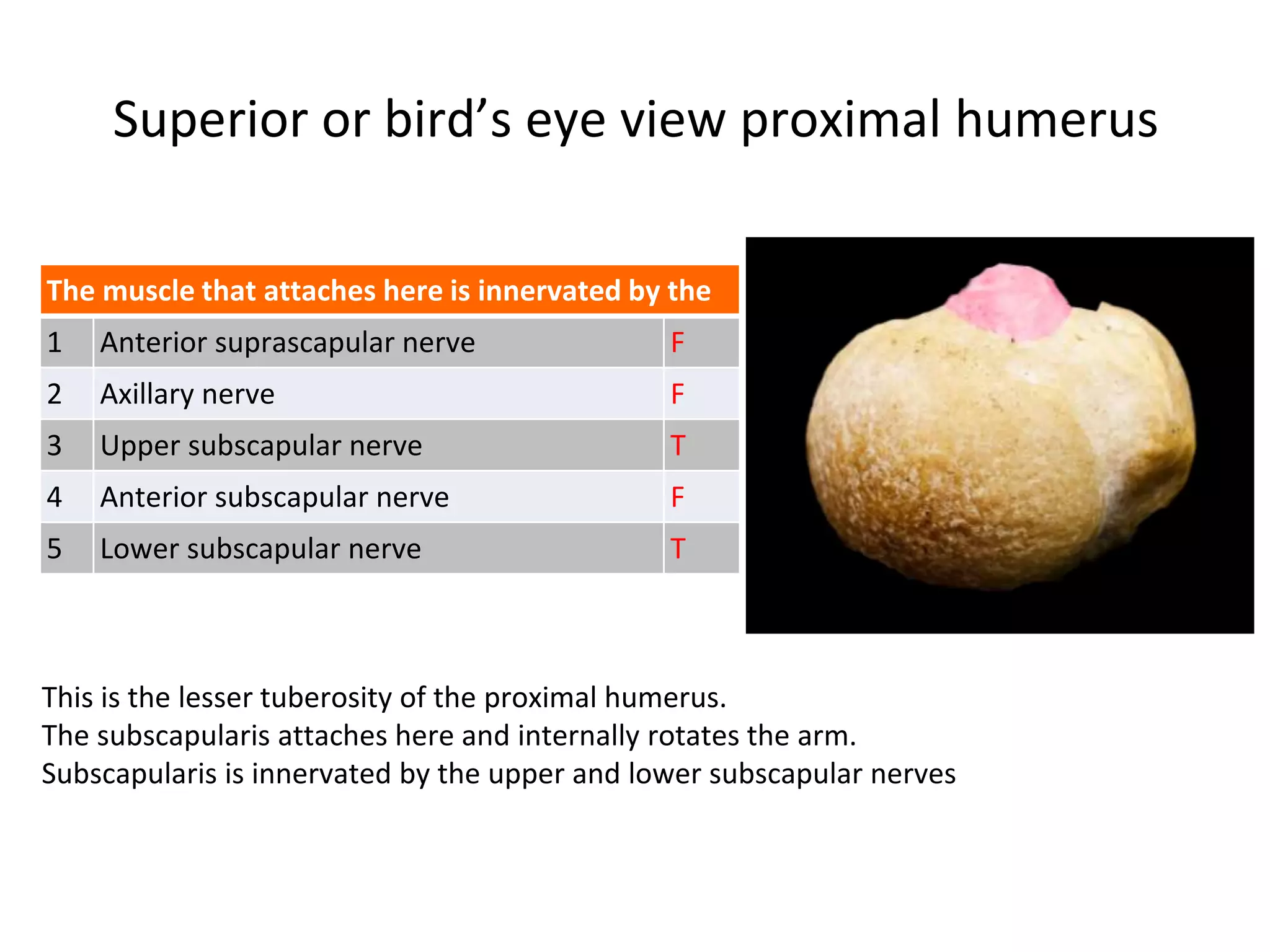
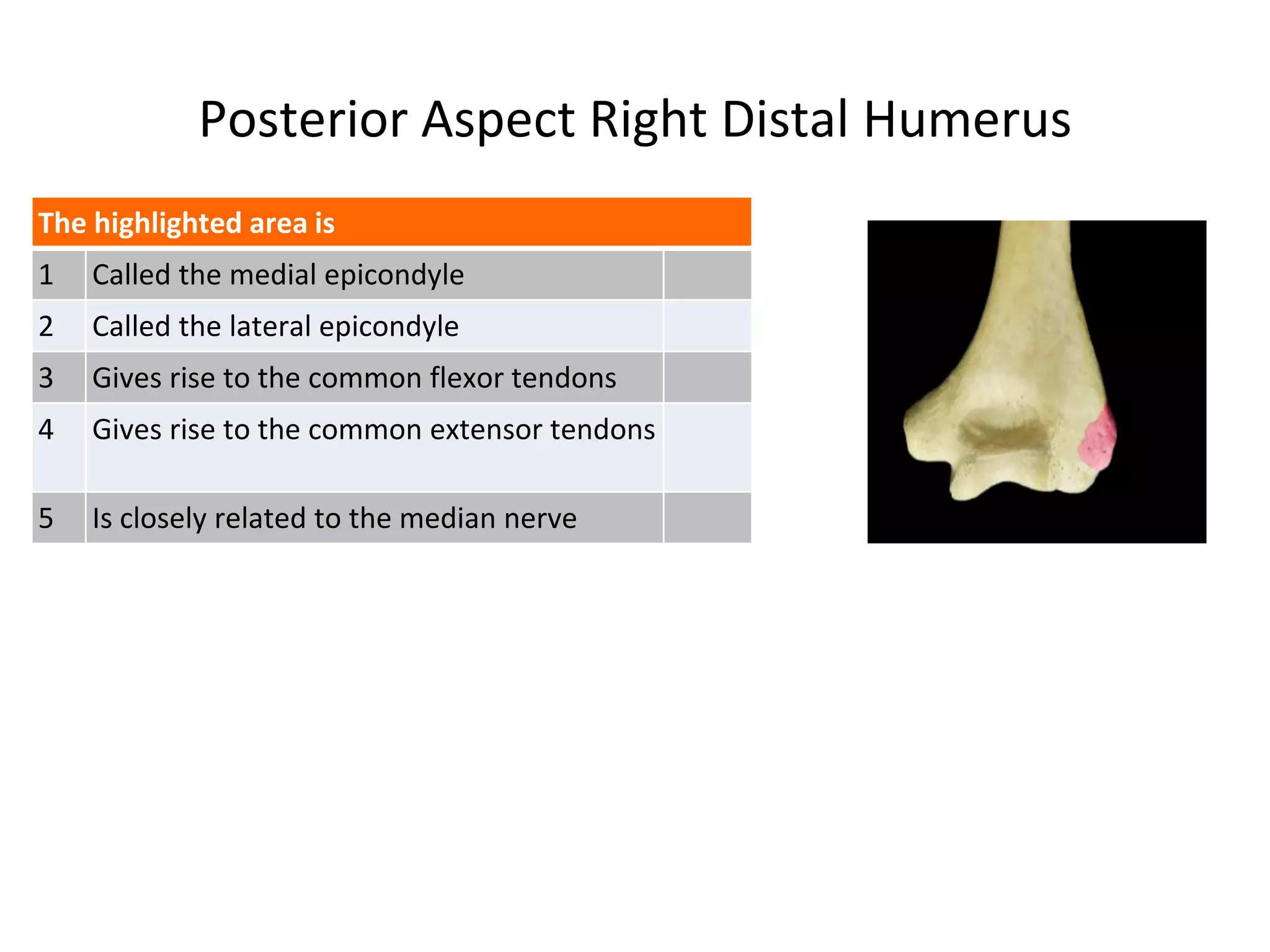
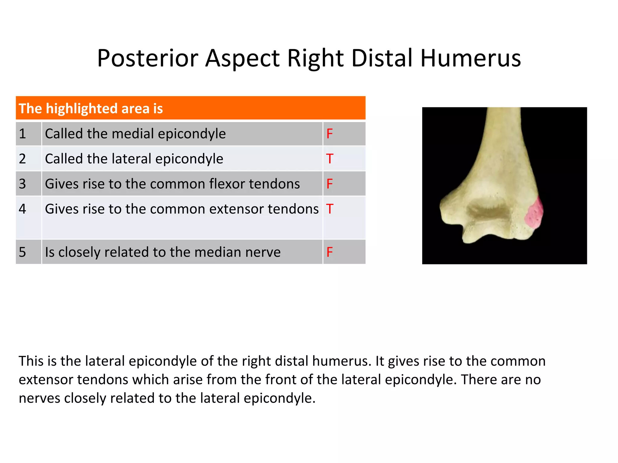
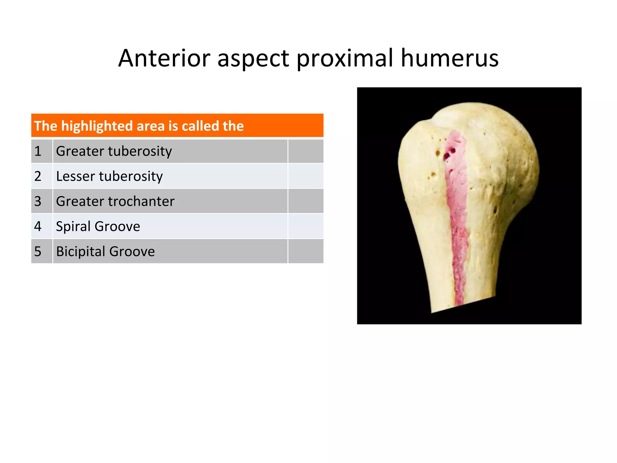


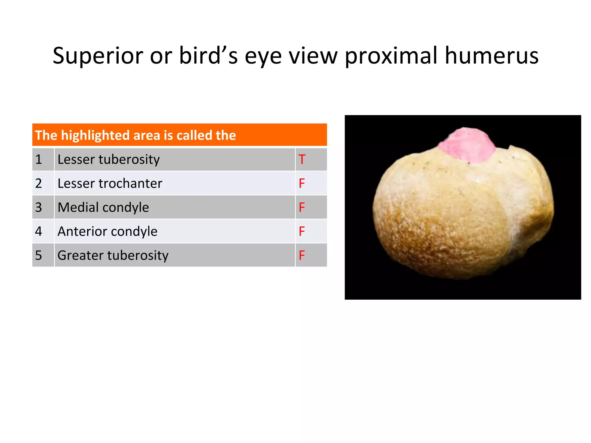
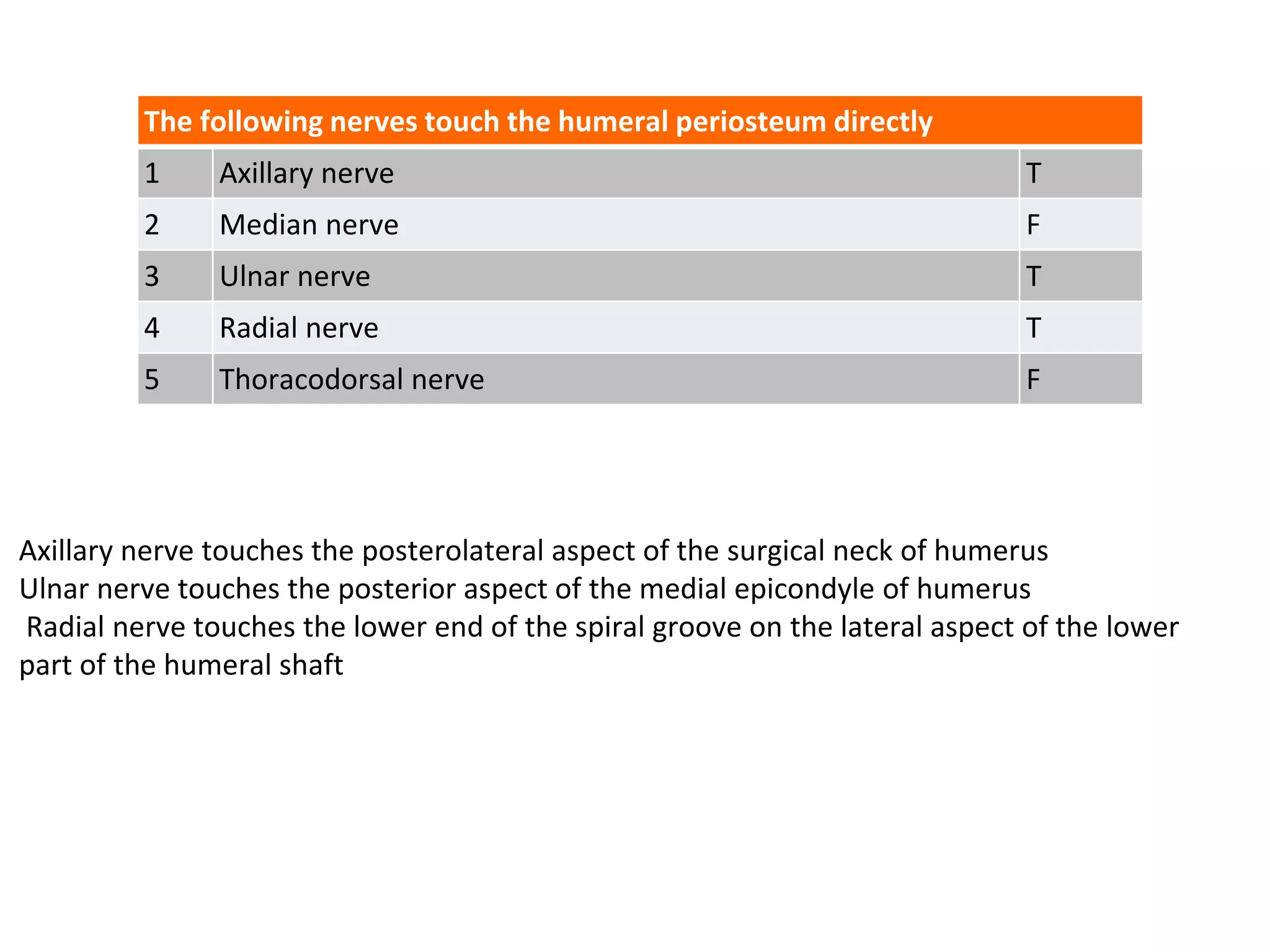
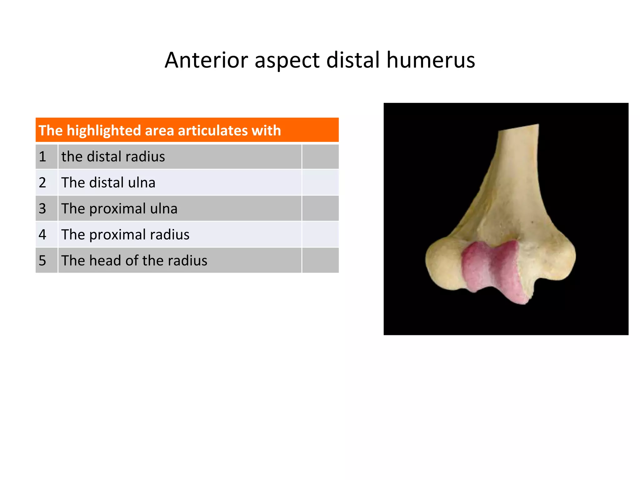
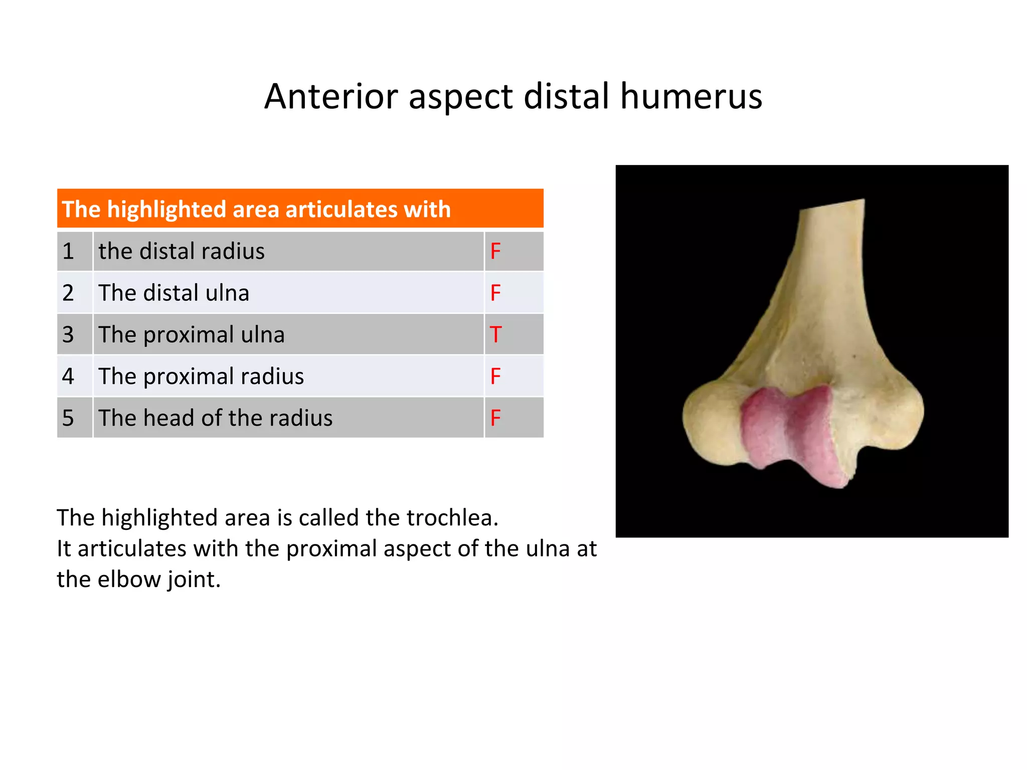

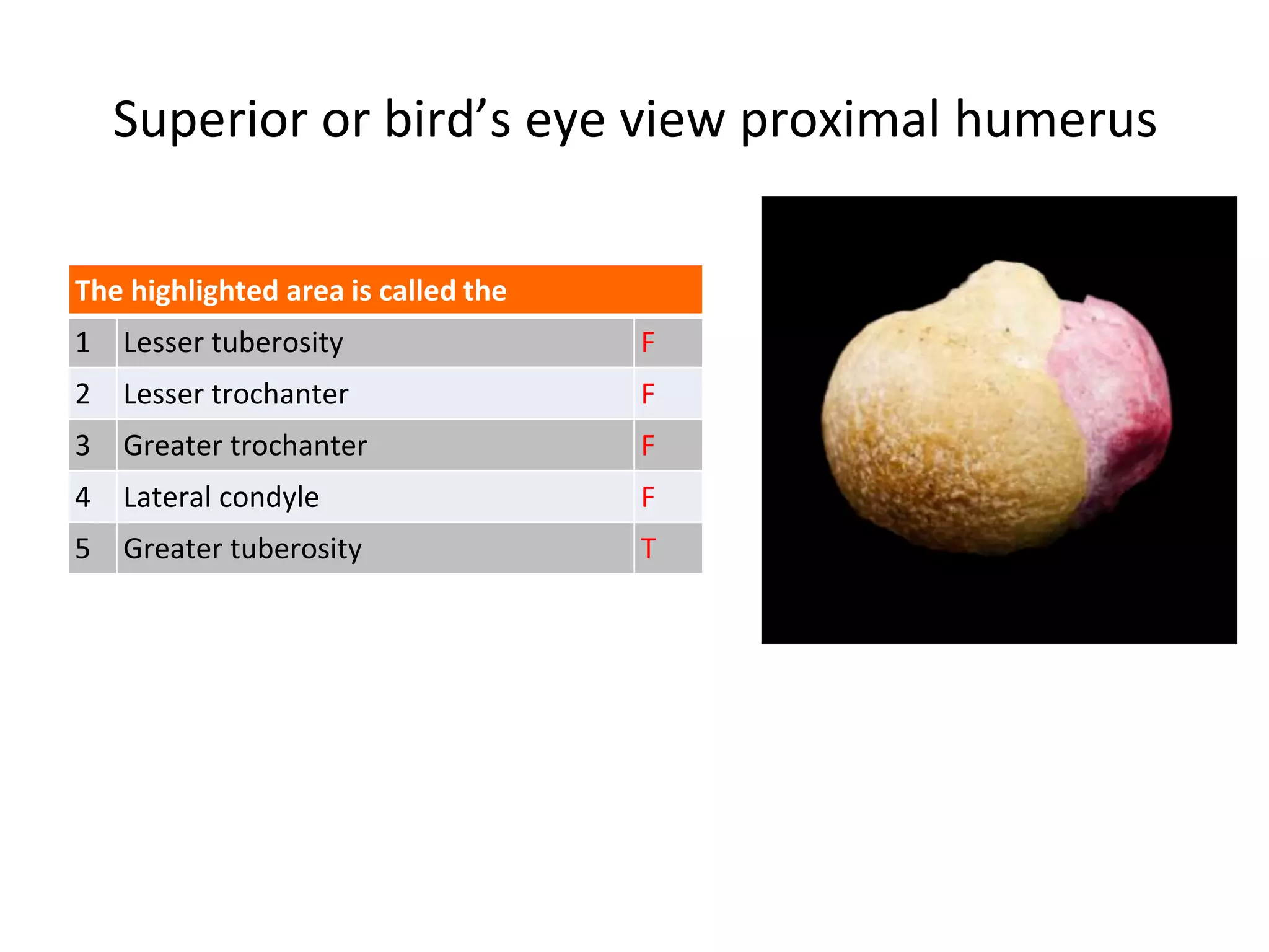

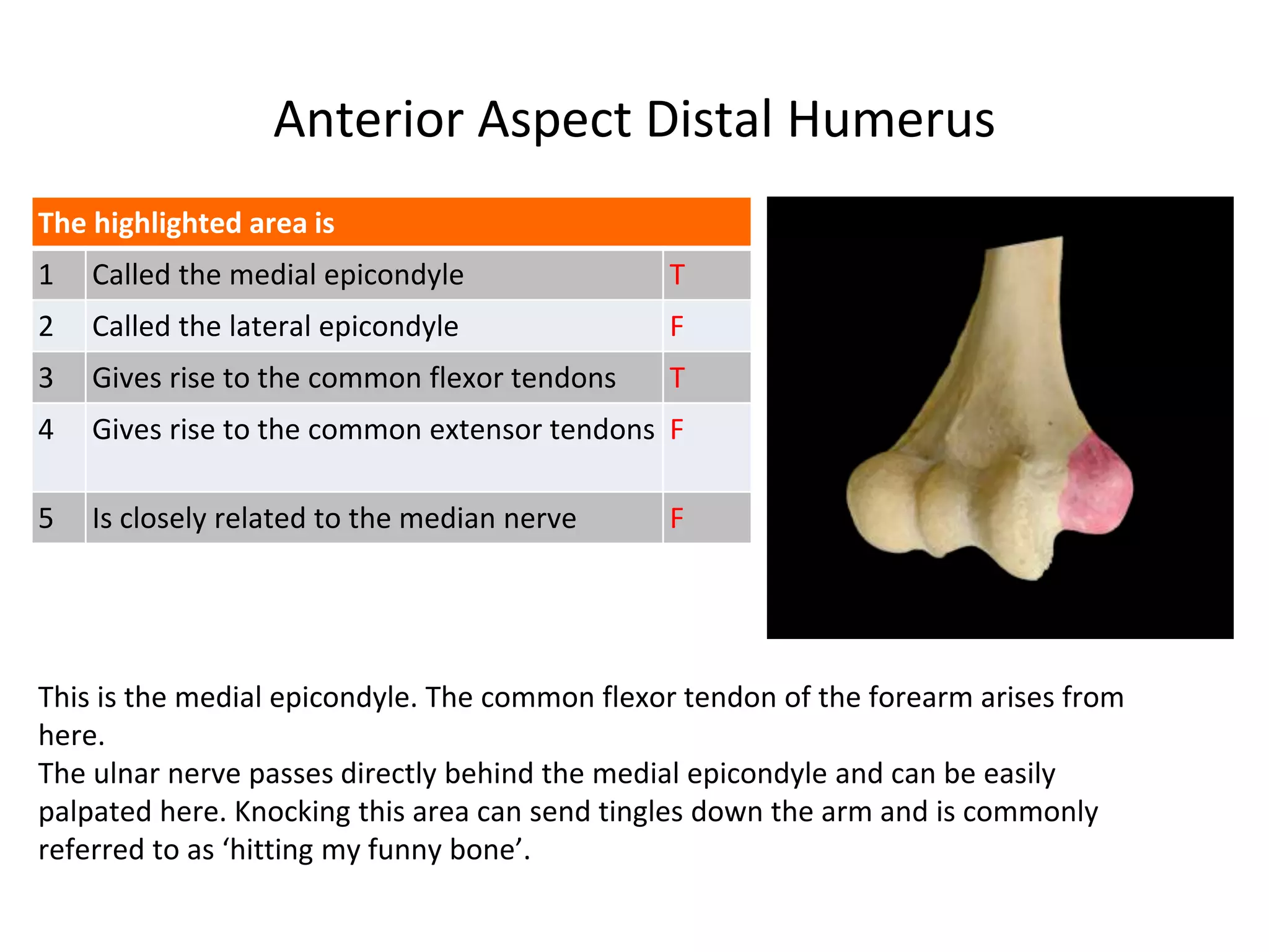
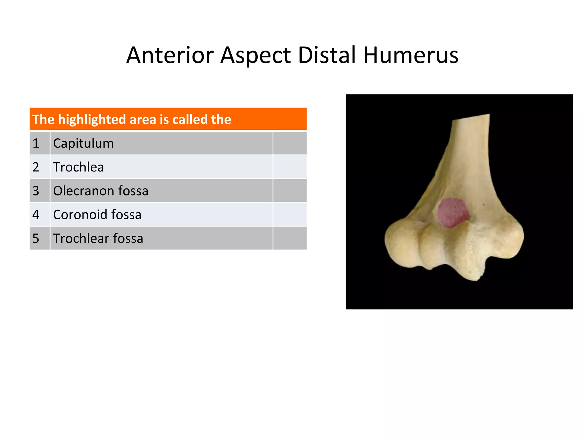


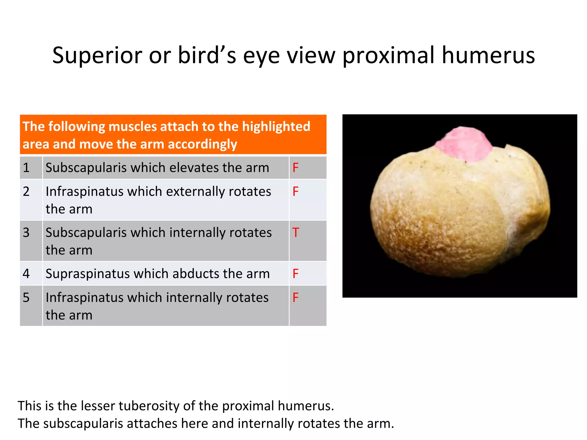
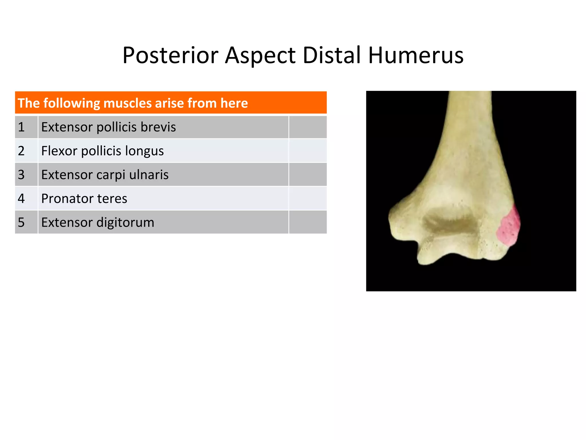


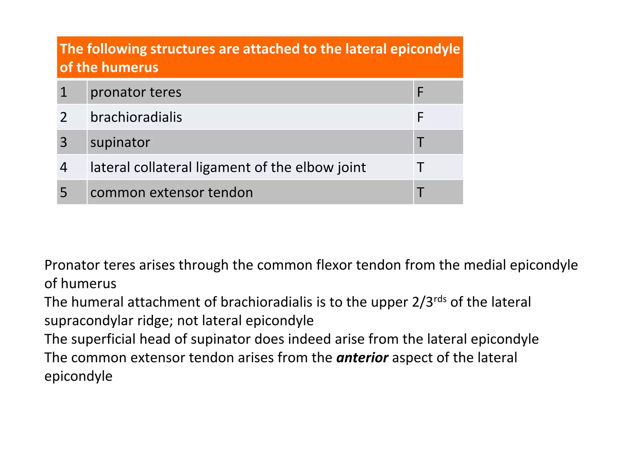

The document provides a detailed anatomical overview of the humerus, categorizing key areas such as the greater and lesser tuberosities, olecranon fossa, and trochlea, along with their functions and the muscles that attach to them. It explains the relationships of various nerves to the humerus and identifies structures important for arm movement and articulation at the elbow joint. Additionally, it clarifies which nerves and muscles are associated with specific regions of the humerus, particularly in terms of their anatomical significance and function.


