Learning outcomes
At theend of the lecture, students should be able to:
2.1 Describe the pectoral, scapular and shoulder
muscles.
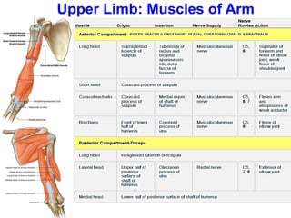
2.2 Describe the boundaries and contents of
quadrangular space. List and explain the muscles of
anterior and posterior compartments of the arm.
2.3 Define cubital fossa and describe its boundaries &
contents. List and describe the muscles of the flexor
and extensor compartment of the forearm.
2.4 Explain the formation, contents of carpal tunnel and
its clinical significance.
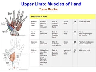
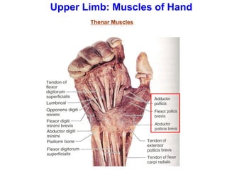
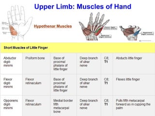
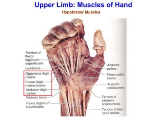
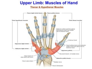
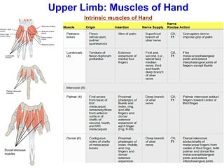
2.5 List and explain the muscles of hand-thenar,
hypothenar, lumbricals and interossei.
3.
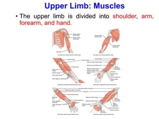
Upper Limb: Muscles
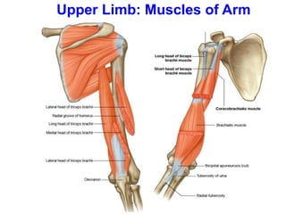
•The upper limb is divided into shoulder, arm,
forearm, and hand.
Upper Limb: ShoulderMuscles
Muscles of the shoulder can be grouped into :
1. Muscles connecting the thoracic wall (axial skeleton)
with scapula, clavicle or to the proximal end of the
humerus - pectoralis major, pectoralis minor,
subclavius, serratus anterior
2. Muscles connecting the vertebral column (axial
skeleton) with scapula and clavicle (pectoral girdle) or
with proximal end of humerus- trapezius, levator
scapulae, rhomboids, latissimus dorsi
3. Muscles connecting the scapula and clavicle to the
humerus - the 4 rotator cuff muscles: subscapularis,
supraspinatus, infraspinatus, teres minor along with
teres major & deltoid
6.
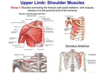
Group 1: Musclesconnecting the thoracic wall (axial skeleton) with scapula,
clavicle or to the proximal end of the humerus
Upper Limb: Shoulder Muscles
7.
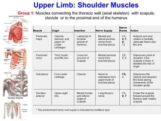
Group 1: Musclesconnecting the thoracic wall (axial skeleton) with scapula,
clavicle or to the proximal end of the humerus
Upper Limb: Shoulder Muscles
8.
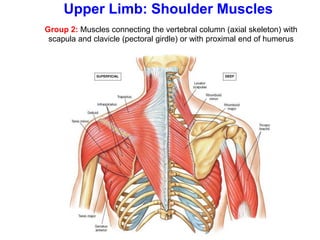
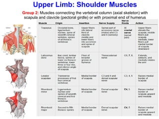
Group 2: Musclesconnecting the vertebral column (axial skeleton) with
scapula and clavicle (pectoral girdle) or with proximal end of humerus
Upper Limb: Shoulder Muscles
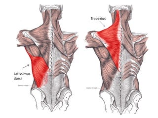
Group 2: Musclesconnecting the vertebral column (axial skeleton) with
scapula and clavicle (pectoral girdle) or with proximal end of humerus
Upper Limb: Shoulder Muscles
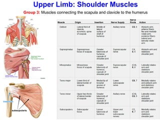
11.
Group 3: Musclesconnecting the scapula and clavicle to the humerus
Upper Limb: Shoulder Muscles
12.
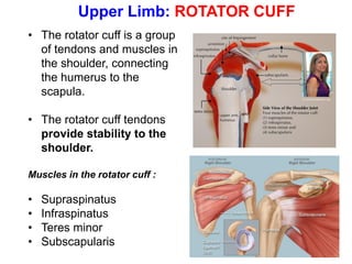
Upper Limb: ROTATORCUFF
• The rotator cuff is a group
of tendons and muscles in
the shoulder, connecting
the humerus to the
scapula.
• The rotator cuff tendons
provide stability to the
shoulder.
Muscles in the rotator cuff :
• Supraspinatus
• Infraspinatus
• Teres minor
• Subscapularis
13.
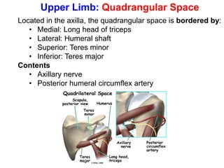
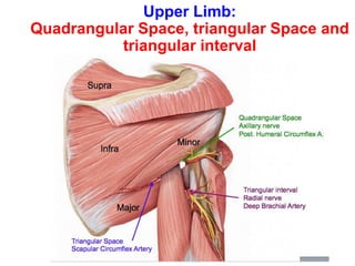
Upper Limb: QuadrangularSpace
Located in the axilla, the quadrangular space is bordered by:
• Medial: Long head of triceps
• Lateral: Humeral shaft
• Superior: Teres minor
• Inferior: Teres major
Contents
• Axillary nerve
• Posterior humeral circumflex artery
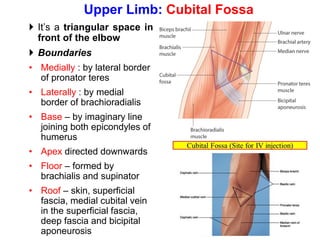
It’s atriangular space in
front of the elbow
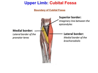
Boundaries
• Medially : by lateral border
of pronator teres
• Laterally : by medial
border of brachioradialis
• Base – by imaginary line
joining both epicondyles of
humerus
• Apex directed downwards
• Floor – formed by
brachialis and supinator
• Roof – skin, superficial
fascia, medial cubital vein
in the superficial fascia,
deep fascia and bicipital
aponeurosis
Upper Limb: Cubital Fossa
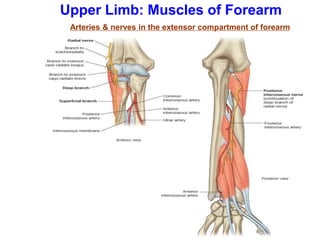
Arteries & nervesin the extensor compartment of forearm
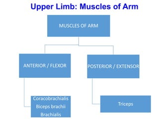
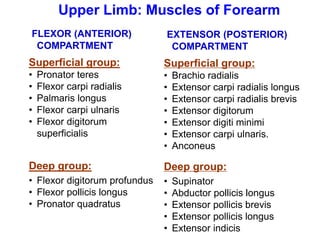
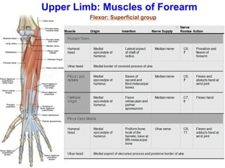
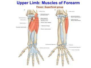
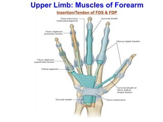
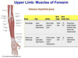
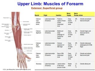
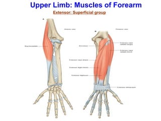
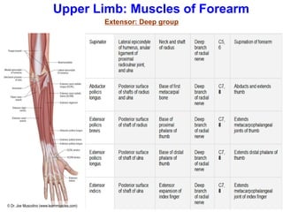
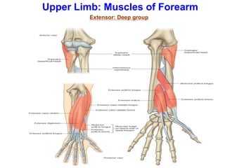
Upper Limb: Muscles of Forearm
35.
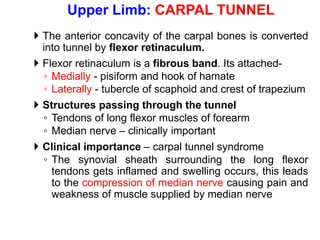
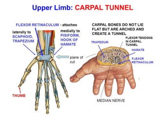
The anteriorconcavity of the carpal bones is converted
into tunnel by flexor retinaculum.
Flexor retinaculum is a fibrous band. Its attached-
◦ Medially - pisiform and hook of hamate
◦ Laterally - tubercle of scaphoid and crest of trapezium
Structures passing through the tunnel
◦ Tendons of long flexor muscles of forearm
◦ Median nerve – clinically important
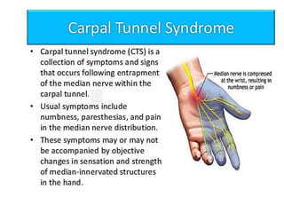
Clinical importance – carpal tunnel syndrome
◦ The synovial sheath surrounding the long flexor
tendons gets inflamed and swelling occurs, this leads
to the compression of median nerve causing pain and
weakness of muscle supplied by median nerve
Upper Limb: CARPAL TUNNEL
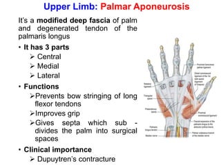
It’s a modifieddeep fascia of palm
and degenerated tendon of the
palmaris longus
• It has 3 parts
➢ Central
➢ Medial
➢ Lateral
• Functions
➢Prevents bow stringing of long
flexor tendons
➢Improves grip
➢Gives septa which sub -
divides the palm into surgical
spaces
• Clinical importance
➢ Dupuytren’s contracture
Upper Limb: Palmar Aponeurosis
46.
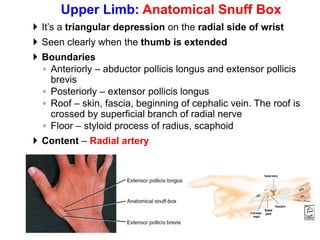
It’s atriangular depression on the radial side of wrist
Seen clearly when the thumb is extended
Boundaries
◦ Anteriorly – abductor pollicis longus and extensor pollicis
brevis
◦ Posteriorly – extensor pollicis longus
◦ Roof – skin, fascia, beginning of cephalic vein. The roof is
crossed by superficial branch of radial nerve
◦ Floor – styloid process of radius, scaphoid
Content – Radial artery
Upper Limb: Anatomical Snuff Box