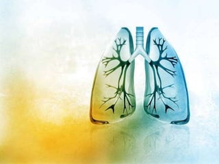
The document describes the anatomy and physiology of the human respiratory system. It discusses the main organs involved, including the nose, pharynx, larynx, lungs, bronchi, bronchioles, alveoli, diaphragm, and intercostal muscles. It explains the functions of these organs in oxygenation of the blood and removal of carbon dioxide through breathing and gas exchange. The primary function of the respiratory system is to oxygenate the blood enabling supply of oxygen to all parts of the body.
The document summarizes the key parts and functions of the human respiratory system including the nasal cavity, pharynx, epiglottis, larynx, trachea, bronchi, bronchioles, alveoli, and diaphragm. It also describes gas exchange that occurs in the lungs where oxygen diffuses into blood cells and carbon dioxide diffuses out. The process of breathing is explained including how inhalation and exhalation are controlled by the diaphragm and intercostal muscles to expand and contract the chest cavity.
The respiratory system is comprised of several organs involved in breathing, including the nose, mouth, pharynx, larynx, trachea, bronchi, bronchioles and lungs. Air enters through the nose or mouth and passes through these structures made of cartilage, muscle and mucous membranes to warm, moisten and filter the air before it reaches the lungs. In the lungs, the air flows through increasingly smaller airways called bronchioles until reaching tiny air sacs called alveoli where oxygen and carbon dioxide are exchanged.
The human respiratory system consists of the nose, pharynx, larynx, trachea, bronchi, bronchioles, lungs, diaphragm and intercostal muscles. Air enters the lungs through the nose and mouth, and passes through the trachea into the bronchi and bronchioles before reaching the alveoli in the lungs. Oxygen and carbon dioxide are passively exchanged between the alveolar air and blood through the process of diffusion. Contraction of the diaphragm and intercostal muscles during inhalation lowers the pressure in the thoracic cavity, drawing air into the lungs. During exhalation, relaxation of these muscles increases pressure and pushes air out.
1) Hemoglobin is more efficient at transporting oxygen than suggested by its dissociation curve because the amount of oxygen it carries depends on both the partial pressure of oxygen and carbon dioxide.
2) At tissues with high carbon dioxide levels, hemoglobin has a lower affinity for oxygen and releases more oxygen, while at the lungs with low carbon dioxide it has a higher affinity and picks up more oxygen.
3) Fetal hemoglobin has a higher affinity for oxygen than adult hemoglobin due to its dissociation curve being shifted left, allowing it to more readily obtain oxygen from the mother's blood through the placenta.
This document discusses the human respiratory system and the process of respiration. It describes the major organs of the respiratory system, including the nose, pharynx, larynx, trachea, bronchi, bronchioles and alveoli. It explains that the respiratory system allows for the intake of oxygen and removal of carbon dioxide. The document outlines the two phases of breathing - inspiration and expiration. It also discusses gas exchange that occurs between the blood and alveoli, and lists some common malfunctions and diseases of the respiratory system like asthma, bronchitis, emphysema and pneumonia.
The human respiratory system allows for gas exchange between the external environment and tissues within the body. It consists of the respiratory tract and respiratory organs. The respiratory tract includes the nose, pharynx, larynx, trachea, bronchi and lungs. The lungs contain alveoli which facilitate the diffusion of oxygen into the bloodstream and carbon dioxide out. Breathing is controlled by the respiratory centers in the medulla and involves inspiration through muscle contraction and expiration through relaxation. Disorders can disrupt gas exchange like asthma and emphysema.
The human respiratory system consists of the upper and lower respiratory tract. The upper tract includes the nose, nasal cavity, sinuses, pharynx and larynx. The lower tract includes the trachea, bronchi, bronchioles and lungs. The nose warms and filters inhaled air. The lungs are the primary organs for gas exchange, extracting oxygen from inhaled air into the bloodstream and releasing carbon dioxide from the bloodstream. Respiration is controlled by respiratory centers in the brainstem which regulate the muscles of breathing.
Anatomy of lungs and development of lungsDrhappyachu
This document provides an overview of lung anatomy and development. It discusses the gross organization of the lungs including surfaces, borders, lobes, and root. It describes the trachea, bronchi, pulmonary arteries and veins. Segmental anatomy and lymphatics are covered. Lung development progresses from embryonic to pseudoglandular, canalicular, saccular and alveolar stages. Pleura and its relations are also summarized.
Anatomy/Physiology Slideshow: The Respiratory SystemLHSprincipal
The respiratory system brings air into the body during breathing. It includes the nose, throat, trachea, and lungs. In the lungs, oxygen from the air is transferred to the bloodstream and distributed to cells throughout the body. The respiratory system has several parts - air enters through the nose and passes through the pharynx and larynx before reaching the trachea. The trachea divides into bronchi that lead into the lungs and branch into smaller bronchioles and alveoli, where oxygen and carbon dioxide are exchanged between the blood and air.
The document summarizes the process of gaseous exchange in humans. It describes the major components of the respiratory system including the nasal cavity, pharynx, larynx, trachea, bronchi, bronchioles and alveoli. It explains how air passes through these structures and the role of vocal cords, diaphragm and ribs in breathing. Gas exchange occurs through the alveoli which are surrounded by capillaries to oxygenate blood and remove carbon dioxide.
The respiratory system allows humans to breathe and obtain oxygen while removing carbon dioxide. It includes the nasal cavity, pharynx, larynx, trachea, bronchi, bronchioles and lungs. The nasal cavity and mouth allow air to enter, which then passes through the pharynx and larynx before entering the trachea and branching bronchi that lead to the lungs. Within the lungs are clusters of tiny alveoli sacs where the exchange of oxygen and carbon dioxide occurs through thin walls, allowing oxygen to enter the bloodstream and carbon dioxide to be removed.
The respiratory system provides oxygen to the body's cells while removing carbon dioxide through the processes of inhalation and exhalation. It consists of an upper respiratory tract including the nose, mouth and larynx, and a lower respiratory tract including the trachea, bronchi, bronchioles and alveoli in the lungs. The lungs contain volumes of air that can be measured including tidal volume, inspiratory reserve volume, expiratory reserve volume and residual volume. Gas exchange occurs through diffusion between the alveoli and blood plasma, and between plasma and tissues throughout the body, facilitated by factors like partial pressures and surface area.
I use this presentation to discuss the parts of the respiratory system to my Grade 9 students. This presentation only shows the major parts and does not discuss everything in great detail. Hope it helps!
The document summarizes key aspects of the respiratory system including:
1. The three major steps of respiration: pulmonary ventilation, external respiration, and internal respiration.
2. The major organs of the respiratory system including the nose, pharynx, larynx, trachea, bronchi, lungs, and alveoli.
3. The processes of breathing including inhaling via the diaphragm and intercostal muscles and exhaling via relaxation of these muscles.
The respiratory system functions to oxygenate the blood and remove carbon dioxide through gas exchange that occurs in the alveoli of the lungs. It is organized into an upper respiratory tract including the nose and pharynx, and a lower respiratory tract including the larynx, trachea, bronchi, and lungs. The lungs are paired organs located in the thoracic cavity that contain alveoli which are the sites of gas exchange between the respiratory and circulatory systems.
Gas exchange occurs through the respiratory system where oxygen enters the body through the lungs and diffuses into blood capillaries, and carbon dioxide diffuses out of the capillaries into the lungs. In humans, air is brought into the lungs through the nasal cavity and travels through the trachea into bronchioles and alveoli, where gas exchange occurs by diffusion across the alveoli walls. Hemoglobin in red blood cells transports oxygen to tissues and transports carbon dioxide back to the lungs to be exhaled. Smoking damages the lungs by destroying protective cells and tissues, increasing risk of cancer and other respiratory diseases.
This document provides an overview of respiratory physiology, covering the definition and function of respiration, the steps of respiration, the structure and function of the conducting and respiratory zones of the lungs, lung volumes and capacities, mechanics of breathing, gas exchange, transport of oxygen and carbon dioxide in the blood and tissues, and control of breathing. Key points include that respiration involves the exchange of oxygen and carbon dioxide between the environment and body cells, occurring in five steps including ventilation, diffusion, transport, and cellular respiration. Gas exchange takes place in the alveoli via differences in partial pressures and is facilitated by a large surface area and thin diffusion barrier. Oxygen is transported in both dissolved and hemoglobin-bound forms in the blood, while carbon
The document provides an overview of the respiratory system and control of respiration. It describes the key organs involved, including the nose, pharynx, larynx, trachea, bronchi, lungs and alveoli. It explains the mechanics of breathing through inspiration and expiration. Gas exchange occurs as oxygen passes from the alveoli into the blood and carbon dioxide passes from the blood into the alveoli to be exhaled. The lungs, diaphragm, ribs and autonomic nervous system work together to regulate breathing and ventilation.
The document discusses the respiratory system and the process of breathing. It describes how breathing takes in oxygen and removes carbon dioxide. It lists the respiratory systems of different living things like fish, amphibians, plants and worms. It then focuses on the human respiratory system, describing how breathing in and out works through the movement of the ribs, diaphragm and lungs. It details the path air takes through the nose, windpipe, bronchi and alveoli where oxygen passes into blood vessels and carbon dioxide passes out of blood vessels and into the air sacs.
The document discusses the process of respiration in four parts:
1. Pulmonary ventilation involves breathing air in and out of the lungs through inhalation and exhalation.
2. External respiration is the exchange of oxygen and carbon dioxide between the alveoli in the lungs and blood in the pulmonary capillaries.
3. Internal respiration is the exchange of gases between blood in the systemic capillaries and tissue cells throughout the body.
4. Respiration is regulated through various pulmonary volumes including tidal volume, vital capacity, functional residual capacity, and total lung capacity.
The document provides information on the functional anatomy and physiology of the respiratory system. It discusses the importance of understanding respiratory anatomy and physiology in fields like pulmonology, anesthesiology and critical care. It then describes the processes of external and internal respiration, the structural and functional unit of alveoli, and the roles and functions of various respiratory structures like the nose, pharynx, larynx, trachea, bronchi, lungs and pleura. It also discusses topics like mucociliary clearance, Kartagener's syndrome, airway innervation, Weibel's model of the tracheobronchial tree and blood supply to the lungs.
The document summarizes the process of gas exchange in the human respiratory system. It describes the major organs involved, including the lungs, diaphragm, ribs, larynx, trachea, bronchi, bronchioles, and alveoli. It explains how breathing in and out works via the contraction and relaxation of the intercostal muscles and diaphragm. The roles of ciliated cells and mucus in protecting the lungs are also outlined. Finally, it provides details on the composition differences between inspired and expired air.
The respiratory system consists of organs and structures that facilitate gas exchange. It includes the lungs, airways, blood vessels, and muscles that allow breathing. The respiratory tract includes the nostrils, nasal cavity, pharynx, larynx, trachea, bronchi, bronchioles and alveoli. Oxygen is inhaled into the lungs and transported via blood to cells, where it is used to produce energy. Carbon dioxide is produced as a waste product and diffused from cells and blood into the lungs to be exhaled out. The respiratory system thus oxygenates the body and removes carbon dioxide through a process of ventilation and gas exchange.
The document describes the structure and function of the respiratory system. It discusses the upper and lower respiratory tract, which are divided into conducting and respiratory portions. The upper tract includes the nose, nasal cavity, sinuses, pharynx and larynx. The lower tract includes the trachea, bronchi and bronchioles which branch into millions of alveoli in the lungs where gas exchange occurs. The document also outlines the processes of breathing, gas exchange, and the roles of various respiratory structures.
The circulatory system transports nutrients, oxygen, water and removes wastes throughout the body. It consists of the heart, blood, and blood vessels. The heart pumps blood through thousands of miles of blood vessels. Blood carries oxygen, nutrients, water and waste products. It contains red blood cells which carry oxygen, white blood cells which fight infection, platelets which help with clotting, and plasma which transports blood cells. The three types of blood vessels are arteries which carry oxygenated blood away from the heart, capillaries which connect arteries and veins and allow exchange of materials, and veins which return deoxygenated blood to the heart.
The document describes the normal respiratory cycle and gas exchange in the lungs. During inhalation, air enters the mouth and flows through the upper airways, lower airways, and into the alveoli. Oxygen diffuses into the blood in the alveoli while carbon dioxide diffuses out. Various volumes are involved including tidal volume, vital capacity, and residual volume. The relationship between these volumes and pressures allows for gas exchange through diffusion while also keeping the alveoli open.
The respiratory system (also respiratory apparatus, ventilatory system) is a biological system consisting of specific organs and structures used for gas exchange in animals and plants. The anatomy and physiology that make this happen varies greatly, depending on the size of the organism, the environment in which it lives and its evolutionary history. In land animals the respiratory surface is internalized as linings of the lungs. Gas exchange in the lungs occurs in millions of small air sacs; in mammals and reptiles these are called alveoli, and in birds they are known as atria. These microscopic air sacs have a very rich blood supply, thus bringing the air into close contact with the blood.These air sacs communicate with the external environment via a system of airways, or hollow tubes, of which the largest is the trachea, which branches in the middle of the chest into the two main bronchi. These enter the lungs where they branch into progressively narrower secondary and tertiary bronchi that branch into numerous smaller tubes, the bronchioles. In birds the bronchioles are termed parabronchi. It is the bronchioles, or parabronchi that generally open into the microscopic alveoli in mammals and atria in birds. Air has to be pumped from the environment into the alveoli or atria by the process of breathing which involves the muscles of respiration.
In most fish, and a number of other aquatic animals (both vertebrates and invertebrates) the respiratory system consists of gills, which are either partially or completely external organs, bathed in the watery environment. This water flows over the gills by a variety of active or passive means. Gas exchange takes place in the gills which consist of thin or very flat filaments and lammelae which expose a very large surface area of highly vascularized tissue to the water.
Other animals, such as insects, have respiratory systems with very simple anatomical features, and in amphibians even the skin plays a vital role in gas exchange. Plants also have respiratory systems but the directionality of gas exchange can be opposite to that in animals. The respiratory system in plants includes anatomical features such as stomata, that are found in various parts of the plant.
Resp-System.ppt human anatomy and physiologyRubikhan18
The human respiratory system consists of the nose, pharynx, larynx, trachea, bronchi, lungs and diaphragm. It takes in oxygen and releases carbon dioxide through external respiration in the lungs and internal respiration in the cells. The respiratory system can be affected by diseases like asthma, COPD, pneumonia and lung cancer.
The respiratory system is the network of organs and tissues that help you breathe. It includes your airways, lungs, and blood vessels. The muscles that power your lungs are also part of the respiratory system. These parts work together to move oxygen throughout the body and clean out waste gases like carbon dioxide.
The respiratory system consists of the airways, lungs, and respiratory muscles that mediate air flow into and out of the body. Within the lungs, oxygen and carbon dioxide are passively exchanged between air and blood through diffusion. The system also helps maintain acid-base balance by efficiently removing carbon dioxide from the blood. Air flows through the upper respiratory tract into the lower respiratory tract and lungs where gas exchange occurs in the alveoli.
Respiratory System - Human Anatomy and Physiology bPharmPankaj Saha
The document summarizes the organization and function of the respiratory system. It describes:
1. The respiratory system consists of an upper respiratory tract that conducts air from the nose to the larynx, and a lower respiratory tract from the trachea onwards. The lower tract is further divided into a conducting zone from the trachea to terminal bronchioles, and a respiratory zone from bronchioles to alveoli where gas exchange occurs.
2. The main functions of the respiratory system are to supply oxygen to the body and remove carbon dioxide, as well as filter air, regulate pH and temperature, and contain smell receptors. Breathing involves cyclic inhalation and exhalation phases.
3. The
The respiratory system allows for gas exchange in animals. It consists of organs and structures used for breathing. In land animals, gas exchange occurs in microscopic air sacs called alveoli in the lungs. Air flows from the nose and pharynx into the trachea, bronchi and bronchioles before reaching the alveoli. External respiration is the exchange of oxygen and carbon dioxide between the lungs and blood. Internal respiration is the exchange between blood and tissues. Normal human respiration is nasal, diaphragmatic, slow at 12 breaths per minute at rest, and imperceptible. Each breath has a tidal volume of 500ml and the respiratory cycle involves inspiration and expiration over 1.5-2 seconds each.
The respiratory system allows for oxygen to enter the body and carbon dioxide to exit through a series of major organs. Air enters through the nose or mouth and passes through the pharynx, larynx, trachea, bronchi and into the lungs where gas exchange occurs in the alveoli. Oxygen then passes into the bloodstream and carbon dioxide passes out of the bloodstream and is exhaled. Breathing is facilitated by the contraction and relaxation of the diaphragm and rib cage which expands and contracts the chest cavity to inhale and exhale air.
The respiratory system allows for gas exchange of oxygen and carbon dioxide. It includes the nose, pharynx, larynx, trachea, bronchi, bronchioles and lungs. The upper respiratory tract includes the nose, nasal cavity and sinuses which warm, moisten and filter inhaled air. The lower respiratory tract includes the lungs and airways inside the chest. The lungs contain bronchioles and alveoli where gas exchange occurs. During respiration, oxygen is absorbed into the blood from the alveoli and carbon dioxide is released, in a continuous cycle of breathing in and out.
The document discusses the key components and functions of the respiratory system. It defines important terms like alveoli, bronchi, epiglottis, larynx, and lists the main parts of the respiratory tract including the nose, mouth, pharynx, larynx, trachea, lungs. It describes gas exchange that occurs in the alveoli and capillaries of the lungs and identifies structures involved in breathing like the ribs and diaphragm. The document also briefly discusses common respiratory diseases like COPD, lung cancer, and pneumonia.
The document provides an overview of the respiratory system, including its anatomy and physiology. It describes the upper respiratory tract including the nose, sinuses, pharynx and larynx. It then describes the lower respiratory tract including the trachea, bronchi, bronchioles, lungs and alveoli. It discusses the mechanisms of ventilation including inspiration and expiration. It also summarizes several diagnostic tests for evaluating respiratory function such as arterial blood gases, pulmonary function tests, sputum analysis, chest imaging and biopsies.
Johny's A&P structure and function of respiratory systemJohny Kutty Joseph
The respiratory system allows for breathing and gas exchange. It is divided into the upper and lower respiratory tract. The upper tract includes the nose, mouth, larynx, and pharynx. The lower tract includes the trachea, bronchi, and lungs. The lungs contain bronchioles and alveoli where gas exchange occurs. During breathing, the diaphragm and intercostal muscles work to expand and contract the lungs and chest cavity to inhale and exhale air.
The respiratory system allows for the intake of oxygen and removal of carbon dioxide through a series of organs. Air enters the nose or mouth and passes through the pharynx, larynx, trachea and bronchi into tiny air sacs in the lungs called alveoli. In the alveoli, oxygen passes into the bloodstream and carbon dioxide passes out of the bloodstream and into the air that is then exhaled. Breathing is facilitated by the contraction and relaxation of the diaphragm and rib cage which expands and contracts the lungs and chest cavity, drawing air in during inhalation and pushing it out during exhalation.
The document summarizes the structure and function of the human respiratory system. It describes that the respiratory system consists of respiratory tract and lungs. The respiratory tract includes the nose, nasal passages, pharynx, larynx, trachea, and bronchial tree. The lungs are located in the thoracic cavity and contain bronchioles and alveoli where gas exchange takes place. The diaphragm and intercostal muscles help in the mechanism of breathing by contracting and relaxing during inhalation and exhalation.
The respiratory system allows for the intake of oxygen and removal of carbon dioxide through a series of organs. Air enters the nose and mouth, then passes through the pharynx, larynx, trachea, bronchi and into air sacs called alveoli in the lungs. In the alveoli, oxygen passes into blood vessels and carbon dioxide passes out of blood vessels, allowing for gas exchange. Breathing is driven by the contraction and relaxation of the diaphragm and rib cage, inhaling air into the lungs and exhaling air out.
The respiratory system allows for the intake and exchange of oxygen and carbon dioxide throughout the body. It consists of the nose, pharynx, larynx, trachea, bronchi and lungs. The lungs contain alveoli where gas exchange takes place between inhaled air and blood in capillaries. During inhalation, contraction of the diaphragm and expansion of the rib cage lower pressure in the chest cavity, drawing air into the lungs. Exhalation occurs passively as the diaphragm and rib cage relax, raising pressure and pushing air out.
The document discusses the structure and function of the respiratory system. It can be summarized as follows:
1) The respiratory system is divided into an upper and lower portion. The upper portion includes the nose, nasal cavity and pharynx, while the lower portion includes the larynx, trachea, bronchi and lungs.
2) Gas exchange occurs in the lungs through diffusion of oxygen and carbon dioxide between the alveoli and blood in the pulmonary capillaries. The partial pressures of oxygen and carbon dioxide drive this diffusion.
3) Respiration is regulated by both nervous and chemical mechanisms. The brain stem contains centers that control automatic breathing, while voluntary control occurs in the cerebral cortex.
The respiratory system allows for oxygen to enter the body and carbon dioxide to be expelled. It includes the nose, pharynx, larynx, trachea, bronchi, lungs and muscles of respiration. The nose warms, filters and humidifies inhaled air. The pharynx is a passageway for air and food. The larynx contains the vocal cords and protects the lungs. The trachea and bronchi form branching airways within the lungs whose alveoli facilitate gas exchange with blood in capillaries. Respiration is driven by the diaphragm and intercostal muscles.
The respiratory system has three main functions: gas exchange, regulating blood pH, and producing sounds. It consists of the upper respiratory tract including the nose and pharynx, and the lower tract including the lungs. Respiration has three steps: pulmonary ventilation, external respiration of gas exchange in the lungs, and internal respiration of gas exchange in tissues. The lungs obtain oxygen and expel carbon dioxide through breathing which involves the muscles and elastic recoil of the lungs and chest wall. The document then discusses various respiratory structures, processes, and disorders in more detail.
The human respiratory system involves the inhaling of oxygen and exhaling of carbon dioxide. It is divided into an upper respiratory tract and lower respiratory tract. The upper tract includes the nose, nasal cavity, throat and voice box. The lower tract contains the lungs, trachea, bronchi and bronchioles. The lungs obtain oxygen and remove carbon dioxide via alveoli, while other parts like the diaphragm and ribs aid in breathing movements.
The lungs are the essential organs for respiration in air-breathing animals. In humans, the trachea divides into the two main bronchi that enter the roots of the lungs, continuing to branch into bronchioles and alveolar sacs made up of clusters of alveoli where gas exchange occurs. The principal function of the lungs is to transport oxygen into the bloodstream and release carbon dioxide, with differences in lobe structure between human and pig lungs.
Drugs affecting the GI system are used in the treatment of gastric acidity, peptic ulcers, and gastroesophageal reflux disease (GERD), bowel motility disorders (gastroparesis [delayed gastric emptying due to partial paralysis of the stomach muscles], constipation, and diarrhea), and for the treatment of nausea and vomiting.
This document discusses antibiotics. It begins by defining antibiotics as chemical compounds that kill or inhibit the growth of bacteria. It then provides a brief history of antibiotics, noting that penicillin was the first antibiotic successfully used to treat bacterial infections. The document goes on to classify antibiotics based on their chemical structure and mechanism of action. It also discusses antibiotic resistance and appropriate antibiotic use.
Non-steroidal anti-inflammatory drugs is a class of analgesic medication that reduces pain, fever and inflammation. Since most episodes of back pain involve inflammation, NSAIDs such as ibuprofen and naproxen are often an effective treatment option.
The document discusses the various roles of pharmacists in the healthcare system. It begins by explaining how the focus of pharmacy has shifted from technical to more patient-oriented services. Pharmacists now take responsibility for patient outcomes related to drug therapy by working with other healthcare providers. The main roles of pharmacists include academic, industrial, community, clinical, and hospital settings. As the first contact of the healthcare system, pharmacists ensure patients receive the right medication in the right dose through the right route. Pharmacists are integral parts of the healthcare system by playing important roles in education, research, quality control, patient counseling, and optimizing drug therapy.
The GI tract contains four layers: the innermost layer is the mucosa, underneath this is the submucosa, followed by the muscularis propria and finally, the outermost layer - the adventitia. The structure of these layers varies, in different regions of the digestive system, depending on their function.
Body fluids are liquids originating from inside the bodies of living humans. They include fluids that are excreted or secreted from the body. Human blood, body fluids, and other body tissues are widely recognised as vehicles for the transmission of human disease.
Human Anatomy and Physiology are the branches of biology that concerns with forms (structures) and functions of human body. ANATOMY- Study of structure of whole body and individual parts (organs) and their correlation with each other.
The new corona virus is a respiratory virus which spreads primarily through droplets generated when an infected person coughs or sneezes, or through droplets of saliva or discharge from the nose. To protect yourself, clean your hands frequently with an alcohol-based hand rub or wash them with soap and water.
oint: The area where two bones are attached for the purpose of permitting body parts to move. A joint is usually formed of fibrous connective tissue and cartilage.
The lymph node is a small bean-shaped object which factors in the body's immune system. Lymph nodes clean out substances that travel through the lymphatic fluid, and they contain white blood cells that are the body's defense when fighting off ailments. There are many lymph nodes found throughout the body.
The document discusses the structure and function of human cells. It begins by defining the cell as the basic structural and functional unit of living things, wrapped in a membrane. There are around 100 trillion cells in the human body, each containing the genetic information to produce a human. The document then discusses the discovery of cells by early scientists like Hooke and van Leeuwenhoek using early microscopes. It provides details on the main parts of the human cell, including the nucleus that houses DNA, organelles like mitochondria and the endoplasmic reticulum, and the cytoplasm. It describes the functions of these various parts and how they work together to keep the cell alive.
This document discusses air pollution, including its causes, effects, and methods of control. It defines air pollution and lists major primary and secondary pollutants such as sulfur oxides, nitrogen oxides, carbon monoxide, and particulate matter. Sources of pollution include both natural sources like volcanoes and human activities like burning fossil fuels. Health effects include respiratory and cardiac issues. Air pollution also negatively impacts economies. Methods of control involve government regulations, green technologies, and individual conservation efforts. Devices like electrostatic precipitators, baghouses, and scrubbers can help remove pollutants from emission streams.
Water is very important. But some peoples can’t understand the importance of water. Who are very rich but they are poor without a water. So let’s give a message’SAVE WATER TO SAVE LIFE’
The document provides information about the human reproductive system. It discusses that reproduction allows organisms to produce offspring similar to themselves. The male and female reproductive systems both contribute gametes - sperm and eggs respectively - which can unite during fertilization. The male system produces sperm in the testes and transports it, while the female system produces eggs and supports gestation and birth. The parts of each system are described in detail, including their functions in fertilization and development.
Blood is a connective tissue composed of formed elements suspended in plasma. It functions to transport oxygen, nutrients, waste products, hormones, and more throughout the body. The formed elements include red blood cells, which carry oxygen, white blood cells which help fight infection, and platelets which help with clot formation. Plasma is mostly water and contains proteins, glucose, clotting factors, electrolytes and more. It helps maintain pH, osmotic pressure, and protects against infection.
This document provides information about antipsychotic drugs. It discusses that antipsychotics are mainly used to treat mental illnesses such as schizophrenia and bipolar disorder. There are two main types of antipsychotics: newer atypical antipsychotics and older typical antipsychotics. Antipsychotics are thought to work by altering chemicals in the brain like dopamine and serotonin. The document also summarizes schizophrenia, bipolar disorder, and different types of antipsychotic drugs.
The document summarizes the human nervous system. It describes that the nervous system conducts stimuli from sensory receptors to the brain and spinal cord, and conducts impulses back to other parts of the body. The nervous system has two main parts - the central nervous system consisting of the brain and spinal cord, and the peripheral nervous system consisting of nerves connecting to the central nervous system. The central nervous system coordinates and integrates the body's activities.
More from Jai Narain Vyas University Jodhpur Rajasthan India 342003 (20)
Macroeconomics- Movie Location
This will be used as part of your Personal Professional Portfolio once graded.
Objective:
Prepare a presentation or a paper using research, basic comparative analysis, data organization and application of economic information. You will make an informed assessment of an economic climate outside of the United States to accomplish an entertainment industry objective.
Introduction to AI for Nonprofits with Tapp NetworkTechSoup
Dive into the world of AI! Experts Jon Hill and Tareq Monaur will guide you through AI's role in enhancing nonprofit websites and basic marketing strategies, making it easy to understand and apply.
How to Add Chatter in the odoo 17 ERP ModuleCeline George
In Odoo, the chatter is like a chat tool that helps you work together on records. You can leave notes and track things, making it easier to talk with your team and partners. Inside chatter, all communication history, activity, and changes will be displayed.
Exploiting Artificial Intelligence for Empowering Researchers and Faculty, In...Dr. Vinod Kumar Kanvaria
Exploiting Artificial Intelligence for Empowering Researchers and Faculty,
International FDP on Fundamentals of Research in Social Sciences
at Integral University, Lucknow, 06.06.2024
By Dr. Vinod Kumar Kanvaria
it describes the bony anatomy including the femoral head , acetabulum, labrum . also discusses the capsule , ligaments . muscle that act on the hip joint and the range of motion are outlined. factors affecting hip joint stability and weight transmission through the joint are summarized.
MATATAG CURRICULUM: ASSESSING THE READINESS OF ELEM. PUBLIC SCHOOL TEACHERS I...NelTorrente
In this research, it concludes that while the readiness of teachers in Caloocan City to implement the MATATAG Curriculum is generally positive, targeted efforts in professional development, resource distribution, support networks, and comprehensive preparation can address the existing gaps and ensure successful curriculum implementation.
2. HUMAN RESPIRATORY SYSTEM
JAI NARAIN VYAS UNIVERSITY, JODHPUR
ASSISTANT PROFESSOR:-ASHWIN SINGH CHOUHAN
DEPARTMENT:- PHARMACOLOGY
E-MAIL:- anshukavya1993@gmail.com
3. RESPIRATORY SYSTEM
ASC JNVU PHARMACY, JODHPUR
The primary function of the respiratory system is to oxygenate the
blood enabling the supply of oxygen to all parts of the body. It is a
complex process involving multiple organs at micro and macro levels.
This chapter describes the anatomy and physiology of the human
respiratory system. A brief description of respiratory volume
parameters, Lung cancer, pulmonary nodules and its causes,
similarities and differences between benign and malignant nodules
are presented. The purpose of the CAD tool being proposed in the
thesis is to identify the morphological changes that happen in the
lungs and correlate them for identification and classification of
abnormalities resulting in lung cancer. Such a tool necessitates the
understanding of the anatomy of the system being diagnosed and in
this thesis lungs are considered.
4. Cells of the body require oxygen for the oxidation of food materials to
obtain energy. CO2 is released as a result of cellular respiration which
combines with water to form carbonic acid. Carbonic acid will lower
the blood pH , so CO2 must be eliminated from the body. Breathing
in and breathing out of air is known as respiration. Respiration
includes Ventilation (or) breathing: Flow of air into and out of lungs.
External respiration: Exchange of air in alveoli and blood in
pulmonary capillaries. Transport of gas: Transport of gas by blood
between lungs and tissues. Internal respiration: Exchange of air or
gas between the blood in systematic capillaries and tissue cells.
FUNCTIONS OF RESPIRATION
Exchange of O2 and CO2 .
Maintenance blood PH by eliminating CO2. 41
Maintains temperature of the body by removing some heat through
exhaled air.
Respiration draws blood from inferior parts of the body to
abdomen.
ASC JNVU PHARMACY, JODHPUR
6. ASC JNVU PHARMACY, JODHPUR
THE RESPIRATORY SYSTEM IS DIVIDED INTO TWO
DIVISIONS
Conducting division contain thick walls (no gas exchange to
capillaries) and including the nasal cavities, pharynx, larynx, trachea,
bronchi, and bronchioles.
Respiratory division containing thin walls (permitting gas exchange
to blood capillaries) and including respiratory bronchioles, alveolar
ducts, artia (space from which the alveoli of the sacs arise), and
alveolar sacs.
7. ASC JNVU PHARMACY, JODHPUR
NOSE
The nose is divided into two parts, external nose and internal nose.
External nose External nose is formed by bones and hyaline
cartilage. The two openings of external nose are called nostrils.
Internal nose It consists of large cavity known as nasal cavity.
Nasal septum divides the nasal cavity into right and left halves. Roof
of the nasal cavity is formed by – sphenoid, ethmoid, frontal and
nasal bones. Floor of the nasal cavity is formed by a hard palate
which contains Palatine bones. Lateral walls of the nasal cavity are
formed by Marilla, ethmoid and inferior nasal conchae. Air filled
cavities called paranasal sinuses opens into nasal cavity.
FUNCTIONS OF NOSE
Warming of air close to body temperature.
Mucous secreted by goblet cells trap the dust particles.
Olfactory epithelium detects the olfactory stimuli.
Paranasal sinuses act as resonance chambers for speech.
8. ASC JNVU PHARMACY, JODHPUR
PHARYNX
It is also known as throat. It lies behind the nasal cavity, oral
cavity and larynx. It is divided into three parts.
1. Nasopharynx It is present behind the nasal cavity. Nasal
cavity opens into nasopharynx through two openings known as
internal nares.
2. Oropharynx It lies behind the oral cavity. It contains palatine
and lingual tonsils.
3. Laryngopharynx It is present behind the larynx.
FUNCTIONS OF PHARYNX
Used for both respiration and digestion.
Tonsils protect against microbes.
Humidify the warm and humid air from the nose.
It consists of olfactory nerve endings which provide taste.
9. ASC JNVU PHARMACY, JODHPUR
LARYNX
It is also called the voice box. It is a tube like structure supported by
cartilages. It consists of following cartilages
a) Thyroid cartilage It is the largest cartilage. It is formed by two
broad plates of nyalin cartilage which are fused incompletely. It
forms the ventral and lateral walls.
b) Cricoid cartilage Behind the thyroid cartilage there is a ring like
structure called cricoid cartilage.
c) Arytenoids cartilage On the dorsal side of larynx a pair of
arytenoids cartilage is present.
d) Corniculate cartilage These are located above the each
arytenoids cartilage.
e) Cuneiform cartilage They are two in number and lie anterior to
circulate cartilage.
the Function of the Larynx
The respiratory and digestive systems separate at the larynx, making
it a vital organ in the function of both. Another primary function of
the voice box is producing sounds and speech.
10. ASC JNVU PHARMACY, JODHPUR
EPIGLOTTIS
It arises from thyroid cartilage. Extending between thyroid and
arytenoids cartilage two fibroclastic strands called vocal cords are
present.
FUNCTIONS OF EPIGLOTTIS
Sound is produced due to vibrations of the vocal cords.
Acts as passage for air.
It filters, warm and humidifies the air.
TRACHEA
It is also called wind pipe. It is a thin walled tube that passes through
the neck on ventral side of oesophagus. It is supported by C-shaped
cartilagenous rings which are incomplete dosally. They keep the
trachea open. Histologically wall of trachea consists of Aaventitia and
Hyaline cartilage followed by sub mucosa and then mucosa.
FUNCTIONS OF TRACHEA
Conduct air between larynx ad bronchi.
C-Shaped rings prevent the collapse of trachea.
11. ASC JNVU PHARMACY, JODHPUR
LUNGS
The lungs are a pair of cone shaped organs placed one on either side
within the thorax, and separated from each other by the heart and
other contents of the mediastinum. The substance of the lung is of a
light, porous, spongy texture; it floats in water, and crepitates when
handled, owing to the presence of air in the alveoli. it is also highly
elastic hence the retracted state of these organs when they are
removed from the closed cavity of the thorax. The surface is smooth,
shining, and marked out into numerous polyhedral areas, indicating
the lobules of the organ. Superior portion of lungs is known as apex
and inferior portion is known as base. Each lung is covered by double
layered peritoneum called pleural membrane. Outer layer is known as
parietal layer and inner layer is called visceral layer. Between the two
layers pleural cavity is 45 present which contains pleural fluid. Right
lung is divided into three lobes and left lung is divided into two lobes.
Inside each lung alveolar ducts ends in alveolar sacs. Each alveolar
sac is formed by alveoli. Each alveolus is lined by simple squamous
epithelium. Simple squamous epithelium of alveolus and one layer of
endothelium and their base membranes form respiratory membrane.
12. ASC JNVU PHARMACY, JODHPUR
The main function of the lungs is the process of gas
exchange called respiration (or breathing). In respiration,
oxygen from incoming air enters the blood, and carbon
dioxide, a waste gas from the metabolism, leaves the blood.
A reduced lung function means that the ability of lungs to
exchange gases is reduced
13. ASC JNVU PHARMACY, JODHPUR
ALVEOLI
The alveoli (alveolus, singular) are tiny round (balloon-like) sacs that
are connected to larger tubes of the lungs by tiny tubes known as
alveolar ducts and bronchioles. The alveoli are so small that there are
billions in adult lungs. This very small size produces a maximum
surface area through which external respiration takes place. External
respiration is the actual exchange of gases between the air in the
alveolar spaces and the adjacent blood capillaries through their walls.
The inner surfaces of the alveoli must be kept wet in order for this
transfer of gases to be possible
FUNCTION OF ALVEOLI
The function of the alveoli is to get oxygen into the blood
stream for transport to the tissues, and to remove carbon
dioxide from the blood stream.
14. ASC JNVU PHARMACY, JODHPUR
BRONCHI AND BRONCHIOLES
Trachea on entering into thorax, it divides into two primary bronchi.
Each primary bronchi on entering into the lung, is divided into
secondary bronchi which is further divided into teritiary bronchi.
Teritiary bronchi divides into many branches called bronchioles. They
are in the order of primary bronchioles, secondary, teritiary, terminal
and respiratory bronchioles.
THE FUNCTION OF THE BRONCHIOLES is to deliver air to a
diffuse network of around 300 million alveoli in the lungs.5As you
inhale, oxygenated air is pulled into the bronchioles. Carbon dioxide
collected by the alveoli is then expelled from the lungs as you exhale.
16. ASC JNVU PHARMACY, JODHPUR
DIAPHRAGM
The diaphragm, the chief muscle of respiration, is a thin, but strong,
domeshaped muscular membrane. It separates the abdominal and
thoracic cavities. The diaphragm is attached to the inferior margin of
the rib cage and to the bodies of the lumbar vertebrae behind. As a
muscular membrane, it domes upward into the thoracic cavity. Upon
contraction, the fibers of the diaphragm shorten and pull downward.
This downward motion produces a piston-like pressure on the
contents of the abdominopelvic cavity.
DIAPHRAGM FUNCTION
The diaphragm is a thin skeletal muscle that sits at the base of the
chest and separates the abdomen from the chest. It contracts and
flattens when you inhale. This creates a vacuum effect that pulls air
into the lungs.
17. ASC JNVU PHARMACY, JODHPUR
INTERCOSTAL MUSCLES
The intercostal spaces are filled by two layers of intercostal
muscles. The intercostal muscles extend from the vertebrae
behind to the sternum in front. A strengthening "plywood effect"
is created by the arrangement of the two layers at a right angle to
each other. These muscles help maintain the "solid-wall" condition
of the thorax. For this reason, a pressure gradient can be
maintained between the inside and outside of the thorax.
The intercostal muscles play a part in the mechanics of breathing.
Quiet breathing takes place due to the alternate contraction and
relaxation of the diaphragm and the internal intercostal muscles.
As an individual breathes in, the diaphragm contracts and, at the
same time, the external intercostal muscles contract causing the
ribs to be pulled upward and the sternum to be pushed forward.
This increases the anterior-posterior diameter of the thoracic
cavity. (The volume of the chest cavity increases.) When the
individual breathes out, the external intercostal muscles relax, the
ribs move downward, and, as the diaphragm relaxes, the thoracic
cage moves upward. These movements decrease the vertical and
anterior-posterior diameters of the thoracic cavity. The thoracic
cavity (smaller in volume) returns to its resting size.
18. ASC JNVU PHARMACY, JODHPUR
PLEURA
Surrounding each lung individually is a serous cavity called the
pleural cavity (figure 1-9). The minute quantity of serous fluid in the
cavity serves as a lubricant. This serves to minimize friction for the
expansion and contraction of the lungs during breathing.
Each lung is covered with a serous membrane called the visceral
pleura. The outer wall of the pleural cavity is lined with another
serous membrane known as the parietal pleura. Areas of the
parietal pleura are variously named according to their location.
The mediastinal pleura form the lateral wall of the mediastinum.
The diaphragmatic pleura cover the superior surface of the
diaphragm. The costal pleura line the inner surface of the rib
cage. The cupolar pleura form a dome-like extension into the root
of the neck. It contains the apex of the lung.
When each lung is in its smaller volume, its corresponding
diaphragmatic pleura lies close to the lower costal pleura. The slit-
like cavity between them is called the costophrenic sinus. Fluids of
each pleural cavity tend to collect in this sinus since it is the
lowest area for each. When the diaphragm contracts and flattens
out, each costophrenic sinus opens up, and the inferior portion of
the expanding lung occupies this space
19. ASC JNVU PHARMACY, JODHPUR
FUNCTION OF PLEURA
The pleural cavity, with its associated pleurae, aids optimal
functioning of the lungs during breathing. The pleural cavity also
contains pleural fluid, which acts as a lubricant and allows the pleurae
to slide effortlessly against each other during respiratory movements.
20. ASC JNVU PHARMACY, JODHPUR
PHYSIOLOGY OF RESPIRATION
Breathing involves taking air into the lungs and sending it out of the
lungs. Thoracic cavity is bound dorsally vertebral column ventrally
sternum, posteriorly dome shaped diaphragm and laterally ribs.
Breathing is brought about by diaphragm and intercostals muscles. It
involves two steps, inspiration and expiration.
INSPIRATION
Taking air into lungs. It is brought about by the contraction of the
muscles of diaphragm and external intercostals muscles. By the
contraction of muscles diaphragm, dome shaped diaphragm becomes
flattened, so, volume increases anteroposteriorly. By the contraction
of external intercostal muscles, rib cage moves forward and
downward so volume of thoracic cavity increases dorsoventrally.
Finally, by the contraction of these muscles volume increases and
then 75% of air enters into the lungs.
EXPIRATION
It is by the relaxation of muscles of diaphragm and external
intercostal muscles. By the relaxation of muscles of diaphragm it
becomes dome shaped, so, volume of thoracic cavity decreases. By
the relaxation of external intercostal muscles, rib cage move to its
original position, so, volume of thoracic cavity decreases and air will
be expelled out.
22. ASC JNVU PHARMACY, JODHPUR
1. Pulmonary gas exchange also called external respiration (P-partial
pressure) Due to difference in PO2 and PCO2 in lungs and
pulmonary capillaries. There is an exchange of O2 from lungs to
pulmonary capillaries and diffusion of CO2 in opposite direction.
2. 2. Systemic gas exchange Exchange of O2 from systemic
capillaries (oxygenated blood) into tissues and diffusion of CO2 in
the opposite direction.
MECHANISM
Respiratory muscles (diaphragm and intercostals muscles)
contract and relax by receiving nerve impulse from respiratory
centre. Respiratory centre is located in medullaoblongata and
pons veroli. Respiratory centre includes Inspiratory centre and
Expiratory centre.
Inspiratory centre sends impulses to diaphragm and external
intercostal areas then contract. As a result volume of thoracic
cavity increases and then inspiration occurs.
Whenever these impulses cease relaxation of diaphragm and
external intercostal muscles result in expiration
23. ASC JNVU PHARMACY, JODHPUR
RESPIRATORY VOLUME PARAMETERS
The parameters of respiration are measurements that indicate the
state of respiratory function, including lung volumes and capacities,
airway resistance, lung compliance and elasticity, and intrathoracic
pressure. Only a portion of air entering the respiratory system
actually reaches the alveoli. The volume of air that is not available for
gas exchange with the blood resides in the conducting spaces. This is
known as dead air and fills dead space, consisting of 150ml . The
instrument used to measure respiration rate is called spirometer or
respirometer.
Tidal volumes
Volume of air inhaled or exhaled during breathing. Normally 500 ml
is tidal volume only 350 ml enter into respiratory zone and remaining
150 ml does not reach respiration zone
Inspiratory Reserved
Volumes (3100 ml - males / 1900 ml - females) Amount of air
inhaled after forceful inspiration.
Expiratory Reserve Volume
(1200 ml in males / 700 ml – in females) Amount of air that can be
exhaled after forceful expiration.
Residual Volume
Volume of air that remains in the lungs after forceful expiration.
24. ASC JNVU PHARMACY, JODHPUR
VITAL CAPACITY
Vital capacity is the sum of the tidal volume, the inspiratory reserve
volume, and the expiratory reserve volume.
Vital capacity = T.V + IRV + ERV
Males = 500 ml + 3100 ml + 1200 ml = 4800 ml
Females = 500 ml + 1900 ml + 700 ml =3100 ml
Inspiratory capacity(IC) Amount of air that can be breathed by
forced inspiration.
IC = Tidal volume + Inspiratory reserve volume.
Total lung capacity It is the sum of vital capacity and residual
volume. It is 6 litres in males and 4.2 litres in females.
26. ASC JNVU PHARMACY, JODHPUR
DISEASE OF RESPIRATORY SYSTEM
(1) Lung cancer:- People inhale many irritating substances as a
part of ordinary breathing. Inhaled smoke and almost all pollutants
have an irritating effect on the bronchial tubes and lungs. These
pollutants act as stresses or irritating stimuli. A common lung cancer,
bronchogenic carcinoma, starts in the walls of the bronchi and is
caused by stress and irritation.
(2) Nasal polyps:- These polyps, protruding growths of mucous
membrane hanging down from the posterior wall of the nasal
septum, are bluish-white tumors. As they become larger, they may
fill the nasopharynx making breathing through the nose difficult. A
doctor can remove these polyps easily.
(3) Bronchial asthma:- Usually, bronchial asthma is caused by an
allergy to edible or air-borne substances--for example, wheat or dust.
Muscles in the walls of the small bronchi and bronchioles go into
spasms caused by the allergy. Also, the smaller bronchi and
bronchioles may be clogged with excessive amounts of mucous
making breathing difficult.
27. ASC JNVU PHARMACY, JODHPUR
(4) Bronchitis. Bronchitis is inflammation of the bronchi. The most
important cause of chronic bronchitis is cigarette smoking. (Chronic
bronchitis is bronchitis that lasts for at least three months of the year
for two successive years.)
(5) Emphysema. In this disease, the alveolar walls lose their
elasticity and remain filled with air during expiration. The word
"emphysema" means "blown up" or "full of air." A person with
emphysema must actively work to exhale. Also, as a result of
damage to the alveolar-capillary membrane, the respiration rate
slows down. Removing the irritating stimuli-- air pollution,
occupational exposure to dust, cigarette smoking--can slow down the
progressive deterioration
(6) Pneumonia. Pneumonia is an acute infection or inflammation of
the alveoli. The amount of air space in the lungs is reduced because
the alveolar sacs fill up with fluid and dead white cells. A bacteria
called pneumococcus bacterium is the most common cause of this
disease, but other bacteria or a fungus may also cause pneumonia.
Several viruses may cause viral pneumonia.
28. ASC JNVU PHARMACY, JODHPUR
(7) Tuberculosis. Tuberculosis is caused by a bacteria--
Mycobacterium tuberculosis--which destroys parts of the lung tissue.
Tuberculosis bacteria are spread by inhaling, can live through some
disinfectants, but are killed by sunlight. This disease is sometimes
associated with crowded, poorly lit housing. A person with
tuberculosis must have rest, sunlight, and good diet.
(8) Coryza (common cold) and Influenza (flu). Common colds
are caused by viruses and typical symptoms include sneezing,
excessive nasal secretion, and congestion. (A fever is not usually one
of the symptoms.) A virus also causes influenza (flu) with
accompanying symptoms of chills, fever (usually higher than 101ºF),
headache, and muscular aches. As the fever subsides, cold-like
symptoms appear.