
Download to read offline

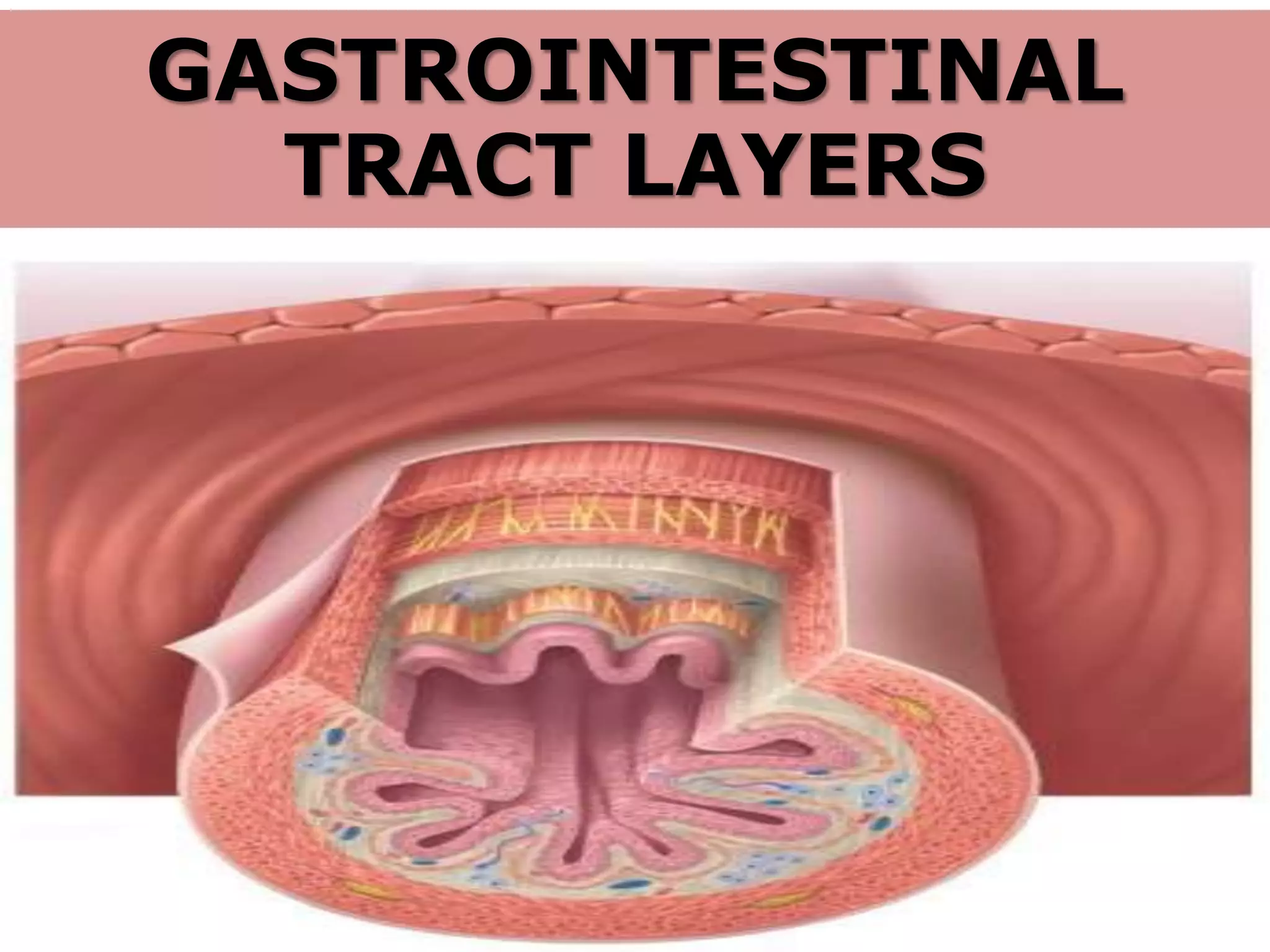
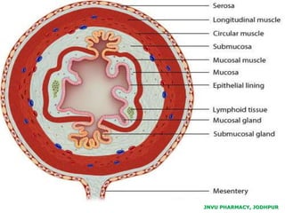
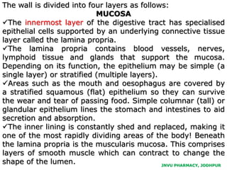
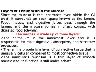
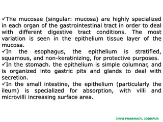
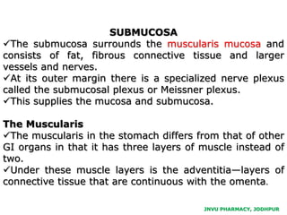

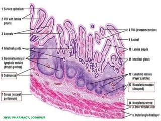

The gastrointestinal tract consists of four main layers: mucosa, submucosa, muscularis externa, and serosa, each with distinct structures and functions. The mucosa has specialized epithelial cells for digestion, while the muscularis externa facilitates movement of food through peristalsis. The serosa, composed of connective tissue and mesothelium, reduces friction between the organs and body cavities.


![GI PHYSIOLOGY new].pptx](https://cdn.slidesharecdn.com/ss_thumbnails/giphysiologynew-230405152805-b9462356-thumbnail.jpg?width=640&height=640&fit=bounds)


