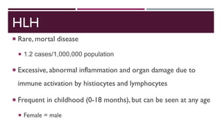
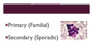
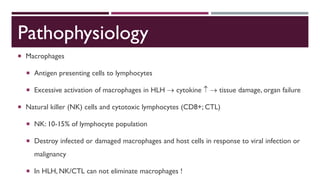
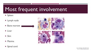
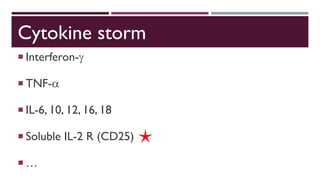
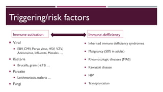
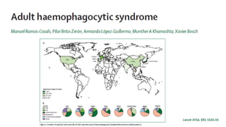
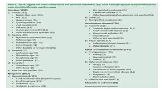
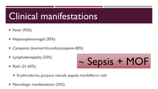
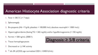
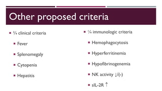
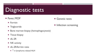
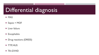
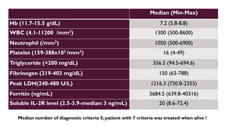
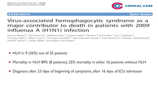
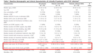
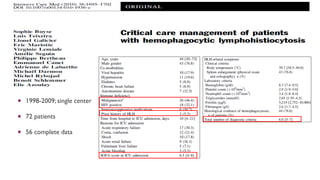
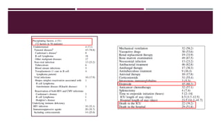
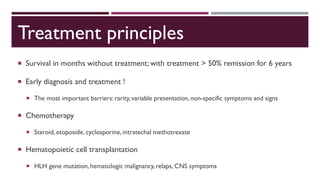
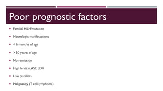
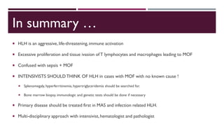
Hemophagocytic lymphohistiocytosis
ArzuTOPELI ISKIT
Hacettepe University Faculty of Medicine, Medical Intensive Care Unit
HLH
 Rare, mortal disease
 1.2 cases/1,000,000 population
 Excessive, abnormal inflammation and organ damage due to
immune activation by histiocytes and lymphocytes
 Frequent in childhood (0-18 months), but can be seen at any age
 Female = male
Primary (Familial)
Secondary (Sporadic)
Primary HLH (familial erythrophagocytic lymphohistiocytosis)
 Hereditary (heterogenous transmission; autosomal
recessive)
 (-) Family history in general
 Consangiunity is frequent
 Infection as a trigerring factor
Secondary HLH (acquired HLH)
Strong immunologic activation
Systemic infection
Immune defficiency
Cancer (lymphoma …)
Macrophage activation syndrome (MAS)
A form of HLH
Reactive hemophagocytic syndrome
Secondary to rheumatologic diseases
Juvenile idiopathic arthritis (Still disease) …
Pathophysiology
 Macrophages
 Antigen presenting cells to lymphocytes
 Excessive activation of macrophages in HLH  cytokine   tissue damage, organ failure
 Natural killer (NK) cells and cytotoxic lymphocytes (CD8+; CTL)
 NK: 10-15% of lymphocyte population
 Destroy infected or damaged macrophages and host cells in response to viral infection or
malignancy
 In HLH, NK/CTL can not eliminate macrophages !
Pathophysiology
 Uncontrolled activation of cellular immune system
 Decreased apoptosis
 T lymphocyte and macrophage activation
 Aggressive proliferation of macrophages and histiocytes; and phagocytosis
of erythrocytes, leukocytes, platelets, etc.
 Not a malignant proliferation
 Not a clonal proliferation (i.e., Langerhans histiocytosis, histiocytosis X)
 Gene mutation
 Mutation can be seen in genes related with other immune defficiency syndromes.
 Perforin gene mutation  apoptosis ↓
 Demonstrated in 20-40% cases with familial HLH
 Perforin is a membranolytic protein expressed in cytoplasmic granules of NK cells and CTL.
 It is responsible for establishing a pore in the membrane by translocating granzyme B from cytotoxic
cells to target cells.
 Granzyme B reaches the target nucleus and induces apoptosis.
 In case of perforin defficiency, there is decreased or no cytolytic effect of CTL and NK cells.
Mainly perforin gene mutation
Apoptosis pathways
Another pathophysiological mechanism
Toll-like receptor activation
 Non-antigen-specific receptor on NK cell surface
 Activated by microorganisms
Most frequent involvement
 Spleen
 Lymph node
 Bone marrow
 Liver
 Skin
 Meninx
 Spinal cord
Cytokine storm
 Interferon-
 TNF-
 IL-6, 10, 12, 16, 18
 Soluble IL-2 R (CD25)
 …
Triggering/risk factors
Immune-activation
 Viral
 EBV, CMV, Parvo virus, HSV, VZV,
Adenovirus, Influenza, Measles …
 Bacteria
 Brucella, gram (-),TB …
 Parazite
 Leishmaniasis, malaria …
 Fungi
Immune-defficiency
 Inherited immune defficiency syndromes
 Malignancy (50% in adults)
 Rheumatologic diseases (MAS)
 Kawasaki disease
 HIV
 Transplantation
Clinical manifestations Ateş
 Fever (93%)
 Hepatosplenomegali (95%)
 Cytopenia (anemia+thrombocytopenia 80%)
 Lymphadenopathy (33%)
 Rash (31-65%)
 Erythroderma, purpura, macule, papule, morbiliform rash
 Neurologic manifestations (33%)
 Sepsis + MOF
American Histiocyte Association diagnostic criteria
1. Fever ≥ 38.5˚C (> 7 days)
2. Splenomegaly
3. Bi-cytopenia (Hb < 9 g/dL; platelets < 100,000 /mcL; absolute neutrophil < 1000 /mcL)
4. Hypertrygliceridemia (fastingTG > 265 mg/dL) and/or hypofibrinogenemia (< 150 mg/dL)
5. Ferritin > 500 ng/mL (3000 ?)
6. Tissue hemophagocytosis
7. Decreased or (-) NK activity
8.  sIL-2R (sCD25; age normalized 2SD+; >2400 IU/mL)
Diagnosis: ≥ 5/8 criteria
Other proposed criteria
 ¾ clinical criteria
 Fever
 Splenomegaly
 Cytopenia
 Hepatitis
 ¼ immunologic criteria
 Hemophagocytosis
 Hyperferritinemia
 Hypofibrinogenemia
 NK activity ↓/(-)
 sIL-2R 
Diagnostic tests
 Fever, MOF
 Ferritin
 Triglyceride
 Bone marrow biopsy (hemophagocytosis)
 Tissue biopsy
 sIL-2R
 NK activity
 sIL-2R/ferritin ratio
  in lymphoma related HLH
 Genetic tests
 Infection screening
Differential diagnosis
 MAS
 Sepsis + MOF
 Liver failure
 Encephalitis
 Drug reactions (DRESS)
 TTP, HUS
 TA-GVHD
Patients
1 2 3 4 5 6 7 8 9 10 Median (min-max)
Age (years) 73 42 21 55 53 73 20 21 22 25 33.5 (20-73)
Gender F F F M F M F M F F -
APACHE II score 15 23 23 22 12 14 24 24 16 37 22.5 (12-37)
Admission SOFA score 15 9 10 8 7 8 14 16 8 14 9.5 (12-37)
Last SOFA score 20 20 14 18 16 17 14 20 20 16 17.5 (14-20)
Co-morbidities HT, DM Breast
cancer
Optic
gliom
GP PU CAD, COPD,
HT,
Hyperthyroid
CVID Renal
trans
CVID,
pregnancy
Pregnancy,
Cholecystitis
LOS (ICU) (days) 12 6 55 9 30 17 4 27 9 42 14.5 (4-55)
LOS (Hospital) (days) 13 29 56 10 49 50 26 36 40 44 38 (10-56)
Viral serology - - - - CMV - Parvo-
EBV
CMV CMV-
EBV
CMV
Nosocomial infection in all; all ex; only 1 was diagnosed when alive and treatment was given !
Median (Min-Max)
Hb (11.7-15.5 g/dL) 7.2 (5.8-8.8)
WBC (4.1-11200 /mm3) 1300 (500-8600)
Neutrophil (/mm3) 1050 (500-6900)
Platelet (159-388x103 /mm3) 16 (4-49)
Triglyceride (<200 mg/dL) 356.5 (94.5-694.6)
Fibrinogen (219-403 mg/dL) 150 (63-788)
Peak LDH(240-480 U/L) 1216.3 (730.8-2355)
Ferritin (ng/mL) 3684.5 (639.8-40316)
Soluble IL-2R level (2.5-3.9-median: 3 ng/mL) 20 (8.6-72.4)
Median number of diagnostic criteria 5; patient with 7 criteria was treated when alive !
 HLH in 9 (36%) out of 25 patients
 Mortality in HLH 89% (8 patients); 25% mortality in other 16 patients without HLH
 Diagnosis after 23 days of beginning of symptoms, after 16 days of ICU admission
 1998-2009; single center
 72 patients
 56 complete data
 Can be missed, delay in diagnosis
 Bi-cytopenia is the most important parameter for awareness
 Bone marrow biopsy is the most important test for confirmation
Treatment principles
 Survival in months without treatment; with treatment > 50% remission for 6 years
 Early diagnosis and treatment !
 The most important barriers: rarity, variable presentation, non-specific symptoms and signs
 Chemotherapy
 Steroid, etoposide, cyclosporine, intratechal methotrexate
 Hematopoietic cell transplantation
 HLH gene mutation, hematologic malignancy, relaps, CNS symptoms
Poor prognostic factors
 Familial HLH/mutation
 Neurologic manifestations
 < 6 months of age
 > 50 years of age
 No remission
 High ferritin,AST, LDH
 Low platelets
 Malignancy (T cell lymphoma)
In summary …
 HLH is an aggressive, life-threatening, immune activation
 Excessive proliferation and tissue ivasion of T lymphocytes and macrophages leading to MOF
 Confused with sepsis + MOF
 INTENSIVISTS SHOULDTHINK OF HLH in cases with MOF with no known cause !
 Splenomegaly, hyperferritinemia, hypertriglyceridemia should be searched for.
 Bone marrow biopsy, immunologic and genetic tests should be done if necessary
 Primary disease should be treated first in MAS and infection related HLH.
 Multi-disciplinary approach with intensivist, hematologist and pathologist
Hl hing

Hl hing

  • 1.
    Hemophagocytic lymphohistiocytosis ArzuTOPELI ISKIT HacettepeUniversity Faculty of Medicine, Medical Intensive Care Unit
  • 3.
    HLH  Rare, mortaldisease  1.2 cases/1,000,000 population  Excessive, abnormal inflammation and organ damage due to immune activation by histiocytes and lymphocytes  Frequent in childhood (0-18 months), but can be seen at any age  Female = male
  • 4.
  • 5.
    Primary HLH (familialerythrophagocytic lymphohistiocytosis)  Hereditary (heterogenous transmission; autosomal recessive)  (-) Family history in general  Consangiunity is frequent  Infection as a trigerring factor
  • 6.
    Secondary HLH (acquiredHLH) Strong immunologic activation Systemic infection Immune defficiency Cancer (lymphoma …)
  • 7.
    Macrophage activation syndrome(MAS) A form of HLH Reactive hemophagocytic syndrome Secondary to rheumatologic diseases Juvenile idiopathic arthritis (Still disease) …
  • 8.
    Pathophysiology  Macrophages  Antigenpresenting cells to lymphocytes  Excessive activation of macrophages in HLH  cytokine   tissue damage, organ failure  Natural killer (NK) cells and cytotoxic lymphocytes (CD8+; CTL)  NK: 10-15% of lymphocyte population  Destroy infected or damaged macrophages and host cells in response to viral infection or malignancy  In HLH, NK/CTL can not eliminate macrophages !
  • 9.
    Pathophysiology  Uncontrolled activationof cellular immune system  Decreased apoptosis  T lymphocyte and macrophage activation  Aggressive proliferation of macrophages and histiocytes; and phagocytosis of erythrocytes, leukocytes, platelets, etc.  Not a malignant proliferation  Not a clonal proliferation (i.e., Langerhans histiocytosis, histiocytosis X)
  • 10.
     Gene mutation Mutation can be seen in genes related with other immune defficiency syndromes.  Perforin gene mutation  apoptosis ↓  Demonstrated in 20-40% cases with familial HLH  Perforin is a membranolytic protein expressed in cytoplasmic granules of NK cells and CTL.  It is responsible for establishing a pore in the membrane by translocating granzyme B from cytotoxic cells to target cells.  Granzyme B reaches the target nucleus and induces apoptosis.  In case of perforin defficiency, there is decreased or no cytolytic effect of CTL and NK cells. Mainly perforin gene mutation
  • 11.
  • 12.
    Another pathophysiological mechanism Toll-likereceptor activation  Non-antigen-specific receptor on NK cell surface  Activated by microorganisms
  • 13.
    Most frequent involvement Spleen  Lymph node  Bone marrow  Liver  Skin  Meninx  Spinal cord
  • 14.
    Cytokine storm  Interferon- TNF-  IL-6, 10, 12, 16, 18  Soluble IL-2 R (CD25)  …
  • 15.
    Triggering/risk factors Immune-activation  Viral EBV, CMV, Parvo virus, HSV, VZV, Adenovirus, Influenza, Measles …  Bacteria  Brucella, gram (-),TB …  Parazite  Leishmaniasis, malaria …  Fungi Immune-defficiency  Inherited immune defficiency syndromes  Malignancy (50% in adults)  Rheumatologic diseases (MAS)  Kawasaki disease  HIV  Transplantation
  • 18.
    Clinical manifestations Ateş Fever (93%)  Hepatosplenomegali (95%)  Cytopenia (anemia+thrombocytopenia 80%)  Lymphadenopathy (33%)  Rash (31-65%)  Erythroderma, purpura, macule, papule, morbiliform rash  Neurologic manifestations (33%)  Sepsis + MOF
  • 19.
    American Histiocyte Associationdiagnostic criteria 1. Fever ≥ 38.5˚C (> 7 days) 2. Splenomegaly 3. Bi-cytopenia (Hb < 9 g/dL; platelets < 100,000 /mcL; absolute neutrophil < 1000 /mcL) 4. Hypertrygliceridemia (fastingTG > 265 mg/dL) and/or hypofibrinogenemia (< 150 mg/dL) 5. Ferritin > 500 ng/mL (3000 ?) 6. Tissue hemophagocytosis 7. Decreased or (-) NK activity 8.  sIL-2R (sCD25; age normalized 2SD+; >2400 IU/mL) Diagnosis: ≥ 5/8 criteria
  • 20.
    Other proposed criteria ¾ clinical criteria  Fever  Splenomegaly  Cytopenia  Hepatitis  ¼ immunologic criteria  Hemophagocytosis  Hyperferritinemia  Hypofibrinogenemia  NK activity ↓/(-)  sIL-2R 
  • 21.
    Diagnostic tests  Fever,MOF  Ferritin  Triglyceride  Bone marrow biopsy (hemophagocytosis)  Tissue biopsy  sIL-2R  NK activity  sIL-2R/ferritin ratio   in lymphoma related HLH  Genetic tests  Infection screening
  • 22.
    Differential diagnosis  MAS Sepsis + MOF  Liver failure  Encephalitis  Drug reactions (DRESS)  TTP, HUS  TA-GVHD
  • 24.
    Patients 1 2 34 5 6 7 8 9 10 Median (min-max) Age (years) 73 42 21 55 53 73 20 21 22 25 33.5 (20-73) Gender F F F M F M F M F F - APACHE II score 15 23 23 22 12 14 24 24 16 37 22.5 (12-37) Admission SOFA score 15 9 10 8 7 8 14 16 8 14 9.5 (12-37) Last SOFA score 20 20 14 18 16 17 14 20 20 16 17.5 (14-20) Co-morbidities HT, DM Breast cancer Optic gliom GP PU CAD, COPD, HT, Hyperthyroid CVID Renal trans CVID, pregnancy Pregnancy, Cholecystitis LOS (ICU) (days) 12 6 55 9 30 17 4 27 9 42 14.5 (4-55) LOS (Hospital) (days) 13 29 56 10 49 50 26 36 40 44 38 (10-56) Viral serology - - - - CMV - Parvo- EBV CMV CMV- EBV CMV Nosocomial infection in all; all ex; only 1 was diagnosed when alive and treatment was given !
  • 25.
    Median (Min-Max) Hb (11.7-15.5g/dL) 7.2 (5.8-8.8) WBC (4.1-11200 /mm3) 1300 (500-8600) Neutrophil (/mm3) 1050 (500-6900) Platelet (159-388x103 /mm3) 16 (4-49) Triglyceride (<200 mg/dL) 356.5 (94.5-694.6) Fibrinogen (219-403 mg/dL) 150 (63-788) Peak LDH(240-480 U/L) 1216.3 (730.8-2355) Ferritin (ng/mL) 3684.5 (639.8-40316) Soluble IL-2R level (2.5-3.9-median: 3 ng/mL) 20 (8.6-72.4) Median number of diagnostic criteria 5; patient with 7 criteria was treated when alive !
  • 26.
     HLH in9 (36%) out of 25 patients  Mortality in HLH 89% (8 patients); 25% mortality in other 16 patients without HLH  Diagnosis after 23 days of beginning of symptoms, after 16 days of ICU admission
  • 28.
     1998-2009; singlecenter  72 patients  56 complete data
  • 31.
     Can bemissed, delay in diagnosis  Bi-cytopenia is the most important parameter for awareness  Bone marrow biopsy is the most important test for confirmation
  • 33.
    Treatment principles  Survivalin months without treatment; with treatment > 50% remission for 6 years  Early diagnosis and treatment !  The most important barriers: rarity, variable presentation, non-specific symptoms and signs  Chemotherapy  Steroid, etoposide, cyclosporine, intratechal methotrexate  Hematopoietic cell transplantation  HLH gene mutation, hematologic malignancy, relaps, CNS symptoms
  • 35.
    Poor prognostic factors Familial HLH/mutation  Neurologic manifestations  < 6 months of age  > 50 years of age  No remission  High ferritin,AST, LDH  Low platelets  Malignancy (T cell lymphoma)
  • 36.
    In summary … HLH is an aggressive, life-threatening, immune activation  Excessive proliferation and tissue ivasion of T lymphocytes and macrophages leading to MOF  Confused with sepsis + MOF  INTENSIVISTS SHOULDTHINK OF HLH in cases with MOF with no known cause !  Splenomegaly, hyperferritinemia, hypertriglyceridemia should be searched for.  Bone marrow biopsy, immunologic and genetic tests should be done if necessary  Primary disease should be treated first in MAS and infection related HLH.  Multi-disciplinary approach with intensivist, hematologist and pathologist