Histochemistry Krishnendu Sinha
Assistant Professor
Department of Zoology
Jhargram Raj College
KS_JRC_Histochemistry
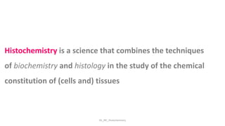
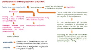
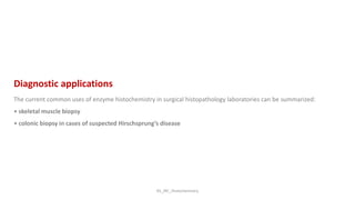
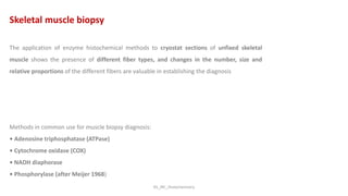
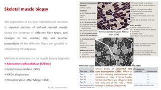
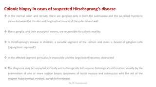
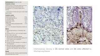
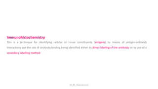
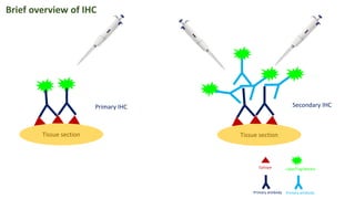
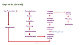
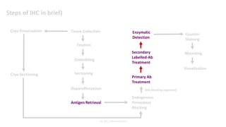
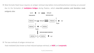
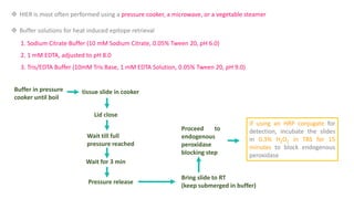
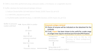
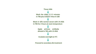
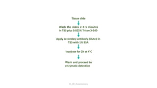
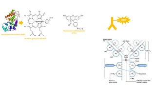
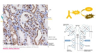
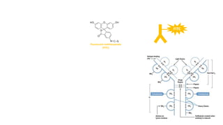
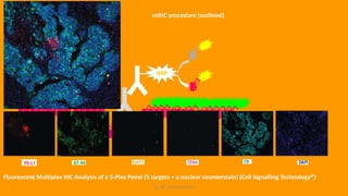
Histochemistry is a science that combines the techniques
of biochemistry and histology in the study of the chemical
constitution of (cells and) tissues
KS_JRC_Histochemistry
Enzyme Histochemistry
Enzymes are labile and their preservation is important
Fixation will destroy many
of the oxidative enzymes
freezing and subsequent
thawing of blocks or sections
damages lysosome
Hydrolytic enzymes show
considerable diffusion if
demonstrated on frozen,
unfixed sections
Tissues to be used for the demonstration
of the majority of hydrolytic enzymes may
be subjected to controlled fixation
decreasing the amount of demonstrable
enzyme within the section, does allow for
a considerably sharper localization of the
remaining enzyme activity
for the demonstration of hydrolytic
enzymes, histochemical techniques are
usually applied to sections that have been
prefixed in cold (4°C) formal calcium
Fixation for enzyme histochemistry
Cryofixation
Chemical fixation
Chemical fixation
Cryofixation
Cryofixation will preserve many
of the oxidative enzymes
Cryosectioning and staining
Mitochondria
Contains most of the oxidative enzymes and
damaged immediately after blood supply cut
Lysosome
Contains most of the hydrolytic enzymes and
damaged on freeze thaw
KS_JRC_Histochemistry
.
Smears
 In enzyme histochemistry, the use of smears for cytochemical identification and
evaluation of cells is used
 Preparation may be achieved in various ways, include blood, bone marrow and
tissue cell suspensions
 Three of the most useful enzymes are non-specific esterase, acid phosphatase
and chloroacetate esterase
 It is usual to fix smears before histochemical staining to preserve cell structure
and enzyme localization
KS_JRC_Histochemistry
Enzyme types
Types of histochemical reactions
1. Simultaneous capture
2. Post incubation coupling
3. Self coloured substrate
4. Intramolecular rearrangement
Classical histochemical reactions are
generally based on one of the 4 principles:
1.Simple ionic interactions
2.Reactions of aldehydes with Schiff’s reagent or silver compounds
3.Coupling of aromatic diazonium salts with aromatic residues on protein
4.Conversion acting on a substrate to form a colored ppt
KS_JRC_Histochemistry
1. Simultaneous capture
-Gomori’s Metal ppt. technique
-Azo dye method
Enzyme Substrate
Diazonium
Salt
Final Reaction
Product (FRP)
(IS)
Primary Reaction
Product (PRP)
Disadvantage
1.Diffusion of PRP
2.Rate of hydrolysis of substrate
3.Diffusion coefficient of the PRP for the buffer
4.Rate of coupling of the PRP and diazo salt
5.Diazo salt and Enzyme same pH
Diazonium
Salt
Advantage
1. A great repository of alternative substrates
2. Highly sensitive
KS_JRC_Histochemistry
2. Post incubation coupling
Enzyme Substrate
Diazonium
Salt
FRP(IS)
(in different
medium)
Primary Reaction
Product (PRP)
Insoluble remain at the
site of formation
Disadvantage
1. PRP is not completely insoluble
2. Diffusion is always there
Advantage
1. Case where long first incubation stage is necessary
2.Optimum pH for enzyme and for diazonium salt separately
KS_JRC_Histochemistry
3. Self coloured substrate
Enzyme
Colored
Soluble
Substrate
Hydrolysis Insoluble FRP
Advantage
1. Diazonium coupling not required
Disadvantage
1. Limited number of substrates are available
KS_JRC_Histochemistry
4. Intramolecular rearrangement
Enzyme
Molecular
Rearrangement
of Substrate
Hydrolysis
Insoluble
colored FRP
Advantage
1. Diazonium coupling not required
Soluble
Substrate
Disadvantage
1. Limited number of substrates are available
KS_JRC_Histochemistry
Diagnostic applications
The current common uses of enzyme histochemistry in surgical histopathology laboratories can be summarized:
• skeletal muscle biopsy
• colonic biopsy in cases of suspected Hirschsprung’s disease
KS_JRC_Histochemistry
The application of enzyme histochemical methods to cryostat sections of unfixed skeletal
muscle shows the presence of different fiber types, and changes in the number, size and
relative proportions of the different fibers are valuable in establishing the diagnosis
Methods in common use for muscle biopsy diagnosis:
• Adenosine triphosphatase (ATPase)
• Cytochrome oxidase (COX)
• NADH diaphorase
• Phosphorylase (after Meijer 1968)
Skeletal muscle biopsy
The application of enzyme histochemical methods
to cryostat sections of unfixed skeletal muscle
shows the presence of different fiber types, and
changes in the number, size and relative
proportions of the different fibers are valuable in
establishing the diagnosis
Methods in common use for muscle biopsy diagnosis:
• Adenosine triphosphatase (ATPase)
• Cytochrome oxidase (COX)
• NADH diaphorase
• Phosphorylase (after Meijer 1968)
Skeletal muscle biopsy
Normal skeletal muscle, ATPase
stain x 100
Muscle biopsy of Congenital fibre
type disproportion (CFTD). ATPase at
pH 4.63, showing predominance and
smallness of type 1 fibres (darker
fibres). Clear fibres are of type 2. Note
that only one of the type 2 fibres
belongs to subtype 2B (asterisk).
KS_JRC_Histochemistry
Colonic biopsy in cases of suspected Hirschsprung’s disease
 In the normal colon and rectum, there are ganglion cells in both the submucosa and the so-called myenteric
plexus between the circular and longitudinal muscle of the outer bowel wall
 These ganglia, and their associated nerves, are responsible for colonic motility
 In Hirschsprung’s disease in children, a variable segment of the rectum and colon is devoid of ganglion cells
(‘aganglionic segment’)
 In the affected segment peristalsis is impossible and the large bowel becomes obstructed
 The diagnosis may be suspected clinically and radiologically but requires histological confirmation, usually by the
examination of one or more suction biopsy specimens of rectal mucosa and submucosa with the aid of the
enzyme histochemical method, acetylcholinesterase.
Cholinesterase staining in (A) normal colon and (B) colon affected by
Hirschsprung disease
KS_JRC_Histochemistry
Immunohistochemistry
KS_JRC_Histochemistry
This is a technique for identifying cellular or tissue constituents (antigens) by means of antigen-antibody
interactions and the site of antibody binding being identified either by direct labeling of the antibody, or by use of a
secondary labelling method
Immunohistochemistry
Tissue section Tissue section
Epitope
Primary antibody
Label/Tag/Marker
Primary IHC Secondary IHC
Brief overview of IHC
Primary antibody
KS_JRC_Histochemistry
Steps of IHC (in brief)
Tissue Collection
Fixation
Embedding
Sectioning
Deparaffinization
Antigen Retrieval
Endogenous
Peroxidase
Blocking
Primary Ab
Treatment
Secondary
Labelled-Ab
Treatment
Counter
Staining
Visualization
Mounting
Cryo-Preservation
BSA blocking (optional)
Cryo-Sectioning
Enzymatic
Detection
KS_JRC_Histochemistry
Steps of IHC in brief)
Tissue Collection
Fixation
Embedding
Sectioning
Deparaffinization
Antigen Retrieval
Endogenous
Peroxidase
Blocking
Primary Ab
Treatment
Secondary
Labelled-Ab
Treatment
Enzymatic
Detection
Counter
Staining
Visualization
Mounting
Cryo-Preservation
Cryo-Sectioning
BSA blocking (optional)
KS_JRC_Histochemistry
Antigen Retrieval (AR) for IHC-P
KS_JRC_Histochemistry
 Most formalin-fixed tissue requires an antigen retrieval step before immunohistochemical staining can proceed
due to the formation of methylene bridges during fixation, which cross-link proteins and therefore mask
antigenic sites
 The two methods of antigen retrieval are
Heat-mediated (also known as heat-induced epitope retrieval, or HIER) and enzymatic
 HIER is most often performed using a pressure cooker, a microwave, or a vegetable steamer
 Buffer solutions for heat induced epitope retrieval
1. Sodium Citrate Buffer (10 mM Sodium Citrate, 0.05% Tween 20, pH 6.0)
2. 1 mM EDTA, adjusted to pH 8.0
3. Tris/EDTA Buffer (10mM Tris Base, 1 mM EDTA Solution, 0.05% Tween 20, pH 9.0)
Buffer in pressure
cooker until boil
tissue slide in cooker
Lid close
Wait till full
pressure reached
Wait for 3 min
Pressure release
Bring slide to RT
(keep submerged in buffer)
Proceed to
endogenous
peroxidase
blocking step
If using an HRP conjugate for
detection, incubate the slides
in 0.3% H2O2 in TBS for 15
minutes to block endogenous
peroxidase
Enzymatic antigen retrieval
 Choice of enzyme will be indicated on the datasheet for the
antibody
 If not, trypsin has been shown to be useful for a wide range
of antigen that require retrieval post formalin/PFA fixation.
KS_JRC_Histochemistry
Primary Ab-treatment
Tissue slide
Wash the slides 2 X 5 minutes
in TBS plus 0.025% Triton X-100
Block in 10% normal serum with 1% BSA
in TBS for 2 hours at room temperature
Apply primary antibody
diluted in TBS with 1% BSA
Incubate overnight at 4°C
Proceed to secondary Ab-treatment
KS_JRC_Histochemistry
Secondary Ab-treatment
KS_JRC_Histochemistry
Tissue slide
Wash the slides 2 X 5 minutes
in TBS plus 0.025% Triton X-100
Apply secondary antibody diluted in
TBS with 1% BSA
Incubate for 2h at 4°C
Wash and proceed to
enzymatic detection
KS_JRC_Histochemistry
Enzymatic Detection
Horseradish Peroxidase (HRP)
Fe-heme group of the HRP
Fluorescein isothiocyanate
(FITC)
HRP
Horseradish Peroxidase (HRP)
Fe-heme group of the HRP
Fluorescein isothiocyanate
(FITC)
FITC
Horseradish Peroxidase (HRP)
Fe-heme group of the HRP
HRP
DAB
DAB
3,3'-Diaminobenzidine (DAB)
Fluorescein isothiocyanate
(FITC)
Histone, Kidney (Abcam)
Fluorescein isothiocyanate
(FITC)
FITC
Fluorescein isothiocyanate
(FITC)
FITC
Cadherin-17 in Human Ascending Colon, Hu (Cadherin, DAPI)
Source: https://www.rndsystems.com/products/human-cadherin-
17-antibody-141713_mab1032
KS_JRC_Histochemistry
SIGNAL AMPLIFICATION
HRP
DAB
DAB
Avidin
Biotin/Streptavidin
20 Ab
10 Ab
Avidin–Biotin Complex (ABC)
and the Labeled/Streptavidin–
Biotin (LSAB) staining methods
Biotin
Biotin, also known as vitamin H, is a small molecule (MW 244.3) that is present in tiny amounts in all living cells and is critical for a number of
biological processes. The valeric acid side chain of the biotin molecule can be derivatized in order to incorporate various reactive groups that
are used to attach biotin to other molecules. In the context of IHC, biotin is conjugated to antibodies or to the enzyme reporters use to
detect target antigens.
Avidin (AV)
The extraordinary affinity of avidin (AV) for biotin allows biotin-containing molecules in a complex mixture to be specifically bound to avidin.
Avidin is a glycoprotein found in the egg white and tissues of birds, reptiles and amphibia. It contains four identical subunits having a
combined mass of 67 to 68 kDa. Each subunit consists of 128 amino acids and binds one molecule of biotin; thus, a total of four biotin
molecules can bind to a single avidin molecule. The extent of glycosylation on avidin is very high; carbohydrates account for about 10% of the
total mass of the tetramer. Avidin has a basic isoelectric point (pI) of 10 to 10.5 and is stable over a wide range of pH and temperatures.
Extensive chemical modification has little effect on the activity of avidin, making it especially useful for protein purification. However,
because of its carbohydrate content and basic pI, avidin exhibits relatively high nonspecific binding properties. Avidin–biotin binding is the
strongest known non-covalent interaction between a protein and ligand. The bond between biotin and avidin is formed very rapidly, and
once formed, is unaffected by extremes in pH, temperature, organic solvents and other denaturing agents. These features of avidin make
detecting or purifying biotin-labeled proteins or other molecules particularly useful for a number of biomedical applications.
Streptavidin (SA)
Streptavidin (SA) is a biotin-binding protein isolated from Streptomyces avidinii, and is similar in size and affinity for biotin. In contrast to
avidin, though, streptavidin is not glycosylated, which makes the protein less prone to nonspecific binding in IHC applications.
ThermoFisher
KS_JRC_Histochemistry
 HRP-polymer secondary antibodies use micropolymer technology to form smaller detection complexes that allow better
tissue penetration and sensitivity
 In addition, HRP-polymer secondaries bind more horseradish peroxidase than standard HRP secondary antibodies,
increasing signal.
HRP-polymer Secondary Antibodies
Abcam
KS_JRC_Histochemistry
Multiplex IHC (mIHC)
Fluorescent multiplex immunohistochemistry (mIHC) is a method that enables simultaneous detection of multiple
proteins of interest in formalin-fixed paraffin-embedded (FFPE) tissue sections. There are various approaches to
fluorescent multiplexing:
1. Direct immunofluorescence: involves the use of multiple antigen-specific primary antibodies conjugated to distinct
fluorophores. The disadvantage of this approach is limited sensitivity for targets of low abundance due to lack of signal
amplification.
2. Indirect immunofluorescence: antigen detection is mediated via conjugated secondary antibodies specific to the
species of the host in which each primary antibody was raised. This approach provides modest signal amplification but is
limited by the number of available host species, e.g. rabbit, mouse, rat, and others.
3. Deposition assays: involve the use of enzyme-labeled antibodies and tyramide-fluorophore conjugates. This approach
is unhindered by host species and isotype concerns, while providing ample signal amplification.
Fluorescent Multiplex Immunohistochemistry with Tyramide Signal Amplification
KS_JRC_Histochemistry
Inactive tyramide
Active tyramide
mIHC procedure (outlined)
HRP
KS_JRC_Histochemistry
HRP
mIHC procedure (outlined)
KS_JRC_Histochemistry
HRP
mIHC procedure (outlined)
Fluorescent Multiplex IHC Analysis of a 5-Plex Panel (5 targets + a nuclear counterstain) (Cell Signalling Technology®)
KS_JRC_Histochemistry
Reference
KS_JRC_Histochemistry

Histochemistry

  • 1.
    Histochemistry Krishnendu Sinha AssistantProfessor Department of Zoology Jhargram Raj College
  • 2.
    KS_JRC_Histochemistry Histochemistry is ascience that combines the techniques of biochemistry and histology in the study of the chemical constitution of (cells and) tissues
  • 3.
  • 4.
    Enzymes are labileand their preservation is important Fixation will destroy many of the oxidative enzymes freezing and subsequent thawing of blocks or sections damages lysosome Hydrolytic enzymes show considerable diffusion if demonstrated on frozen, unfixed sections Tissues to be used for the demonstration of the majority of hydrolytic enzymes may be subjected to controlled fixation decreasing the amount of demonstrable enzyme within the section, does allow for a considerably sharper localization of the remaining enzyme activity for the demonstration of hydrolytic enzymes, histochemical techniques are usually applied to sections that have been prefixed in cold (4°C) formal calcium Fixation for enzyme histochemistry Cryofixation Chemical fixation Chemical fixation Cryofixation Cryofixation will preserve many of the oxidative enzymes Cryosectioning and staining Mitochondria Contains most of the oxidative enzymes and damaged immediately after blood supply cut Lysosome Contains most of the hydrolytic enzymes and damaged on freeze thaw
  • 5.
    KS_JRC_Histochemistry . Smears  In enzymehistochemistry, the use of smears for cytochemical identification and evaluation of cells is used  Preparation may be achieved in various ways, include blood, bone marrow and tissue cell suspensions  Three of the most useful enzymes are non-specific esterase, acid phosphatase and chloroacetate esterase  It is usual to fix smears before histochemical staining to preserve cell structure and enzyme localization
  • 6.
  • 7.
    Types of histochemicalreactions 1. Simultaneous capture 2. Post incubation coupling 3. Self coloured substrate 4. Intramolecular rearrangement Classical histochemical reactions are generally based on one of the 4 principles: 1.Simple ionic interactions 2.Reactions of aldehydes with Schiff’s reagent or silver compounds 3.Coupling of aromatic diazonium salts with aromatic residues on protein 4.Conversion acting on a substrate to form a colored ppt KS_JRC_Histochemistry
  • 8.
    KS_JRC_Histochemistry 1. Simultaneous capture -Gomori’sMetal ppt. technique -Azo dye method Enzyme Substrate Diazonium Salt Final Reaction Product (FRP) (IS) Primary Reaction Product (PRP) Disadvantage 1.Diffusion of PRP 2.Rate of hydrolysis of substrate 3.Diffusion coefficient of the PRP for the buffer 4.Rate of coupling of the PRP and diazo salt 5.Diazo salt and Enzyme same pH Diazonium Salt Advantage 1. A great repository of alternative substrates 2. Highly sensitive
  • 9.
    KS_JRC_Histochemistry 2. Post incubationcoupling Enzyme Substrate Diazonium Salt FRP(IS) (in different medium) Primary Reaction Product (PRP) Insoluble remain at the site of formation Disadvantage 1. PRP is not completely insoluble 2. Diffusion is always there Advantage 1. Case where long first incubation stage is necessary 2.Optimum pH for enzyme and for diazonium salt separately
  • 10.
    KS_JRC_Histochemistry 3. Self colouredsubstrate Enzyme Colored Soluble Substrate Hydrolysis Insoluble FRP Advantage 1. Diazonium coupling not required Disadvantage 1. Limited number of substrates are available
  • 11.
    KS_JRC_Histochemistry 4. Intramolecular rearrangement Enzyme Molecular Rearrangement ofSubstrate Hydrolysis Insoluble colored FRP Advantage 1. Diazonium coupling not required Soluble Substrate Disadvantage 1. Limited number of substrates are available
  • 12.
    KS_JRC_Histochemistry Diagnostic applications The currentcommon uses of enzyme histochemistry in surgical histopathology laboratories can be summarized: • skeletal muscle biopsy • colonic biopsy in cases of suspected Hirschsprung’s disease
  • 13.
    KS_JRC_Histochemistry The application ofenzyme histochemical methods to cryostat sections of unfixed skeletal muscle shows the presence of different fiber types, and changes in the number, size and relative proportions of the different fibers are valuable in establishing the diagnosis Methods in common use for muscle biopsy diagnosis: • Adenosine triphosphatase (ATPase) • Cytochrome oxidase (COX) • NADH diaphorase • Phosphorylase (after Meijer 1968) Skeletal muscle biopsy
  • 14.
    The application ofenzyme histochemical methods to cryostat sections of unfixed skeletal muscle shows the presence of different fiber types, and changes in the number, size and relative proportions of the different fibers are valuable in establishing the diagnosis Methods in common use for muscle biopsy diagnosis: • Adenosine triphosphatase (ATPase) • Cytochrome oxidase (COX) • NADH diaphorase • Phosphorylase (after Meijer 1968) Skeletal muscle biopsy Normal skeletal muscle, ATPase stain x 100 Muscle biopsy of Congenital fibre type disproportion (CFTD). ATPase at pH 4.63, showing predominance and smallness of type 1 fibres (darker fibres). Clear fibres are of type 2. Note that only one of the type 2 fibres belongs to subtype 2B (asterisk). KS_JRC_Histochemistry
  • 15.
    KS_JRC_Histochemistry Colonic biopsy incases of suspected Hirschsprung’s disease  In the normal colon and rectum, there are ganglion cells in both the submucosa and the so-called myenteric plexus between the circular and longitudinal muscle of the outer bowel wall  These ganglia, and their associated nerves, are responsible for colonic motility  In Hirschsprung’s disease in children, a variable segment of the rectum and colon is devoid of ganglion cells (‘aganglionic segment’)  In the affected segment peristalsis is impossible and the large bowel becomes obstructed  The diagnosis may be suspected clinically and radiologically but requires histological confirmation, usually by the examination of one or more suction biopsy specimens of rectal mucosa and submucosa with the aid of the enzyme histochemical method, acetylcholinesterase.
  • 16.
    KS_JRC_Histochemistry Colonic biopsy incases of suspected Hirschsprung’s disease  In the normal colon and rectum, there are ganglion cells in both the submucosa and the so-called myenteric plexus between the circular and longitudinal muscle of the outer bowel wall  These ganglia, and their associated nerves, are responsible for colonic motility  In Hirschsprung’s disease in children, a variable segment of the rectum and colon is devoid of ganglion cells (‘aganglionic segment’)  In the affected segment peristalsis is impossible and the large bowel becomes obstructed  The diagnosis may be suspected clinically and radiologically but requires histological confirmation, usually by the examination of one or more suction biopsy specimens of rectal mucosa and submucosa with the aid of the enzyme histochemical method, acetylcholinesterase.
  • 17.
    KS_JRC_Histochemistry Colonic biopsy incases of suspected Hirschsprung’s disease  In the normal colon and rectum, there are ganglion cells in both the submucosa and the so-called myenteric plexus between the circular and longitudinal muscle of the outer bowel wall  These ganglia, and their associated nerves, are responsible for colonic motility  In Hirschsprung’s disease in children, a variable segment of the rectum and colon is devoid of ganglion cells (‘aganglionic segment’)  In the affected segment peristalsis is impossible and the large bowel becomes obstructed  The diagnosis may be suspected clinically and radiologically but requires histological confirmation, usually by the examination of one or more suction biopsy specimens of rectal mucosa and submucosa with the aid of the enzyme histochemical method, acetylcholinesterase.
  • 18.
    KS_JRC_Histochemistry Colonic biopsy incases of suspected Hirschsprung’s disease  In the normal colon and rectum, there are ganglion cells in both the submucosa and the so-called myenteric plexus between the circular and longitudinal muscle of the outer bowel wall  These ganglia, and their associated nerves, are responsible for colonic motility  In Hirschsprung’s disease in children, a variable segment of the rectum and colon is devoid of ganglion cells (‘aganglionic segment’)  In the affected segment peristalsis is impossible and the large bowel becomes obstructed  The diagnosis may be suspected clinically and radiologically but requires histological confirmation, usually by the examination of one or more suction biopsy specimens of rectal mucosa and submucosa with the aid of the enzyme histochemical method, acetylcholinesterase.
  • 19.
    KS_JRC_Histochemistry Colonic biopsy incases of suspected Hirschsprung’s disease  In the normal colon and rectum, there are ganglion cells in both the submucosa and the so-called myenteric plexus between the circular and longitudinal muscle of the outer bowel wall  These ganglia, and their associated nerves, are responsible for colonic motility  In Hirschsprung’s disease in children, a variable segment of the rectum and colon is devoid of ganglion cells (‘aganglionic segment’)  In the affected segment peristalsis is impossible and the large bowel becomes obstructed  The diagnosis may be suspected clinically and radiologically but requires histological confirmation, usually by the examination of one or more suction biopsy specimens of rectal mucosa and submucosa with the aid of the enzyme histochemical method, acetylcholinesterase.
  • 20.
    KS_JRC_Histochemistry Colonic biopsy incases of suspected Hirschsprung’s disease  In the normal colon and rectum, there are ganglion cells in both the submucosa and the so-called myenteric plexus between the circular and longitudinal muscle of the outer bowel wall  These ganglia, and their associated nerves, are responsible for colonic motility  In Hirschsprung’s disease in children, a variable segment of the rectum and colon is devoid of ganglion cells (‘aganglionic segment’)  In the affected segment peristalsis is impossible and the large bowel becomes obstructed  The diagnosis may be suspected clinically and radiologically but requires histological confirmation, usually by the examination of one or more suction biopsy specimens of rectal mucosa and submucosa with the aid of the enzyme histochemical method, acetylcholinesterase.
  • 21.
    Cholinesterase staining in(A) normal colon and (B) colon affected by Hirschsprung disease
  • 22.
  • 23.
    KS_JRC_Histochemistry This is atechnique for identifying cellular or tissue constituents (antigens) by means of antigen-antibody interactions and the site of antibody binding being identified either by direct labeling of the antibody, or by use of a secondary labelling method Immunohistochemistry
  • 24.
    Tissue section Tissuesection Epitope Primary antibody Label/Tag/Marker Primary IHC Secondary IHC Brief overview of IHC Primary antibody
  • 25.
    KS_JRC_Histochemistry Steps of IHC(in brief) Tissue Collection Fixation Embedding Sectioning Deparaffinization Antigen Retrieval Endogenous Peroxidase Blocking Primary Ab Treatment Secondary Labelled-Ab Treatment Counter Staining Visualization Mounting Cryo-Preservation BSA blocking (optional) Cryo-Sectioning Enzymatic Detection
  • 26.
    KS_JRC_Histochemistry Steps of IHCin brief) Tissue Collection Fixation Embedding Sectioning Deparaffinization Antigen Retrieval Endogenous Peroxidase Blocking Primary Ab Treatment Secondary Labelled-Ab Treatment Enzymatic Detection Counter Staining Visualization Mounting Cryo-Preservation Cryo-Sectioning BSA blocking (optional)
  • 27.
  • 28.
    KS_JRC_Histochemistry  Most formalin-fixedtissue requires an antigen retrieval step before immunohistochemical staining can proceed due to the formation of methylene bridges during fixation, which cross-link proteins and therefore mask antigenic sites  The two methods of antigen retrieval are Heat-mediated (also known as heat-induced epitope retrieval, or HIER) and enzymatic
  • 29.
     HIER ismost often performed using a pressure cooker, a microwave, or a vegetable steamer  Buffer solutions for heat induced epitope retrieval 1. Sodium Citrate Buffer (10 mM Sodium Citrate, 0.05% Tween 20, pH 6.0) 2. 1 mM EDTA, adjusted to pH 8.0 3. Tris/EDTA Buffer (10mM Tris Base, 1 mM EDTA Solution, 0.05% Tween 20, pH 9.0) Buffer in pressure cooker until boil tissue slide in cooker Lid close Wait till full pressure reached Wait for 3 min Pressure release Bring slide to RT (keep submerged in buffer) Proceed to endogenous peroxidase blocking step If using an HRP conjugate for detection, incubate the slides in 0.3% H2O2 in TBS for 15 minutes to block endogenous peroxidase
  • 30.
     HIER ismost often performed using a pressure cooker, a microwave, or a vegetable steamer  Buffer solutions for heat induced epitope retrieval 1. Sodium Citrate Buffer (10 mM Sodium Citrate, 0.05% Tween 20, pH 6.0) 2. 1 mM EDTA, adjusted to pH 8.0 3. Tris/EDTA Buffer (10mM Tris Base, 1 mM EDTA Solution, 0.05% Tween 20, pH 9.0) Buffer in pressure cooker until boil tissue slide in cooker Lid close Wait till full pressure reached Wait for 3 min Pressure release Bring slide to RT (keep submerged in buffer) Proceed to endogenous peroxidase blocking step If using an HRP conjugate for detection, incubate the slides in 0.3% H2O2 in TBS for 15 minutes to block endogenous peroxidase Enzymatic antigen retrieval  Choice of enzyme will be indicated on the datasheet for the antibody  If not, trypsin has been shown to be useful for a wide range of antigen that require retrieval post formalin/PFA fixation.
  • 31.
  • 32.
    Tissue slide Wash theslides 2 X 5 minutes in TBS plus 0.025% Triton X-100 Block in 10% normal serum with 1% BSA in TBS for 2 hours at room temperature Apply primary antibody diluted in TBS with 1% BSA Incubate overnight at 4°C Proceed to secondary Ab-treatment
  • 33.
  • 34.
    KS_JRC_Histochemistry Tissue slide Wash theslides 2 X 5 minutes in TBS plus 0.025% Triton X-100 Apply secondary antibody diluted in TBS with 1% BSA Incubate for 2h at 4°C Wash and proceed to enzymatic detection
  • 35.
  • 36.
    Horseradish Peroxidase (HRP) Fe-hemegroup of the HRP Fluorescein isothiocyanate (FITC) HRP
  • 37.
    Horseradish Peroxidase (HRP) Fe-hemegroup of the HRP Fluorescein isothiocyanate (FITC) FITC
  • 38.
    Horseradish Peroxidase (HRP) Fe-hemegroup of the HRP HRP DAB DAB 3,3'-Diaminobenzidine (DAB) Fluorescein isothiocyanate (FITC) Histone, Kidney (Abcam)
  • 39.
  • 40.
    Fluorescein isothiocyanate (FITC) FITC Cadherin-17 inHuman Ascending Colon, Hu (Cadherin, DAPI) Source: https://www.rndsystems.com/products/human-cadherin- 17-antibody-141713_mab1032
  • 41.
  • 42.
    SIGNAL AMPLIFICATION HRP DAB DAB Avidin Biotin/Streptavidin 20 Ab 10Ab Avidin–Biotin Complex (ABC) and the Labeled/Streptavidin– Biotin (LSAB) staining methods
  • 43.
    Biotin Biotin, also knownas vitamin H, is a small molecule (MW 244.3) that is present in tiny amounts in all living cells and is critical for a number of biological processes. The valeric acid side chain of the biotin molecule can be derivatized in order to incorporate various reactive groups that are used to attach biotin to other molecules. In the context of IHC, biotin is conjugated to antibodies or to the enzyme reporters use to detect target antigens. Avidin (AV) The extraordinary affinity of avidin (AV) for biotin allows biotin-containing molecules in a complex mixture to be specifically bound to avidin. Avidin is a glycoprotein found in the egg white and tissues of birds, reptiles and amphibia. It contains four identical subunits having a combined mass of 67 to 68 kDa. Each subunit consists of 128 amino acids and binds one molecule of biotin; thus, a total of four biotin molecules can bind to a single avidin molecule. The extent of glycosylation on avidin is very high; carbohydrates account for about 10% of the total mass of the tetramer. Avidin has a basic isoelectric point (pI) of 10 to 10.5 and is stable over a wide range of pH and temperatures. Extensive chemical modification has little effect on the activity of avidin, making it especially useful for protein purification. However, because of its carbohydrate content and basic pI, avidin exhibits relatively high nonspecific binding properties. Avidin–biotin binding is the strongest known non-covalent interaction between a protein and ligand. The bond between biotin and avidin is formed very rapidly, and once formed, is unaffected by extremes in pH, temperature, organic solvents and other denaturing agents. These features of avidin make detecting or purifying biotin-labeled proteins or other molecules particularly useful for a number of biomedical applications. Streptavidin (SA) Streptavidin (SA) is a biotin-binding protein isolated from Streptomyces avidinii, and is similar in size and affinity for biotin. In contrast to avidin, though, streptavidin is not glycosylated, which makes the protein less prone to nonspecific binding in IHC applications. ThermoFisher
  • 44.
  • 45.
     HRP-polymer secondaryantibodies use micropolymer technology to form smaller detection complexes that allow better tissue penetration and sensitivity  In addition, HRP-polymer secondaries bind more horseradish peroxidase than standard HRP secondary antibodies, increasing signal. HRP-polymer Secondary Antibodies Abcam
  • 46.
    KS_JRC_Histochemistry Multiplex IHC (mIHC) Fluorescentmultiplex immunohistochemistry (mIHC) is a method that enables simultaneous detection of multiple proteins of interest in formalin-fixed paraffin-embedded (FFPE) tissue sections. There are various approaches to fluorescent multiplexing: 1. Direct immunofluorescence: involves the use of multiple antigen-specific primary antibodies conjugated to distinct fluorophores. The disadvantage of this approach is limited sensitivity for targets of low abundance due to lack of signal amplification. 2. Indirect immunofluorescence: antigen detection is mediated via conjugated secondary antibodies specific to the species of the host in which each primary antibody was raised. This approach provides modest signal amplification but is limited by the number of available host species, e.g. rabbit, mouse, rat, and others. 3. Deposition assays: involve the use of enzyme-labeled antibodies and tyramide-fluorophore conjugates. This approach is unhindered by host species and isotype concerns, while providing ample signal amplification.
  • 47.
    KS_JRC_Histochemistry Multiplex IHC (mIHC) Fluorescentmultiplex immunohistochemistry (mIHC) is a method that enables simultaneous detection of multiple proteins of interest in formalin-fixed paraffin-embedded (FFPE) tissue sections. There are various approaches to fluorescent multiplexing: 1. Direct immunofluorescence: involves the use of multiple antigen-specific primary antibodies conjugated to distinct fluorophores. The disadvantage of this approach is limited sensitivity for targets of low abundance due to lack of signal amplification. 2. Indirect immunofluorescence: antigen detection is mediated via conjugated secondary antibodies specific to the species of the host in which each primary antibody was raised. This approach provides modest signal amplification but is limited by the number of available host species, e.g. rabbit, mouse, rat, and others. 3. Deposition assays: involve the use of enzyme-labeled antibodies and tyramide-fluorophore conjugates. This approach is unhindered by host species and isotype concerns, while providing ample signal amplification. Fluorescent Multiplex Immunohistochemistry with Tyramide Signal Amplification
  • 48.
  • 49.
  • 50.
    KS_JRC_Histochemistry HRP mIHC procedure (outlined) FluorescentMultiplex IHC Analysis of a 5-Plex Panel (5 targets + a nuclear counterstain) (Cell Signalling Technology®)
  • 51.
  • 52.