Medulla Neurons Related to Gastric Lesion in Stressed Rats
•
0 likes•86 views

This study examined neurons in the medulla oblongata related to gastric mucosal lesions in rats subjected to restraint water-immersion stress (RWIS). The study found that compared to controls, RWIS rats had: 1) increased cholinergic neurons in the dorsal motor nucleus of the vagus and nucleus ambiguous, and increased catecholaminergic neurons in the nucleus of the solitary tract; 2) increased oxytocin receptor- and vasopressin 1b receptor-expressing neurons in the dorsal motor nucleus and nucleus of the solitary tract; but 3) no difference in methionine-enkephalin-expressing neurons in the nucleus of the solitary tract. This suggests hyperactivity of cholinergic and cate

Recommended
Recommended
More Related Content
What's hot
What's hot (20)
Similar to Medulla Neurons Related to Gastric Lesion in Stressed Rats
Similar to Medulla Neurons Related to Gastric Lesion in Stressed Rats (20)
More from ANALYTICAL AND QUANTITATIVE CYTOPATHOLOGY AND HISTOPATHOLOGY
More from ANALYTICAL AND QUANTITATIVE CYTOPATHOLOGY AND HISTOPATHOLOGY (20)
Recently uploaded
Recently uploaded (20)
Medulla Neurons Related to Gastric Lesion in Stressed Rats
- 1. 127 OBJECTIVE: Restraint water-immersion stress (RWIS) can induce a gastric mucosal lesion. Our objective was to detect the phenotype of activated neurons in the medulla oblongata of rats subjected to RWIS. STUDY DESIGN: Male Wistar rats were divided into 2 groups in accordance with the duration of RWIS: a control group and the RWIS group. The brain sections were treated with a dual immunohistochemistry of Fos and choline acetyltransferase (ChAT) or tyrosine hy droxylase (TH) or oxytocin receptor (OTR) or vaso- pressin 1b receptor (V1bR) or methionine-enkephalin (M-ENK). RESULTS: Compared to the control group, the number of Fos+ChAT-immunoreactivity (IR) neurons in the dorsal motor nucleus of the vagus (DMV) and nucleus ambiguous (NA) and that of Fos+TH-IR neurons in the nucleus of the solitary tract (NTS) were evidently in- creased in the RWIS group; meanwhile, more OTR-IR and V1bR-IR neurons in the DMV and NTS were ac- tivated. A few M-ENK-IR perikarya were observed only in the NTS, with no difference between the RWIS and control groups. CONCLUSION: Data suggest that the hyperactivity of cholinergic neurons in the DMV and NA and catechol- amine neurons in the NTS is one of the main mechanisms of gastric mucosal lesions induced by RWIS; the OT- sensitive and AVP-sensitive neurons, excepting M-ENK ones, are involved in the modulation of the gastric mu cosal lesion during RWIS. (Anal Quant Cytopathol Histpathol 2019;41:127–138) Keywords: acute gastric mucosal lesion; disease models, animal; gastric mucosa/pathology; gastric ulcer; medulla oblongata; neurons; neurotransmit- ter; rats, Wistar; restraint water-immersion stress; stomach ulcer; stress; stress, psychological; stress gastric mucosal lesion. Stress can cause behavior, immune, and neural responses, and intense and persistent stress re- sponses can lead to a wide range of physiological and psychological dysfunctions in the body.1-3 Re- straint water-immersion stress (RWIS) can induce a gastric mucosal lesion, one of the most common visceral complications after wound. Exploring the pathomechanism of the occurrence and develop- ment of stress gastric mucosal lesions to find suit- able treatment measures has become a focus in Analytical and Quantitative Cytopathology and Histopathology® 0884-6812/19/4104-0127/$18.00/0 © Science Printers and Publishers, Inc. Analytical and Quantitative Cytopathology and Histopathology® Neurons in the Medulla Oblongata Related to Gastric Mucosal Lesion of Rats Subjected to Restraint Water-Immersion Stress Dong-qin Zhao, Ph.D., Yan-jiao Bi, M.D., Sheng-nan Gong, M.D., and Peng-fei Li, M.D. From the Key Laboratory of Animal Resistance of Shandong Province, School of Life Sciences, Shandong Normal University, and Shan- dong Academy for Environmental Planning, Jinan, Shandong Province, P.R. China. Dr. Zhao is Professor, Key Laboratory of Animal Resistance of Shandong Province, School of Life Sciences, Shandong Normal University. Drs. Bi and Gong are Researchers, Key Laboratory of Animal Resistance of Shandong Province, School of Life Sciences, Shandong Nor- mal University. Dr. Li is Researcher, Shandong Academy for Environmental Planning. This research was supported by grants from the National Science Foundation of China (No. 31501861) and the Natural Science Founda- tion of Shandong Province, China (No. ZR2015CM013). Address correspondence to: Dong-qin Zhao, Ph.D., College of Life Sciences, Shandong Normal University, Jinan 250014, Shandong Province, P.R. China (109127@sdnu.edu.cn). Financial Disclosure: The authors have no connection to any companies or products mentioned in this article.
- 2. current biology research. The expression of c-Fos in the dorsal motor nucleus of the vagus (DMV), the nucleus of the solitary tract (NTS), the area postrema (AP), and the nucleus ambiguous (NA) were significantly increased with different dura- tions of RWIS and peaked at 1 hour of the stress, indicating that the neuronal hyperactivity of the DMV, NTS, AP, and NA plays important roles in the gastric dysfunction induced by RWIS.4 Subdi- aphragmatic vagotomy reduced the degree of the gastric mucosal lesion upon RWIS, which indicated that the peripheral regulation mechanism of stress gastric mucosal lesion is due to the enhancement of vagal parasympathetic activity.5-7 A variety of methods such as basic physiology chemistry, elec- trophysiology, immunocytochemistry, pharmacol ogy, and proteomics have been used to explore the central mechanism of stress gastric mucosal lesion from neurotransmitters, receptors, agonists, and blockers.8-11 Synapses are the junctions of information com munication between neurons. The chemical syn apses in the mammalian nervous system account for about 99%. Acetylcholine (ACh) is the first classical neurotransmitter to be found with excite- ment, and it is widely distributed in the brain. In- travenous injection of ACh can cause severe gas- tric mucosal injury in rats.12 Catecholamine, one of the classical neurotransmitters in the central nervous system, is the generic term of norepi nephrine (NE), adrenaline (E), and dopamine (DA) and widely exists in the peripheral and central nervous system. Numerous morphological and electrophysiological studies have shown that cat- echolamine is an important endogenous inhibito- ry neurotransmitter that protects the integrity of gastric mucosa during stress.13 Studies have found that oxytocin and arginine vasopressin (AVP) neurons in the central nervous system have the effect of inhibiting gastric motility and promot- ing gastric secretion through the vagus nerve.14,15 The activity of oxytocin and AVP in the central nervous system is mediated by the oxytocin re- ceptor (OTR) and AVP receptor (V1aR, V1bR, and V2).16 Endogenous opioids are neuropeptides with multifunctionality and widespread distribution.17 Methionine-enkephalin (M-ENK), a member of the opioid neuropeptides family, is distributed in the striatum, hypothalamus, interventricular brain, brain stem, and many parts of the spinal cord. In- traperitoneal injection of M-ENK analogues could prevent gastric mucosal injury induced by cold restraint stress.18 To elucidate the central regulato- ry mechanism of the gastric dysfunction induced by RWIS, the phenotype of activated neurons in the medullary gastrointestinal center of rats dur ing RWIS were studied by immunohistochemical double labeling technique for collocations of cho- line acetyltransferase (ChAT), tyrosine hydroxy- lase (TH), OTR, V1bR, and M-ENK with Fos, which was used as a marker of neuronal activity.19 Materials and Methods Animals Male Wistar rats (Experimental Animal Center of Shandong University, Jinan, China) weighing 220–250 g were housed 2 per cage in a room with controlled ambient temperature of 22±2°C and hu midity of 45–55% under a normal day/night cycle for at least 7 days with food and water available ad libitum before initiation of the RWIS. Before stress, the rats were fasted for 24 hours but allowed free access to water. Grouping and Stress Protocols Rats were randomly divided into 2 groups des- ignated according to their duration of RWIS: the RWIS group (n=5) and the control group (n=5). RWIS was performed as previously described,13 and rats of the control group were not stressed but were kept under otherwise identical condi- tions. To avoid the effect of diurnal variations on the Fos expression, the experiment was performed between 9:00 and 11:00 a.m. All surgeries and post- operative care procedures were conducted in ac cordance with the National Institutes of Health Guidelines for the Care and Use of Laboratory Animals and were approved by the Institution- al Animal Care and Use Committee at Shandong Normal University (Jinan, People’s Republic of China, AEECSDNU2018003). Evaluation of Gastric Mucosal Damage At the end of the procedure, rats were sacrificed with an intraperitoneal pentobarbital sodium in jection (100 mg/kg body weight). Stomachs were removed into 1% formaldehyde solution for 30 minutes, incised along the greater curvature, and rinsed with normal saline. Gastric mucosal lesions were identified with a magnifying lens and mea- sured using the erosion index.20 Tissue Processing Rats were then perfused via the ascending aorta 128 Analytical and Quantitative Cytopathology and Histopathology® Zhao et al
- 3. with 200 mL 0.01 mol/L PBS (pH 7.4) followed by 500 mL 4% paraformaldehyde in 0.1 mol/L phos- phate buffer (pH 7.4), and brains were removed and post-fixed at 4°C for 4 hours in the same fix- ative, and infiltrate with 20% sucrose in 0.1 mol/L phosphate buffer for 48 hours at 4°C. Series of frozen coronal sections of the hypothalamus and medulla oblongata were cut at 30 μm in a cryostat and collected into 0.01 mol/L PBS. Immunohistochemistry The immunoreaction of Fos plus ChAT, TH, M-ENK, OTR, and V1b was detected by a dual SP (streptavidin-biotin-peroxidase) immunohisto chemical technique.21 Briefly, the double-labeling procedure was as follows: free-floating sections were preincubated in methanolic 3% H2O2 for 30 minutes at room temperature followed by an in cubation with blocking buffer containing 5% nor- mal goat serum and 0.3% Triton X-100 in 0.01 mol/L PBS for 30 minutes. Sections were then incubated with rabbit anti-c-Fos antibody (sc-52, Santa Cruz Biotechnology Inc., USA), diluted 1:2000 in 0.01 mol/L PBS containing 3% normal goat serum (NGS) and 0.3% Triton X-100 for 24 hours at 4°C. After incubating with the biotiny lated goat anti-rabbit IgG and streptavidin-biotin- horseradish peroxidase complex (Zymed Labora t ories Inc., USA) for 1 hour at room temperature respectively, the sections were submitted to a dia minobenzidine hydrochloride (DAB, Sigma Chem- ical Co., St. Louis, Missouri, USA), intensified with 0.05% cobalt chloride and 0.05% nickel ammoni- um sulfate for 4–5 minutes. This method produces a blue-black nuclear reaction product. The Fos- immunoreactive sections were incubated for 24– 36 hours with rabbit anti-ChAT (1:1000), M-ENK (1:2000), V1bR (1:200, International Laboratory USA), or OTR (1:200, USCN Life Science, Inc., Houston, Texas, USA), and mouse anti-TH (1:2000, Abcam plc, Cambridge, UK) in 3% NGS and 0.3% Triton X-100 for 48 hours at 4°C. Incubated with the bio tinylated goat anti-rabbit IgG and streptavidin- biotin-horseradish peroxidase complex for 1 hour at room temperature respectively, the visualiza- tion of the immunoreactive products was obtained by reaction with unintensified DAB that produces a brown reaction product. Lastly, the free-floating sections were mounted on gelatin-coated glass slides, air-dried overnight, dehydrated in a series of alcohols, cleared in xylene, and placed under a coverslip with Permount. Evaluation of Immunostaining Pictures of brain sections were taken under iden- tical conditions with a BX51 Olympus micro- scope (Olympus Corporation, Japan) coupled to an Olympus DP70 camera. The nomenclature and nuclear boundaries defined in the rat brain ste- reotaxic atlas of Paxinos and Watson22 were used in this study. For quantitative assessment, the number of immunoreactive neurons using Image- Pro Plus 6.0 (Media Cybernetics Inc., USA) was counted at one level. For the NTS, DMV, and AP: (Bregma, −13.80–14.04 mm), while NA: (−13.32– 14.52 mm). Fos immunoreactivity (Fos-IR) was identified as dark blue-black dots deposited in the nuclei, and others appeared in the cytoplasm as brown in color, and the Fos and neurotransmitter double-labeled neurons were the brown perikar ya with a dark blue-black nucleus. In each nucle- us, 3 kinds of neurons were counted, which were Fos-IR nuclei, V1bR or OTR-IR neurons, and Fos+ V1bR or OTR -IR neurons. The number of immunoreactive neurons was counted in 3 adjacent sections per animal, and the average values of them in 0.01 mm2 are reported as the number of immunoreactivity. Statistical Analysis Counting was performed in 5 rats for each condi- tion, and the data obtained from each animal were used to calculate group means±SD. The statistical procedures were performed with SPSS 20.0 soft- ware (SPSS Inc., Chicago, Illinois, USA). Statisti- cal analysis on the data between groups was per- formed using Student’s t test, while the absolute value between the 2 groups of a same neurotrans- mitters variance in different nuclei were analyzed by ANOVA. The statistical significance level was set at p<0.05. Results Gastric Mucosal Damage Induced by RWIS Macroscopic analysis showed that there was no gastric mucosal damage in the control group, while scattered lesion spots were observed in the muco- sa of the RWIS group (Figure 1). The damage index was significantly different between the control (0.0±0.0) and RWIS groups (12.3±1.1) (p<0.05). Expression of Fos Expressions of Fos in DMV, NTS, AP, and NA were increased significantly between the control group and RWIS group, but in the different nuclei, en- Volume 41, Number 4/August 2019 129 Neurons in the Medulla Oblongata
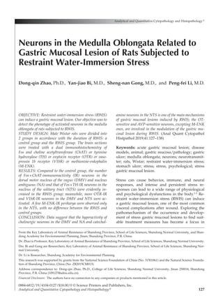
- 4. hancement degree of Fos had no difference (p> 0.05). Expression of ChAT ChAT-IR neurons (per 0.01 mm2) in the DMV were larger and round or fusiform with a few shorter processes, while in the NTS they were smaller with- out processes. ChAT-IR neurons (per 0.01 mm2) in the AP were round but too small and closely dis- tributed to count, while in the NA they were larg- er, fusiform, or polygonal with obvious processes (Figure 2A–D). As compared with the control group, Fos+ ChAT-IR neurons (per 0.01 mm2) in the DMV and NA in the RWIS group were significantly increased (p<0.05), while there was no difference in the NTS (p>0.05) (Figure 2E). The absolute values between the 2 groups of ChAT-IR, Fos+ChAT-IR, the percentage of Fos+ ChAT-IR in the total Fos-IR, or ChAT-IR neurons were significantly different in the different nu- clei (p<0.01) (Figure 3). The percentages of Fos+ ChAT-IR neurons in Fos-IR neurons in the RWIS group were 64.4% and 89.2%, respectively, in the DMV and NA. Fos+ChAT-IR neurons in response to RWIS represented in 21.1% and 36.1% of Fos+ ChAT-IR neurons in the DMV and NA, respec tively—significantly different from the control group (p<0.05) (Table I), but there was no sig- nificant difference in the NTS (p=0.431). These data indicate that ACh neurons in the DMV and NA but not NTS were the major ones related to RWIS regulation. Expression of TH TH-IR neurons (per 0.01 mm2) were small, round, or fusiform with shorter processes in the DMV, while in the NTS they were large, round, fusiform, or triangular with long processes. There were only processes in the NA, while only perikarya in the AP (Figure 4A–D). As compared with the control group, Fos+TH-IR neurons (per 0.01 mm2) in the DMV, NTS, and AP in the RWIS group were sig- nificantly increased (p<0.01) (Figure 4E). The absolute value between the 2 groups of TH-IR neurons in AP was significantly higher from the other nuclei (p<0.01) (Figure 3). The percent- ages of Fos+TH-IR neurons in Fos-IR neurons in the RWIS group were 17.3%, 12.9%, and 22.4% in the DM, NTS, and AP. Fos+TH-IR neurons in re- sponse to RWIS represented in 38.0%, 34.4%, and 18.6% of TH-IR neurons in the DMV, NTS, and AP, respectively, significantly different from the con- trol group (p<0.01) (Table I). That indicated that during the stress process, catecholamine neurons in the DMV, NTS, and AP were involved in RWIS regulation. In the NA, there was a large number of TH- positive terminals not only in the RWIS group but also in the control group, which indicated that neurons in the NA, such as ACh neurons, might accept the projection of fibers from catecholamine neurons in other nuclei. Expression of OTR OTR-IR neurons in the DMV were large, while those in the interior part were fusiform profiles with obvious neuronal processes and those in the lateral part were round profiles with few num- bers of neuronal processes. OTR-IR neurons were smaller but more uniform in size with a scanty cytoplasm in the NTS, which in the DMV were fu- 130 Analytical and Quantitative Cytopathology and Histopathology® Zhao et al Figure 1 Gastric mucosal damage induced by restraint water- immersion stress (RWIS). (A) Control group. (B) RWIS group.
- 5. siform profiles without any neuronal processes. OTR-IR neurons in the NA were larger and round or fusiform with a few shorter processes, while in the AP they were small and densely distributed (Figure 5A–D). As compared with the control group, Fos+ OTR-IR neurons in the DMV, NTS, AP, and NA in the RWIS group were significantly increased (p<0.01) (Figure 5E). The absolute values between the 2 groups of OTR-IR and Fos+OTR-IR neurons in AP were sig- nificantly higher than from the other nuclei (p< 0.01) (Figure 3). Fos+OTR-IR neurons in response to RWIS represented in 10.4%, 9.7%, 9.7%, and Volume 41, Number 4/August 2019 131 Neurons in the Medulla Oblongata Figure 2 Double immunohistochemical staining of Fos and ChAT (A–D) and the cell count (cells per 0.01 mm2) (E) in the medulla oblongata in the control (A, C) and RWIS 1h (B, D). Fos immunoreactivity was identified as dark blue- black dots deposited in the nuclei, and ChAT-IR neurons appeared in the cytoplasm as brown in color. 4V = 4th ventricle, CC = central canal. Scale bar=100 μm. *p<0.05, **p<0.01, control vs. RWIS.
- 6. 18.6% of OTR-IR neurons in the DMV, NTS, AP, and NA, respectively, significantly different from the control group (p<0.05) (Table I). The percent- age of Fos+OTR-IR neurons in Fos-IR neurons was 55.7%, 47.0%, 95.0%, and 100.0% in the DMV, NTS, AP, and NA in the RWIS group (p>0.05) (Table I). These indicated that oxytocin-sensitive neurons in the DMV, NTS, AP, and NA were one of the major activated neuron types involved in RWIS regulation. Expression of V1bR V1bR-IR neurons in the DMV were smaller than those in the NTS, mostly round and a few spin- dled, almost without any processes, while those in the AP were densely packed to count (Figure 5A–B). V1bR-IR neurons in the NA were large, while those in the interior part were fusiform profiles with obvious neuronal processes (Figure 6C–D). Fos+V1bR-IR neurons (cells per 0.01 mm2) in the DMV, NTS, AP, and NA in the RWIS group were significantly increased as compared to those in the control group (p<0.01) (Figure 6E). The absolute values between the 2 groups of V1bR-IR in AP and the percentage of Fos+V1bR-IR 132 Analytical and Quantitative Cytopathology and Histopathology® Zhao et al Table I Percentage of Double-Labeled Neurons to Fos or Neurotransmitter Immunoreactivity Ones in the Medulla Oblongata Gastrointestinal Central (%) DMV NTS AP NA Category Control vs. RWIS Control vs. RWIS Control vs. RWIS Control vs. RWIS Fos+ChAT-IR/Fos-IR 50.3±4.1 vs. 64.4±7.5 38.0±6.4 vs. 9.6±2.2** Too dense to count 82.5±11.8 vs. 79.2±6.8 Fos+ChAT-IR/ChAT-IR 7.2±4.3 vs. 21.1±2.4** 38.3±4.2 vs. 35.6±4.2 Too dense to count 7.5±2.4 vs. 36.1±9.3** Fos+TH-IR/Fos-IR 6.2±2.2 vs. 17.3±4.5* 4.5±0.6 vs. 12.9±0.8** 16.4±5.8 vs. 22.4±7.4 No perikarya Fos+TH-IR/TH-IR 14.3±4.5 vs. 38.0±5.3** 9.7±1.6 vs. 34.4±2.4** 4.5±1.3 vs. 18.6±6.1** No perikarya Fos+OTR-IR/Fos-IR 67.1±10.1 vs. 55.7±6.1 44.0±3.6 vs. 47.0±1.9 98.6±1.5 vs. 95.0±1.0 100.0±0.0 vs. 100.0±0.0 Fos+OTR-IR/OTR-IR 3.1±1.0 vs. 10.4±1.4** 5.3±0.9 vs. 9.7±0.9** 2.3±0.7 vs. 9.7±1.6** 7.4±1.3 vs. 18.6±3.2* Fos+V1bR-IR/Fos-IR 51.3±10.2 vs. 72.2±3.8* 27.0±2.9 vs. 52.3±4.8** 96.4±2.2 vs. 57.5±11.8* 90.0±10.0 vs. 98.6±1.4 Fos+V1bR-IR/V1bR-IR 2.9±1.1 vs. 10.7±1.6** 3.6±0.4 vs. 8.3±0.7** 2.3±0.6 vs. 4.5±1.1 7.0±1.1 vs. 28.5±3.8** *p<0.05, **p<0.01 control vs. RWIS. Figure 3 ANOVA of the absolute value between the 2 groups of ChAT, TH, OTR, and V1bR variance in different nuclei. In the same neurotransmitter, column with a same letter, p>0.05; column with a different lower case letter, p<0.05; column with a different upper case letter, p<0.01.
- 7. neurons in V1bR-IR neurons in NA were signifi cantly higher from the other nuclei (p<0.05) (Fig- ure 3). Fos+V1bR-IR neurons in response to RWIS represented in 10.7%, 8.3%, and 3.8% of V1bR-IR neurons in the DMV, NTS, and NA, respective- ly—significantly different from the control group (p<0.01) (Table I). The percentage of Fos+V1bR-IR neurons in Fos-IR neurons in the RWIS group was increased in the DMV and NTS, while de- creased in AP (p<0.05) (Table I). These data in- dicated that AVP-sensitive neurons in the DMV, NTS, and NA were the major type of the activated neuron during RWIS as well. Expression of M-ENK M-ENK neurons—large, round, or spindle with prominent processes—were present in the NTS but no Fos+M-ENK-IR were found, while only positive terminals in the DMV, NA, and AP (Figure 7). That indicated that M-ENK neurons of the medulla oblongata were not involved in RWIS regulation. Volume 41, Number 4/August 2019 133 Neurons in the Medulla Oblongata Figure 4 Double immunohistochemical staining of Fos and TH (A–D) and the cell count (cells per 0.01 mm2) (E) in the medulla oblongata in the control (A, C) and RWIS 1h (B, D). Fos immunoreactivity was identified as dark blue-black dots deposited in the nuclei, and TH-IR neurons appeared in the cytoplasm as brown in color. 4V = 4th ventricle, CC = central canal. Scale bar=100 μm in A and B, while 50 μm in C and D. *p<0.05, **p<0.01, control vs. RWIS.
- 8. Discussion The vagus nerve fibers regulating the function of the stomach originate from the DMV, NA, NTS, the nucleus intercalatus, and the nucleus retro ambigualis.23 RWIS evoked marked neuronal acti- vation in the DMV, NTS, AP, and NA assessed by 134 Analytical and Quantitative Cytopathology and Histopathology® Zhao et al Figure 5 Double immunohistochemical staining of Fos and OTR (A–D) and the cell count (cells per 0.01 mm2) (E) in the medulla oblongata in the control (A, C) and RWIS 1h (B, D). Fos immunoreactivity was identified as dark blue-black dots deposited in the nuclei, and OTR-IR neurons appeared in the cytoplasm as brown in color. 4V = 4th ventricle, CC = central canal. Scale bar=100 μm. *p<0.05, **p<0.01, control vs. RWIS.
- 9. Volume 41, Number 4/August 2019 135 Neurons in the Medulla Oblongata The Phenotypic Nature of Activated Neurons in the Medullary Visceral Zone Were AVP-Sensitive or Oxytocin-Sensitive Neurons Oxytocin, AVP neurons within the central ner- vous system, have the effect of inhibiting gas- tric motility, promoting gastric secretion, and this effect is achieved through the vagus nerve. By in- fecting rat paraventricular hypothalamic nucleus (PVN) and supraoptic nucleus (SON) hypotha- lamic nuclei with a virus construct, an extensive network of oxytocin fibers in various areas of the brain was revealed including NTS, DMV, and NA.28 When oxytocin receptor blockers are in- jected into the DMV, gastric acid secretion is inhibited and gastric motility is strengthened, while when oxytocin is injected into the DMV, the opposite effect is observed.29 Unilateral in- jection of AVP V1 receptor blockers into the NTS can eliminate the damage to gastric ischemia- reperfusion injury by electrical stimulation of PVN, and microinjection of AVP into the DMV can inhibit gastric motility and promote gastric acid secretion.30 In our study the occurrence of OTR and V1bR was nearly evenly on the mem- brane of neurons of the DMV, NTS, AP, and NA. The significant difference of the ratio of Fos+ OTR-IR, Fos+V1bR-IR neurons to OTR-IR, Fos+ V1bR-IR between the RWIS and control groups indicates that the oxytocin-sensitive and AVP- sensitive neurons are involved in the modula- tion of the gastric dysfunction during RWIS. In addition, in the RWIS group the percentages of Fos+OTR-IR and Fos+V1bR-IR neurons in Fos-IR nuclei were more than 58% and 72% in the DMV and 45% and 52% in the NTS, which indicated that oxytocin and AVP mediate the activity of these neurons mainly by OTR and V1bR, particu- larly the V1b receptor. M-ENK Neurons Have No Obvious Relation with Gastric Mucosal Injury Caused by RWIS Previous studies found that peripheral intravenous injection of M-ENK analogue could attenuate gas- tric mucosal injury induced by cold-restraint stress. At the observation of the hypothalamus and me dulla oblongata, M-ENK-IR perikaryon was only found in the NTS, while only M-ENK nerve end- ings in the DMV and NA and no M-ENK–positive neurons and fiber terminals in the PVN and SON were observed, the M-ENK neurons are not obvi- ously related to gastric mucosal injury caused by RWIS, which is consistent with previous studies.18 Fos expression.4 DMV and NA is the visceral para- sympathetic efferent nucleus, so the gastric mu- cosal lesion induced by RWIS was predominantly because of the hyperactivity of vagal parasympa- thetic efferents. Hyperactivity of Cholinergic Neurons in the DMV and NA but Not the NTS Might Be One of the Primary Central Mechanisms of the Gastric Dysfunctions Induced by RWIS ChAT-IR neurons were all distributed in the DMV, NTS, AP, and NA. In the DMV and NA, the proportion of Fos+ChAT-IR neurons to ChAT- IR neurons in the RWIS group was significantly higher than that in the control group, and 64.4% and 89.2% Fos-IR neurons were ChAT-IR, indi cating that the hyperactivity of cholinergic neu rons in the DMV and NA is one of the main mechanisms of gastric mucosal injury induced by RWIS, which is consistent with the results of continuous intravenous injection of ACh to cause severe gastric mucosal injury in rats.12 In the NTS, there was no significant difference in the ratio of Fos+ChAT-IR neurons to ChAT-IR neu- rons, which indicates that cholinergic neurons in the NTS may not participate in RWIS regu- lation. However, studies have shown that the injection of ACh into the NTS also can signifi- cantly increase the protective effect of electro acupuncture on gastric mucosal injury.24 Possibly it is the different regulation mode of different stress models, but the specifics need to be further studied. Catecholaminergic Neurons in the Medulla Oblongata Gastric Nerve Center May Be Related to the Stress-Responsive Signal Transduction Numerous morphological and electrophysiolog- ical studies have demonstrated that NE and DA are important endogenous inhibitory neurotrans- mitters that play a protective role in the integrity of gastric mucosa during stress.25 Central admin istration of DA antagonists aggravates stress- induced gastric mucosal injury, whereas DA ag- onists attenuate stress-induced gastric mucosal injury.26,27 The higher proportion of Fos+TH-IR neurons to TH-IR or Fos-IR neurons in the RWIS group than that in the control group indicates that catecholamine neurons in the medulla oblon- gata participate in the regulation of gastric dys function induced by RWIS, though maybe not the major neuron phenotype.
- 10. 136 Analytical and Quantitative Cytopathology and Histopathology® Zhao et al sensitive or oxytocin-sensitive neurons, while cat- echolamine neurons in the medulla oblongata are intermediate neurons. In summary, activated ACh neurons in the medulla oblongata are the vagal presympathet- ic preganglionic neurons, most of which are AVP- Figure 6 Double immunohistochemical staining of Fos and V1bR (A–D) and the cell count (cells per 0.01mm2) (E) in the medulla oblongata in the control (A, C) and RWIS 1h (B, D). Fos immunoreactivity was identified as dark blue- black dots deposited in the nuclei, and V1bR-IR neurons appeared in the cytoplasm as brown in color. 4V = 4th ventricle, CC = central canal. Scale bar=100 μm. *p<0.05, **p<0.01, control vs. RWIS.
- 11. Volume 41, Number 4/August 2019 137 Neurons in the Medulla Oblongata water‑immersion stress. Mol Med Rep 2019;20:2303-2315 10. Fan F, Li L, Liu W, Yang M, Ma X, Sun H: Astrocytes and neu- rons in locus coeruleus mediate restraint water immersion stress-induced gastric mucosal damage through the ERK1/2 signaling pathway. Neurosci Lett 2018;675:95-102 11. Xu Y, Jia J, Xie C, Wu Y, Tu W: Transient receptor potential Ankyrin 1 and Substance P mediate the development of gas- tric mucosal lesions in a water immersion restraint stress rat model. Digestion 2018;97:228-239 12. Gatón J, Fernández de la Gándara F, Velasco A: The role of the neurotransmitters acetylcholine and noradrenaline in the pathogenesis of stress ulcers. Comp Biochem Physiol 1993;106(1):125-129 13. Zhao DQ, Lu CL, Ai HB: The role of catecholaminergic neu- rons in the hypothalamus and medullary visceral zone in re- sponse to restraint water-immersion stress in rats. J Physiol Sci 2011;61(1):37-45 14. Pirnik Z, Kiss A: Fos expression variances in mouse hypo- thalamus upon physical and osmotic stimuli: Co-staining with vasopressin, oxytocin, and tyrosine hydroxylase. Brain Res Bull 2005;65:423-431 15. Llewellyn-Smith IJ, Kellett DO, Jordan D, Browning KN, Travagli RA: Oxytocin-immunoreactive innervation of iden- tified neurons in the rat dorsal vagal complex. Neurogastro- enterol Motil 2012;24(3):e136-46 16. Young WS, Li J, Wersinger SR, Palkovit MR: The vasopres- sin 1b receptor is prominent in the hippocampal area CA2 where it is unaffected by restraint stress or adrenalectomy. Neuroscience 2006;143(4):1031-1039 References 1. Yang G, Guo H, Li H, Shan S, Zhang X, Rombout JH, An L: Molecular characterization of LEAP-2 cDNA in common carp (Cyprinus carpio L.) and the differential expression upon a Vibrio anguillarum stimulus; indications for a sig- nificant immune role in skin. Fish Shellfish Immunol 2014; 37:22-29 2. Rombout JH, Yang G, Kiron V: Adaptive immune responses at mucosal surfaces of teleost fish. Fish Shellfish Immunol 2014;40:634-643 3. Yang HT, Zou SS, Zhai LJ, Wang Y, Zhang FM, An LG, Yang GW: Pathogen invasion changes the intestinal microbiota composition and induces innate immune responses in the zebrafish intestine. Fish Shellfish Immunol 2017;71:35-42 4. Zhang YY, Cao GH, Zhu WX, Cui XY, Ai HB: Comparative study of c-Fos expression in the rat dorsal vagal complex and nucleus ambiguous induced by different durations of restraint water-immersion stress. J Chin Physio 2009;52(3): 143-150 5. Liu M, Xie S, Zhou J: Use of animal models for the imaging and quantification of angiogenesis. Exp Anim 2018;67:1-6 7. Kobayashi K, Kashimak K, Higuchi K, Arakawa T: The mech- anisms of gastro-intestinal mucosal injury repair. Nippon Rinsho 1998;56(9):2215-2222 8. Gong SN, Zhu JP, Ma YJ, Zhao DQ: Proteomics of the medi- odorsal thalamic nucleus of rats with stress-induced gastric ulcer. World J Gastroenterol 2019;25(23):2911-2923 9. Zhao DQ, Gong SN, Ma YJ, Zhu JP: Medial prefrontal cor- tex exacerbates gastric dysfunction of rats upon restraint Figure 7 Immunohistochemical staining of M-ENK-IR neurons in the NTS and DMV induced by control (A, B) and RWIS for 1h (C, D). M-ENK immunoreactivity neurons appeared in the cytoplasm as brown in color. NTS = nucleus of solitary tract, DMV = dorsal motor nucleus of the vagus, 4V = 4th ventricle. Magnification ×200, scale bar=100 μm in A and C while ×400, scale bar=50 μm in B and D.
- 12. 138 Analytical and Quantitative Cytopathology and Histopathology® Zhao et al 17. Arida RM, Gomes da Silva S, de Almeida AA, Cavalheiro EA, Zavala-Tecuapetla C, Brand S, Rocha L: Differential effects of exercise on brain opioid receptor binding and activation in rats. J Neurochem 2015;132(2):206-217 18. Chen QS, Dai YL: Role of morphine and methy-enkephalin in stress-induced gastric mucosal lesions. Journal of Nanjing Medical University 1990;10(3):196-198 19. Herrera DG, Robertson HA: Activation of c-Fos in the brain. Prog Neuro Bio 1996;l50:83-107 20. Guth PH, Aures D, Paulsen G: Topical aspirin plus HCl gas- tric lesions in the rat: Cytoprotective effect of prostaglandin, cimetidine, and probanthine. Gastroenterology 1979;76:88-93 21. Ding NZ, Qi QR, Gu XW, Zuo RJ, Liu J, Yang ZM: De novo synthesis of sphingolipids is essential for decidualization in mice. Theriogenology 2018;106:227-236 22. Paxinos G, Watson C: The Rat Brain in Stereotaxic Coordi- nates. Fifth edition. Burlington, Massachusetts, Elsevier Ac- ademic Press, 2005 23. Hayakawa T, Takanaga A, Tanaka K, Maeda S, Seki M: Cells of origin of vagal motor neurons projecting to different parts of the stomach in the rat: Confocal laser scanning and elec- tron microscopic study. Anat Embryol (Berl) 2003;207:289-297 24. Zhou YU, Wang Pan, Tan XQ, Li CW, Huang BL: The effects of microinjection of acetylcholine into nucleus tractus soli- tarious on electroacupuncture against stress gastric muco- sa injury in rats. Journal of Xianning College (Medical Sci) 2007;21(3):34-36 25. Glavin GB, Szabo S: Dopamine in gastrointestinal disease. Dig Dis Sci 1990;35:1153-1155 26. Glavin GB: Central dopamine involvement in experimental gastrointestinal injury. Prog Neuro-psychopharmacol Biol Psychiatry 1992;16(2):217-221 27. Glavin GB: Dopamine: A stress modulator in the brain and gut. Gen Pharmacol 1992;23(6):1023-1026 28. Knobloch HS, Charlet A, Hoffmann LC, Eliava M, Khrulev S, Cetin AH, Osten P, Schwarz MK Seeburg PH, Stoop R, and Grinevich V: Evoked axonal oxytocin release in the central amygdala attenuates fear response. Neuron 2012;73:553-566 29. Rogers RC, Hermann GE: Oxytocin, oxytocin antagonist, TRH, and hypothalamic paraventricular nucleus stimula- tion effects on gastric motility. Peptides 1987;8:505-513 30. Zhu J, Chang L, Xie J, Ai H: Arginine vasopressin injected into the dorsal motor nucleus of the vagus inhibits gastric motility in rats. Gastroenterol Res Pract 2016;2016:4618672