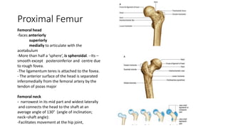
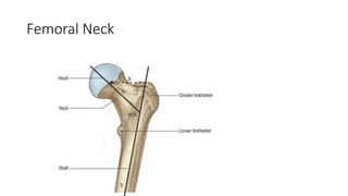
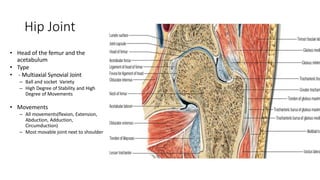
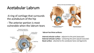
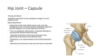
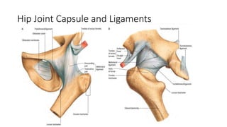
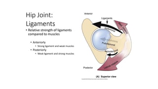
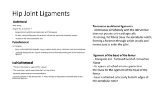
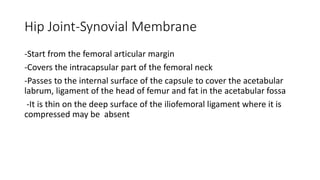
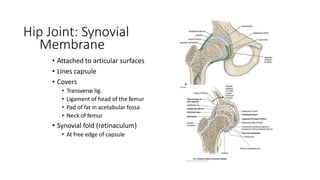
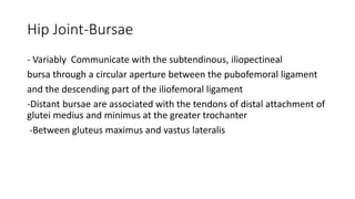
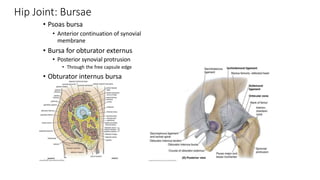
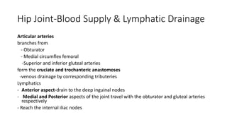
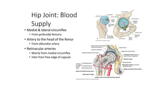
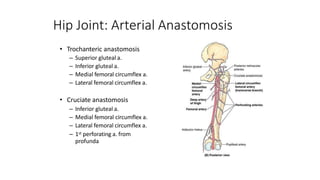
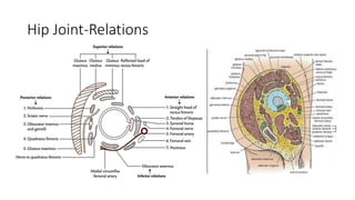
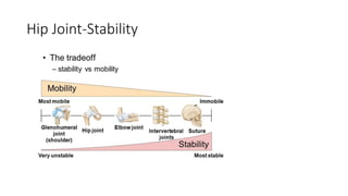
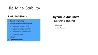
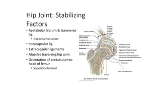
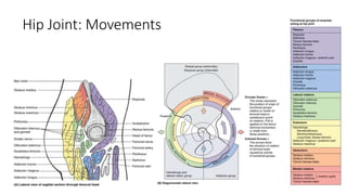
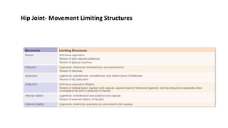
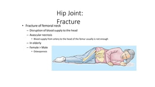
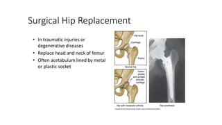
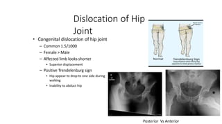
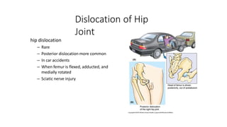
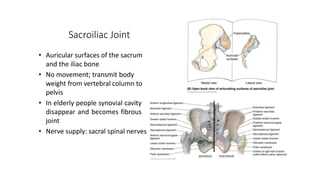
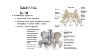
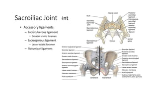
Hip Joint
Dr A J Paranagama
MBBS(Colombo),MD(Orthopaedic Surgery)
DIT(UCSC),HDIT(UCSC)
Acting Consultant Orthopaedic Surgeon
Learning Outcomes
• Describe the components of the hip joint.
• List the ligaments associated with the hip joint and their attachments.
• Describe the muscles acting on the hip joint according to the type and movement they
perform.
• Describe the bursae in relation to the hip joint.
• Describe the stability of the hip joint.
• Describe the blood supply and nerve supply of the hip joint.
• Describe the major palpable bony prominences of the hip joint.
• Discuss the associated clinicals of the hip joint
Acetabulum
-An approximately hemispherical cavity situated about -At
the centre of the lateral aspect of the hip bone
-Faces anteroinferiorly (In abduction and Inversion)
- Circumscribed by an irregular margin deficient inferiorly at
the acetabular notch.
- The acetabular fossa forms the central floor and is rough
and non-articular.
- The articular surface- Lunate surface is widest above (the
‘dome’), where weight is transmitted to the femur
• All three components of the hip bone contribute to the
acetabulum
The pubis- forms the anterosuperior fifth of the articular
surface
The ischium- forms the floor of the fossa and more than the
posteroinferior two-fifths of the articular
Surface
The ilium- postero-Superior two fifth
Acetabulum
Acetabulum
Lunate surface
- Articular part
Acetabular fossa
- No articular part
Acetabular rim
-Acetabular notch
Acetabulum
1, ilium; 2, part of triradiate growth cartilage; 3, superior
femoral epiphysis; 4, cartilaginous growth plates; 5, ossifying
greater
trochanter; 6, ischium; 7, pubis; 8, cartilage between inferior
pubic and
ischial rami.
At Birth
11 Years
Proximal Femur
Femoral head
-faces anteriorly
superiorly
medially to articulate with the
acetabulum
-More than half a ‘sphere’, is spheroidal. --Its –
smooth except posteroinferior and centre due
to rough fovea.
-The ligamentum teres is attached to the fovea.
- The anterior surface of the head is separated
inferomedially from the femoral artery by the
tendon of psoas major
Femoral neck
- narrowest in its mid part and widest laterally
and connects the head to the shaft at an
average angle of 130° (angle of inclination;
neck–shaft angle):
-Facilitates movement at the hip joint,
Femoral Neck
Hip Joint
• Head of the femur and the
acetabulum
• Type
• - Multiaxial Synovial Joint
– Ball and socket Variety
– High Degree of Stability and High
Degree of Movements
• Movements
– All movements(flexion, Extension,
Abduction, Adduction,
Circumduction)
– Most movable joint next to shoulder
Acetabular Labrum
-A ring of cartilage that surrounds
the acetabulum of the hip
- The anterior portion is most
vulnerable when the labrum tears
labrum has three surfaces:
Internal articular surface - adjacent to the joint (avascular)
External articular surface - contacting the joint capsule (vascular)
Basal surface - attached to the acetabular bone and ligaments
Hip Joint – Capsule
-Strong and dense
-Attached superiorly to the acetabular margin 5–6 mm
medial to the
labral attachment
- Anteriorly to the outer labral aspect and, near the
acetabular notch, to the transverse acetabular ligament
and the adjacent rim of the obturator foramen
- From its acetabular attachment, it extends laterally to
surround the femoral head and neck
- Attached anteriorly to the intertrochanteric line,
superiorly to the base of the femoral neck
- Posteriorly 1 cm superomedial to the intertrochanteric
crest
- Inferiorly to the femoral neck near the lesser trochanter
Hip Joint Capsule and Ligaments
Hip Joint:
Ligaments
• Relative strength of ligaments
compared to muscles
• Anteriorly
• Strong ligament and weak muscles
• Posteriorly
• Weak ligament and strong muscles
Hip Joint Ligaments
Iliofemoral
-very strong
-shaped like an inverted Y
- lying anteriorly and intimately blended with the capsule
- Its apex is attached between the anterior inferior iliac spine and acetabular margin
- Its base to the intertrochanteric line.
Pubofemoral
-is triangular,
- base is attached to the iliopubic ramus, superior pubic ramus, obturator crest and membrane.
- It blends distally with the capsule and deep surface of the descending part of the iliofemoral
ligament
Ischiofemoral
- Thickens the posterior aspect of the capsule
- The central part spirals superolaterally from the ischium
-Attached posteroinferior to the acetabulum
- courses posterior to the femoral neck to attach distally to the greater trochanter deep to the
iliofemoral ligament
Transverse acetabular ligaments
-continuous peripherally with the labrum but
does not possess any cartilage cells
-Its strong, flat fibres cross the acetabular notch,
forming a foramen through which vessels and
nerves pass to enter the joint.
ligament of the head of the femur
- triangular and flattened band of connective
Tissue
- Its apex is attached anterosuperiorly in
the fovea for the ligament of the head of the
femur
- base is attached principally to both edges of
the acetabular notch
Hip Joint-Synovial Membrane
-Start from the femoral articular margin
-Covers the intracapsular part of the femoral neck
-Passes to the internal surface of the capsule to cover the acetabular
labrum, ligament of the head of femur and fat in the acetabular fossa
-It is thin on the deep surface of the iliofemoral ligament where it is
compressed may be absent
Hip Joint: Synovial
Membrane
• Attached to articular surfaces
• Lines capsule
• Covers
• Transverse lig.
• Ligament of head of the femur
• Pad of fat in acetabular fossa
• Neck of femur
• Synovial fold (retinaculum)
• At free edge of capsule
Hip Joint-Bursae
- Variably Communicate with the subtendinous, iliopectineal
bursa through a circular aperture between the pubofemoral ligament
and the descending part of the iliofemoral ligament
-Distant bursae are associated with the tendons of distal attachment of
glutei medius and minimus at the greater trochanter
-Between gluteus maximus and vastus lateralis
Hip Joint: Bursae
• Psoas bursa
• Anterior continuation of synovial
membrane
• Bursa for obturator externus
• Posterior synovial protrusion
• Through the free capsule edge
• Obturator internus bursa
Hip Joint-Blood Supply & Lymphatic Drainage
Articular arteries
branches from
- Obturator
- Medial circumflex femoral
-Superior and inferior gluteal arteries
form the cruciate and trochanteric anastomoses
-venous drainage by corresponding tributeries
Lymphatics
- Anterior aspect-drain to the deep inguinal nodes
- Medial and Posterior aspects of the joint travel with the obturator and gluteal arteries
respectively
- Reach the internal iliac nodes
Hip Joint: Blood
Supply
• Medial & lateral circumflex
• From profundal femoris
• Artery to the head of the femur
• From obturator artery
• Retinacular arteries
• Mainly from medial circumflex
• Inter from free edge of capsule
Hip Joint: Arterial Anastomosis
• Trochanteric anastomosis
– Superior gluteal a.
– Inferior gluteal a.
– Medial femoral circumflex a.
– Lateral femoral circumflex a.
• Cruciate anastomosis
– Inferior gluteal a.
– Medial femoral circumflex a.
– Lateral femoral circumflex a.
– 1st perforating a. from
profunda
Hip Joint-Innervation
The anterior capsule of the hip joint is innervated by the
- Femoral
-Obturator and accessory obturator nerves.
The posterior capsule is innervated by the
-Sciatic
- Superior gluteal nerves
-Nerve to quadratus femoris
Hip Joint-Relations
Hip Joint-Relations
The joint capsule is surrounded by muscles
Anteriorly
-lateral fibres of pectineus separate the capsule from the femoral vein.
-Lateral to this, the tendon of psoas major, with iliacus lateral to it, descends to the
lesser trochanter, partly separated from the capsule by a bursa.
-The femoral artery is anterior to the tendon of psoas major, and the femoral nerve lies
deep in a groove between the tendon and iliacus.
- Further laterally the straight head of rectus femoris crosses the joint with a deep layer
of the iliotibial tract
blends with the capsule under the lateral border of the muscle.
Superiorly
-The reflected head of rectus femoris contacts the capsule medially, while gluteus minimus
covers it laterally
Hip Joint-Relations
Inferiorly
-medial fibres of pectineus close to the the capsule
postero-inferiorly obturator externus spirals obliquely to its posterior aspect.
Posteroinferiorly- the capsule is covered by the tendon of obturator externus
separating it from quadratus femoris and accompanied
by an ascending branch of the medial circumflex femoral artery.
Posterosuperiorly- The tendon of obturator internus and the gmelli
contact the joint capsule, separating it from the sciatic nerve.
The nerve to quadratus femoris is deep to the
obturator internus tendon and descends medially on the capsule.
Superior to this- the posterior surface of the joint is crossed by piriformis
Hip Joint-Stability
Hip Joint- Stability
Static Stabilizers Dynamic Stabilizers
-Muscles around
Iliopsoas
Rectus Femoris
Hip Joint: Stabilizing
Factors
• Acetabular labrum & transverse
lig.
• Deepens the socket
• Intracapsular lig.
• Extracapsular ligaments
• Muscles traversing hip joint
• Orientation of acetabulum to
head of femur
• Superiorly located
Hip Joint: Movements
Hip Joint- Movement Limiting Structures
Hip Joint:
Fracture
• Fracture of femoral neck
– Disruption of blood supply to the head
– Avascular necrosis
• Blood supply from artery to the head of the femur usually is not enough
– In elderly
– Female > Male
• Osteoporosis
Surgical Hip Replacement
• In traumatic injuries or
degenerative diseases
• Replace head and neck of femur
• Often acetabulum lined by metal
or plastic socket
Dislocation of Hip
Joint
• Congenital dislocation of hip joint
– Common 1.5/1000
– Female > Male
– Affected limb looks shorter
• Superior displacement
– Positive Trendelenburg sign
• Hip appear to drop to one side during
walking
• Inability to abduct hip
Posterior Vs Anterior
Dislocation of Hip
Joint
hip dislocation
– Rare
– Posterior dislocation more common
– In car accidents
– When femur is flexed, adducted, and
medially rotated
– Sciatic nerve injury
Sacroiliac Joint
• Auricular surfaces of the sacrum
and the iliac bone
• No movement; transmit body
weight from vertebral column to
pelvis
• In elderly people synovial cavity
disappear and becomes fibrous
joint
• Nerve supply: sacral spinal nerves
Sacroiliac
Joint
• Associated ligaments
– Posterior sacroiliac ligament
– Interosseous sacroiliac ligament (between
tuberosities of sacrum and iliac bone)
– Anterior sacroiliac ligament
Sacroiliac Joint
• Accessory ligaments
– Sacrotuberous ligament
• Greater sciatic foramen
– Sacrospinous ligament
• Lesser sciatic foramen
– Iliolumbar ligament
Thank You

Hip Joint.pptx

  • 1.
    Hip Joint Dr AJ Paranagama MBBS(Colombo),MD(Orthopaedic Surgery) DIT(UCSC),HDIT(UCSC) Acting Consultant Orthopaedic Surgeon
  • 2.
    Learning Outcomes • Describethe components of the hip joint. • List the ligaments associated with the hip joint and their attachments. • Describe the muscles acting on the hip joint according to the type and movement they perform. • Describe the bursae in relation to the hip joint. • Describe the stability of the hip joint. • Describe the blood supply and nerve supply of the hip joint. • Describe the major palpable bony prominences of the hip joint. • Discuss the associated clinicals of the hip joint
  • 3.
    Acetabulum -An approximately hemisphericalcavity situated about -At the centre of the lateral aspect of the hip bone -Faces anteroinferiorly (In abduction and Inversion) - Circumscribed by an irregular margin deficient inferiorly at the acetabular notch. - The acetabular fossa forms the central floor and is rough and non-articular. - The articular surface- Lunate surface is widest above (the ‘dome’), where weight is transmitted to the femur • All three components of the hip bone contribute to the acetabulum The pubis- forms the anterosuperior fifth of the articular surface The ischium- forms the floor of the fossa and more than the posteroinferior two-fifths of the articular Surface The ilium- postero-Superior two fifth
  • 4.
    Acetabulum Acetabulum Lunate surface - Articularpart Acetabular fossa - No articular part Acetabular rim -Acetabular notch
  • 5.
    Acetabulum 1, ilium; 2,part of triradiate growth cartilage; 3, superior femoral epiphysis; 4, cartilaginous growth plates; 5, ossifying greater trochanter; 6, ischium; 7, pubis; 8, cartilage between inferior pubic and ischial rami. At Birth 11 Years
  • 6.
    Proximal Femur Femoral head -facesanteriorly superiorly medially to articulate with the acetabulum -More than half a ‘sphere’, is spheroidal. --Its – smooth except posteroinferior and centre due to rough fovea. -The ligamentum teres is attached to the fovea. - The anterior surface of the head is separated inferomedially from the femoral artery by the tendon of psoas major Femoral neck - narrowest in its mid part and widest laterally and connects the head to the shaft at an average angle of 130° (angle of inclination; neck–shaft angle): -Facilitates movement at the hip joint,
  • 7.
  • 8.
    Hip Joint • Headof the femur and the acetabulum • Type • - Multiaxial Synovial Joint – Ball and socket Variety – High Degree of Stability and High Degree of Movements • Movements – All movements(flexion, Extension, Abduction, Adduction, Circumduction) – Most movable joint next to shoulder
  • 9.
    Acetabular Labrum -A ringof cartilage that surrounds the acetabulum of the hip - The anterior portion is most vulnerable when the labrum tears labrum has three surfaces: Internal articular surface - adjacent to the joint (avascular) External articular surface - contacting the joint capsule (vascular) Basal surface - attached to the acetabular bone and ligaments
  • 10.
    Hip Joint –Capsule -Strong and dense -Attached superiorly to the acetabular margin 5–6 mm medial to the labral attachment - Anteriorly to the outer labral aspect and, near the acetabular notch, to the transverse acetabular ligament and the adjacent rim of the obturator foramen - From its acetabular attachment, it extends laterally to surround the femoral head and neck - Attached anteriorly to the intertrochanteric line, superiorly to the base of the femoral neck - Posteriorly 1 cm superomedial to the intertrochanteric crest - Inferiorly to the femoral neck near the lesser trochanter
  • 11.
    Hip Joint Capsuleand Ligaments
  • 12.
    Hip Joint: Ligaments • Relativestrength of ligaments compared to muscles • Anteriorly • Strong ligament and weak muscles • Posteriorly • Weak ligament and strong muscles
  • 13.
    Hip Joint Ligaments Iliofemoral -verystrong -shaped like an inverted Y - lying anteriorly and intimately blended with the capsule - Its apex is attached between the anterior inferior iliac spine and acetabular margin - Its base to the intertrochanteric line. Pubofemoral -is triangular, - base is attached to the iliopubic ramus, superior pubic ramus, obturator crest and membrane. - It blends distally with the capsule and deep surface of the descending part of the iliofemoral ligament Ischiofemoral - Thickens the posterior aspect of the capsule - The central part spirals superolaterally from the ischium -Attached posteroinferior to the acetabulum - courses posterior to the femoral neck to attach distally to the greater trochanter deep to the iliofemoral ligament Transverse acetabular ligaments -continuous peripherally with the labrum but does not possess any cartilage cells -Its strong, flat fibres cross the acetabular notch, forming a foramen through which vessels and nerves pass to enter the joint. ligament of the head of the femur - triangular and flattened band of connective Tissue - Its apex is attached anterosuperiorly in the fovea for the ligament of the head of the femur - base is attached principally to both edges of the acetabular notch
  • 14.
    Hip Joint-Synovial Membrane -Startfrom the femoral articular margin -Covers the intracapsular part of the femoral neck -Passes to the internal surface of the capsule to cover the acetabular labrum, ligament of the head of femur and fat in the acetabular fossa -It is thin on the deep surface of the iliofemoral ligament where it is compressed may be absent
  • 15.
    Hip Joint: Synovial Membrane •Attached to articular surfaces • Lines capsule • Covers • Transverse lig. • Ligament of head of the femur • Pad of fat in acetabular fossa • Neck of femur • Synovial fold (retinaculum) • At free edge of capsule
  • 16.
    Hip Joint-Bursae - VariablyCommunicate with the subtendinous, iliopectineal bursa through a circular aperture between the pubofemoral ligament and the descending part of the iliofemoral ligament -Distant bursae are associated with the tendons of distal attachment of glutei medius and minimus at the greater trochanter -Between gluteus maximus and vastus lateralis
  • 17.
    Hip Joint: Bursae •Psoas bursa • Anterior continuation of synovial membrane • Bursa for obturator externus • Posterior synovial protrusion • Through the free capsule edge • Obturator internus bursa
  • 18.
    Hip Joint-Blood Supply& Lymphatic Drainage Articular arteries branches from - Obturator - Medial circumflex femoral -Superior and inferior gluteal arteries form the cruciate and trochanteric anastomoses -venous drainage by corresponding tributeries Lymphatics - Anterior aspect-drain to the deep inguinal nodes - Medial and Posterior aspects of the joint travel with the obturator and gluteal arteries respectively - Reach the internal iliac nodes
  • 19.
    Hip Joint: Blood Supply •Medial & lateral circumflex • From profundal femoris • Artery to the head of the femur • From obturator artery • Retinacular arteries • Mainly from medial circumflex • Inter from free edge of capsule
  • 20.
    Hip Joint: ArterialAnastomosis • Trochanteric anastomosis – Superior gluteal a. – Inferior gluteal a. – Medial femoral circumflex a. – Lateral femoral circumflex a. • Cruciate anastomosis – Inferior gluteal a. – Medial femoral circumflex a. – Lateral femoral circumflex a. – 1st perforating a. from profunda
  • 21.
    Hip Joint-Innervation The anteriorcapsule of the hip joint is innervated by the - Femoral -Obturator and accessory obturator nerves. The posterior capsule is innervated by the -Sciatic - Superior gluteal nerves -Nerve to quadratus femoris
  • 22.
  • 23.
    Hip Joint-Relations The jointcapsule is surrounded by muscles Anteriorly -lateral fibres of pectineus separate the capsule from the femoral vein. -Lateral to this, the tendon of psoas major, with iliacus lateral to it, descends to the lesser trochanter, partly separated from the capsule by a bursa. -The femoral artery is anterior to the tendon of psoas major, and the femoral nerve lies deep in a groove between the tendon and iliacus. - Further laterally the straight head of rectus femoris crosses the joint with a deep layer of the iliotibial tract blends with the capsule under the lateral border of the muscle. Superiorly -The reflected head of rectus femoris contacts the capsule medially, while gluteus minimus covers it laterally
  • 24.
    Hip Joint-Relations Inferiorly -medial fibresof pectineus close to the the capsule postero-inferiorly obturator externus spirals obliquely to its posterior aspect. Posteroinferiorly- the capsule is covered by the tendon of obturator externus separating it from quadratus femoris and accompanied by an ascending branch of the medial circumflex femoral artery. Posterosuperiorly- The tendon of obturator internus and the gmelli contact the joint capsule, separating it from the sciatic nerve. The nerve to quadratus femoris is deep to the obturator internus tendon and descends medially on the capsule. Superior to this- the posterior surface of the joint is crossed by piriformis
  • 25.
  • 26.
    Hip Joint- Stability StaticStabilizers Dynamic Stabilizers -Muscles around Iliopsoas Rectus Femoris
  • 27.
    Hip Joint: Stabilizing Factors •Acetabular labrum & transverse lig. • Deepens the socket • Intracapsular lig. • Extracapsular ligaments • Muscles traversing hip joint • Orientation of acetabulum to head of femur • Superiorly located
  • 28.
  • 29.
    Hip Joint- MovementLimiting Structures
  • 30.
    Hip Joint: Fracture • Fractureof femoral neck – Disruption of blood supply to the head – Avascular necrosis • Blood supply from artery to the head of the femur usually is not enough – In elderly – Female > Male • Osteoporosis
  • 31.
    Surgical Hip Replacement •In traumatic injuries or degenerative diseases • Replace head and neck of femur • Often acetabulum lined by metal or plastic socket
  • 32.
    Dislocation of Hip Joint •Congenital dislocation of hip joint – Common 1.5/1000 – Female > Male – Affected limb looks shorter • Superior displacement – Positive Trendelenburg sign • Hip appear to drop to one side during walking • Inability to abduct hip Posterior Vs Anterior
  • 33.
    Dislocation of Hip Joint hipdislocation – Rare – Posterior dislocation more common – In car accidents – When femur is flexed, adducted, and medially rotated – Sciatic nerve injury
  • 34.
    Sacroiliac Joint • Auricularsurfaces of the sacrum and the iliac bone • No movement; transmit body weight from vertebral column to pelvis • In elderly people synovial cavity disappear and becomes fibrous joint • Nerve supply: sacral spinal nerves
  • 35.
    Sacroiliac Joint • Associated ligaments –Posterior sacroiliac ligament – Interosseous sacroiliac ligament (between tuberosities of sacrum and iliac bone) – Anterior sacroiliac ligament
  • 36.
    Sacroiliac Joint • Accessoryligaments – Sacrotuberous ligament • Greater sciatic foramen – Sacrospinous ligament • Lesser sciatic foramen – Iliolumbar ligament
  • 37.