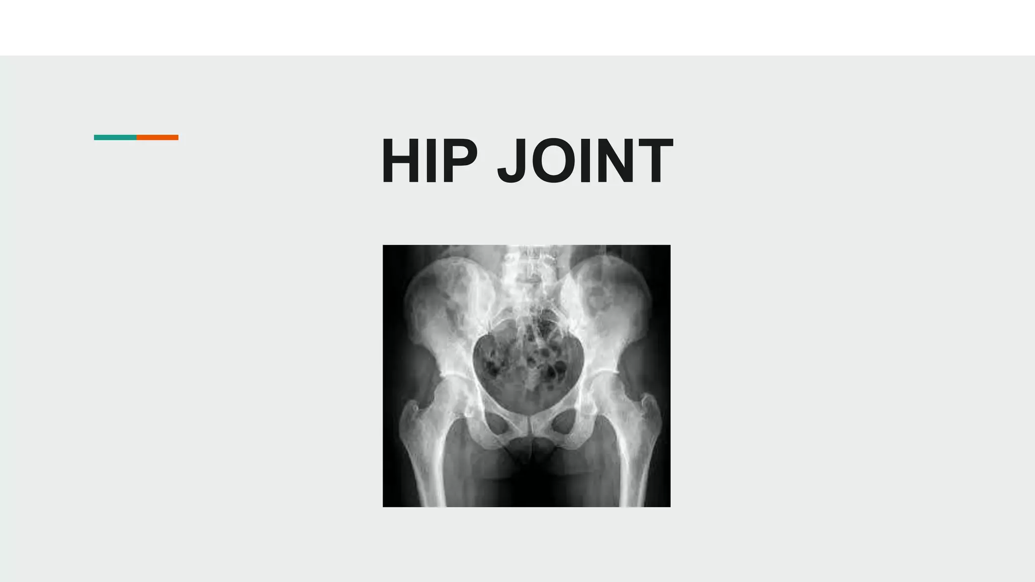
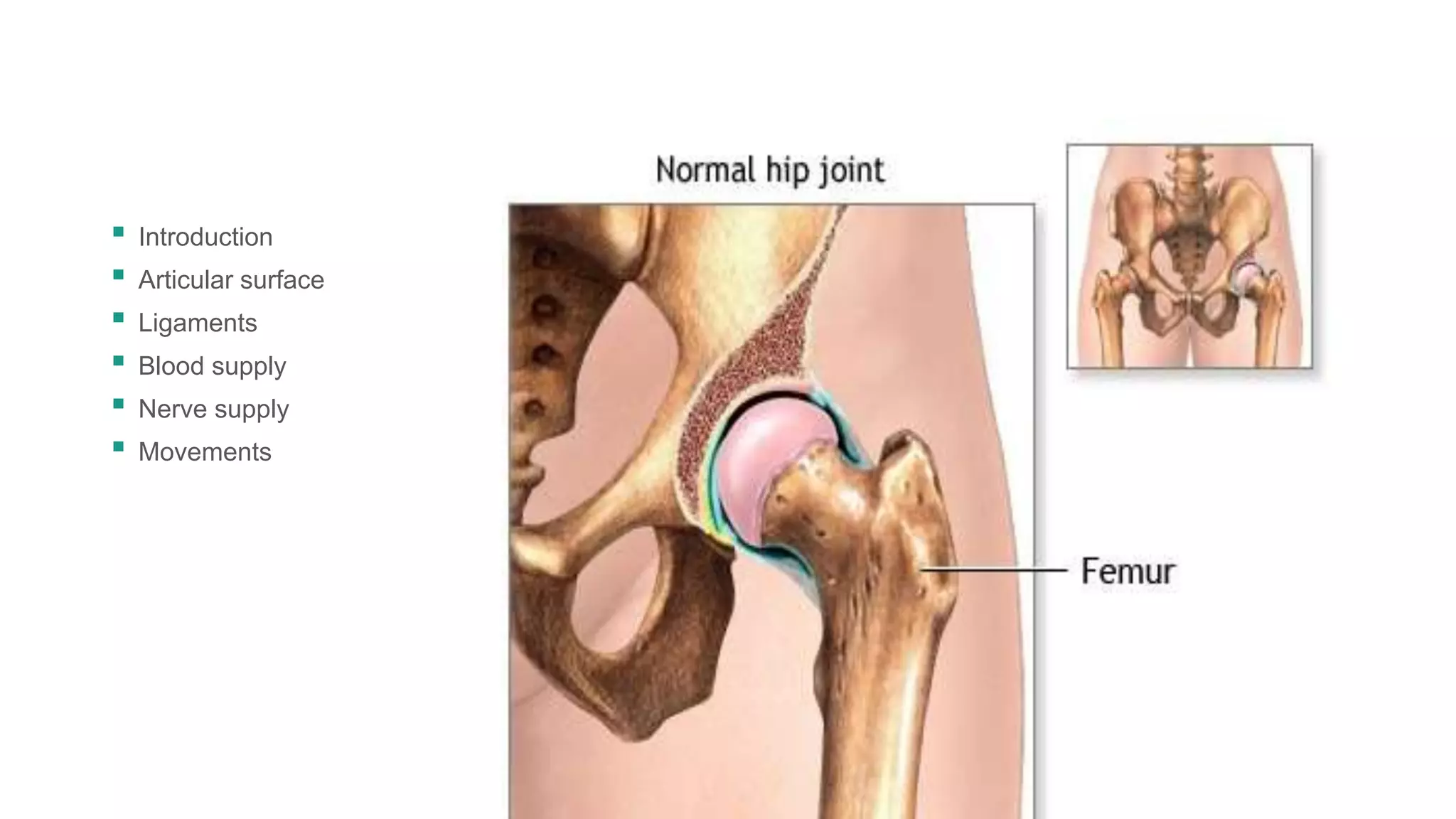
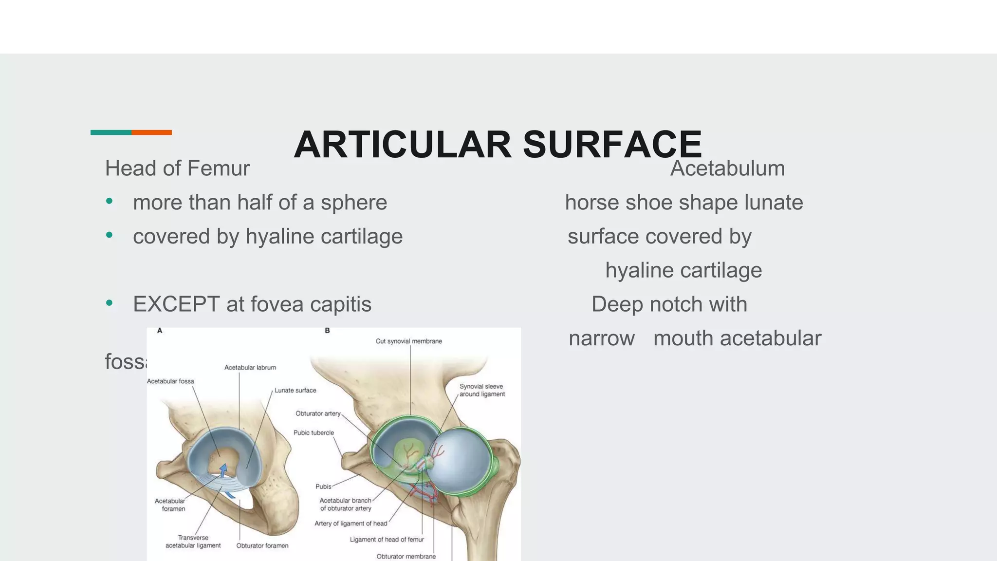
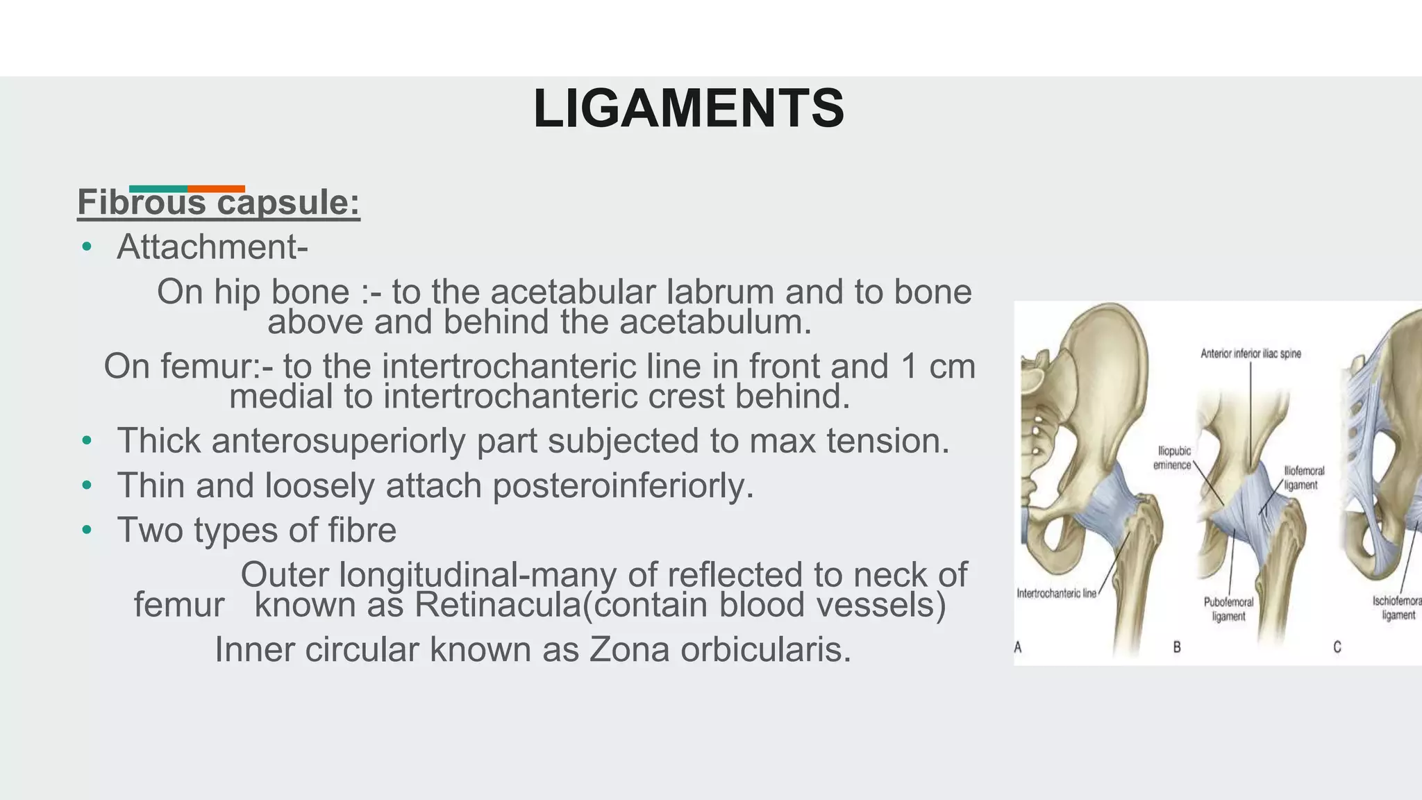
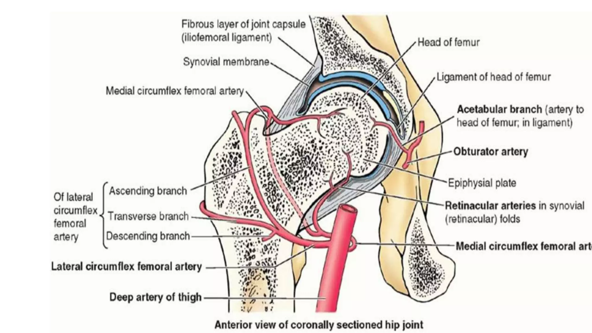
HIP JOINT
▪ Introduction
▪ Articular surface
▪ Ligaments
▪ Blood supply
▪ Nerve supply
▪ Movements
INTRODUCTION
• It is a Ball and Socket variety of synovial joint.
• Multiaxial
• It is a high mobility with high stability joint.
ARTICULAR SURFACE
Head of Femur Acetabulum
• more than half of a sphere horse shoe shape lunate
• covered by hyaline cartilage surface covered by
hyaline cartilage
• EXCEPT at fovea capitis Deep notch with
narrow mouth acetabular
fossa
LIGAMENTS
Fibrous capsule:
• Attachment-
On hip bone :- to the acetabular labrum and to bone
above and behind the acetabulum.
On femur:- to the intertrochanteric line in front and 1 cm
medial to intertrochanteric crest behind.
• Thick anterosuperiorly part subjected to max tension.
• Thin and loosely attach posteroinferiorly.
• Two types of fibre
Outer longitudinal-many of reflected to neck of
femur known as Retinacula(contain blood vessels)
Inner circular known as Zona orbicularis.
ILIOFEMORAL LIGAMENT
●Inverted Y shaped triangular ligament
●Ligament of BIGELOW
●Strongest ligament of body
●Apex- lower half of ASIS
●Base- intertrochanteric line
●The upper and lower fibers are thick strong band
while the middle fibers are tgin and weak.
PUBOFEMOARAL
LIGAMENT
• Support the joint inferomedially
• Triangular in shape
• Superiorly it is attached to the ilio pubic eminence,
obturator crest.
• Inferiorly it merges with the anteroinferior part of
capsule and lower band of iliofemoral ligament.
ISCHIOFEMORAL LIGAMENT
• Comparitively weak and covers the joint posteriorly
• Fibers are twisted and extended from ischium
posteroinferiorly to the acetabulum and the few
fibers to the greater trochanter.
LIGAMENT OF HEAD OF FEMUR
• ROUND LIGAMENT or LIGAMENTUM TERES
• Flat, triangular ligament
• Apex – at fovea capitis
• Base – at transverse ligament and margins of
acetabular notch
• Transmits arteries to the head of femur
ACETABULAR LABRUM
• Cotyloid ligament
• Fibrocartilagenous rim attached to the margins of acetabulum
• Narrows the mouth of acetabulum which helps in holding the head of femur
in position
TRANSVERSE LIGAMNET OF ACETABULUM
• A part of the labrum which bridges the
acetabular notch but it has to cartilage cells
• The notch thus converted into a foramen
transmits vessels and nerves to the joint
SYNOVIAL MEMBRANE
• Lines the fibrous capsule
• Intra capsular portion of neck of femur
• Both surface of acetabular labrum
• Transverse ligament
• Fat in acetabular fossa
• Round ligament of head of femur
BLOOD SUPPLY
• Obturator artery
• Two circumflex femoral artery
• Two gluteal arteries
• Medial and lateral circumflex femoral arteries
form an arterial circle around the capsular attachment
on the neck of femur
• Retiocular arteries arise from the circle and supply
the intracapsular neck and greater part of the head of femur
• A small part of the head near the fovea capitis is supplied by
acetabular branch of obturator and medial circumflex femoral
arteries
NERVE SUPPLY
Hip joint is supplied by:
1. The femoral nerve through the nerves to
rectus femoris
2. Anterior division of obturator nerve
3. Accessory obturator nerve
4. Nerve to quadratus femoris
5. The superior gluteal nerve
MOVEMENTS
• Flexion and extension occur around a transverse axis
• Adduction and abduction occurs around anteroposterior axis
• Medial and lateral rotation occurs around a vertical axis
• Circumduction is a combination of forgoing movement
HIP JOINT(Final).pptx

HIP JOINT(Final).pptx

  • 1.
  • 2.
    ▪ Introduction ▪ Articularsurface ▪ Ligaments ▪ Blood supply ▪ Nerve supply ▪ Movements
  • 3.
    INTRODUCTION • It isa Ball and Socket variety of synovial joint. • Multiaxial • It is a high mobility with high stability joint.
  • 4.
    ARTICULAR SURFACE Head ofFemur Acetabulum • more than half of a sphere horse shoe shape lunate • covered by hyaline cartilage surface covered by hyaline cartilage • EXCEPT at fovea capitis Deep notch with narrow mouth acetabular fossa
  • 5.
    LIGAMENTS Fibrous capsule: • Attachment- Onhip bone :- to the acetabular labrum and to bone above and behind the acetabulum. On femur:- to the intertrochanteric line in front and 1 cm medial to intertrochanteric crest behind. • Thick anterosuperiorly part subjected to max tension. • Thin and loosely attach posteroinferiorly. • Two types of fibre Outer longitudinal-many of reflected to neck of femur known as Retinacula(contain blood vessels) Inner circular known as Zona orbicularis.
  • 6.
    ILIOFEMORAL LIGAMENT ●Inverted Yshaped triangular ligament ●Ligament of BIGELOW ●Strongest ligament of body ●Apex- lower half of ASIS ●Base- intertrochanteric line ●The upper and lower fibers are thick strong band while the middle fibers are tgin and weak.
  • 7.
    PUBOFEMOARAL LIGAMENT • Support thejoint inferomedially • Triangular in shape • Superiorly it is attached to the ilio pubic eminence, obturator crest. • Inferiorly it merges with the anteroinferior part of capsule and lower band of iliofemoral ligament.
  • 8.
    ISCHIOFEMORAL LIGAMENT • Comparitivelyweak and covers the joint posteriorly • Fibers are twisted and extended from ischium posteroinferiorly to the acetabulum and the few fibers to the greater trochanter.
  • 9.
    LIGAMENT OF HEADOF FEMUR • ROUND LIGAMENT or LIGAMENTUM TERES • Flat, triangular ligament • Apex – at fovea capitis • Base – at transverse ligament and margins of acetabular notch • Transmits arteries to the head of femur
  • 10.
    ACETABULAR LABRUM • Cotyloidligament • Fibrocartilagenous rim attached to the margins of acetabulum • Narrows the mouth of acetabulum which helps in holding the head of femur in position
  • 11.
    TRANSVERSE LIGAMNET OFACETABULUM • A part of the labrum which bridges the acetabular notch but it has to cartilage cells • The notch thus converted into a foramen transmits vessels and nerves to the joint
  • 12.
    SYNOVIAL MEMBRANE • Linesthe fibrous capsule • Intra capsular portion of neck of femur • Both surface of acetabular labrum • Transverse ligament • Fat in acetabular fossa • Round ligament of head of femur
  • 13.
    BLOOD SUPPLY • Obturatorartery • Two circumflex femoral artery • Two gluteal arteries • Medial and lateral circumflex femoral arteries form an arterial circle around the capsular attachment on the neck of femur • Retiocular arteries arise from the circle and supply the intracapsular neck and greater part of the head of femur • A small part of the head near the fovea capitis is supplied by acetabular branch of obturator and medial circumflex femoral arteries
  • 15.
    NERVE SUPPLY Hip jointis supplied by: 1. The femoral nerve through the nerves to rectus femoris 2. Anterior division of obturator nerve 3. Accessory obturator nerve 4. Nerve to quadratus femoris 5. The superior gluteal nerve
  • 16.
    MOVEMENTS • Flexion andextension occur around a transverse axis • Adduction and abduction occurs around anteroposterior axis • Medial and lateral rotation occurs around a vertical axis • Circumduction is a combination of forgoing movement