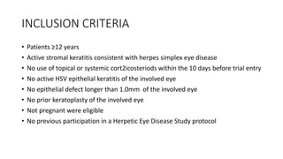
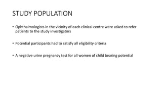
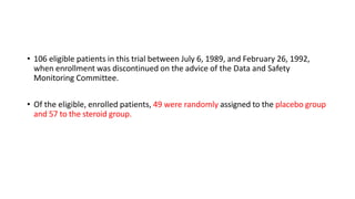
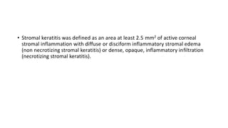
Herpetic Eye Disease study- A
controlled trial of topical corticosteroids for herpes
simplex stromal keratitis
DR.KRISHNAPRIYA K
INTRODUCTION
• Herpes simplex keratitis is a leading cause of corneal opacification in the
United States, other industrialized countries, and developing nations
throughout the world.
• Despite the availability of antiviral agents that are effective in treating herpes
simplex epithelial keratitis, stromal inflammation that can lead to corneal
scarring and visual impairment develops in many patients
• The Herpetic Eye Disease Study (HERS) comprises a series of placebo-
controlled clinical trials designed to evaluate the acute treatment of herpes
simplex stromal keratitis or uveitis.
• The HEDS trial described here compared the effects of tritluridine plus either
topical corticosteroid or placebo in patients with herpes simplex stromal
keratitis who were not receiving a corticosteroid at randomization.
OBJECTIVE
• To evaluate the efficacy of topical corticosteroids in treating herpes
simplex stromal keratitis.
STUDY DESIGN
• Randomised ,double masked ,placebo controlled ,multicentre clinical trial
INCLUSION CRITERIA
• Patients ≥12 years
• Active stromal keratitis consistent with herpes simplex eye disease
• No use of topical or systemic cort2icosteriods within the 10 days before trial entry
• No active HSV epithelial keratitis of the involved eye
• No epithelial defect longer than 1.0mm of the involved eye
• No prior keratoplasty of the involved eye
• Not pregnant were eligible
• No previous participation in a Herpetic Eye Disease Study protocol
STUDY POPULATION
• Ophthalmologists in the vicinity of each clinical centre were asked to refer
patients to the study investigators
• Potential participants had to satisfy all eligibility criteria
• A negative urine pregnancy test for all women of child bearing potential
• 106 eligible patients in this trial between July 6, 1989, and February 26, 1992,
when enrollment was discontinued on the advice of the Data and Safety
Monitoring Committee.
• Of the eligible, enrolled patients, 49 were randomly assigned to the placebo group
and 57 to the steroid group.
• Stromal keratitis was defined as an area at least 2.5 mm2 of active corneal
stromal inflammation with diffuse or disciform inflammatory stromal edema
(non necrotizing stromal keratitis) or dense, opaque, inflammatory infiltration
(necrotizing stromal keratitis).
METHOD
• Patients were assigned to receive either topical prednisolone phosphate or
placebo in a double masked fashion using a random permuted block design
• Eyedropper bottles were packaged by the manufacturer & labelled with a
reference number & instructions for use
• The corticosteroid ophthalmic solution was formulated as 1%pedisolone sodium
phosphate & as 0.125%prednisolne sodium phosphate in buffered isotonic
aqueous solutions
• The same manufacturer provided placebo solutions containing a similar
formulations of vehicles & preservatives without active drug in identical
containers
• Trifluridine 1% ophthalmic solution was prepared in aqueous solution with acetic
acid ,sodium acetate ,sodium chloride & 0.001% thiomersol
• The protocol involved 10 weeks of treatment with the weekly examination ,
discontinuing the treatment regimen at the end of 10 weeks followed by 6 weeks
of biweekly visits & a final visit at 6 months
• Patients assigned to prednisolone phosphate & placebo followed the same dosing
schedule
• Patients with a non severe adverse reaction attributable to trifluridine at the 7
week visit or later remained in the study if another antiviral agent was not used
during the remaining study interval
• Patients received a cycloplegic agent such as 0.25% scopolamine solution at the
the discretion of ophthalmologist examiner
• Each patient was given a medication diary with week specific cards as a reminder
of the treatment regimen
• Compliance monitored by weighing the eyedropper bottles at each visit
CLINCAL EVALUATION
• A complete eye examination was performed at randomization , & re examination
were performed at scheduled visits
• After refinement of the subjective refraction , standardized best corrected visual
acuity ( measured in a trial frame or phoropter at a 4m test distance using
modified Bailey –lovie charts that were retroilluminated at 1025+/-50 lux in
general room illumination)was recorded at randomization , halfway through the
treatment regimen (at the 5 week visit), at the 16 week visit & or on the day of
withdrawl from the trial & at the final 6 month visit
• Slit lamp biomicroscope was performed at each study visit
• The area of stromal keratitis was calculated by multiplying the mean three
readings of the greatest diameter by means of 3 readings of greatest
perpendicular width
• For multiple lesions , each lesion was measured separatetly, & the total area used
was the sum of the individual areas
• At each study visit , the area was compared with the area measured at the initial
examination & with the area measured at the previous visit
• The affected area also measured in terms of depth, density, thickness, stromal
thinning, & endothelial dysfunction
• Anterior chamber cells were counted using a 1.0 * 0.5 mm sli beam at 45 degree
axis
• Intraocular pressure was determined at each visit by applanation or
pneumotonometry
• Photomicrography was performed on the day of randomization , at the 5 week
visit, & at either 16 week visit or the day of withdrawl from the study
• Treatment failure was defined as worsening of stromal keratitis or uveitis at any
scheduled visit , no change in stromal inflammation in the first 2 weeks or 3 later
consecutive weeks or occurrence of an adverse event
• Increased severity of stromal keratitis was defied as any one of the following
Development of a new zone of non necrotizing or necrotizing stromal keratitis
of 15mm2 or greater
Increase in total area of stromal keratitis of 7.5 mm2 or by 75% of the previous
area depending on the size of previous lesion
• Increase in total area of stromal keratitis of 10 mm2 or by 100% of the initial area,
depending on the size of the initial lesion.
• No change was defined as less than a 10% change in the area of stromal
keratitis.
• Increased severity of iridocyclitis was defined as a two-step or greater increase of
cells in the anterior chamber compared with the best attained measurement or
3+ cells or more in the anterior chamber for 2 weeks
• Significant vision decrease was defined as deterioration of visual acuity by four
lines or more from baseline as measured on the Bailey-Louie chart.
• Treatment success was defined as completion of the 10-week regimen of study
medications, resolution of active inflammation during the 16-week
treatment/observation interval, and no reactivation or recurrence for 6 weeks
without medication.
• An overall assessment was made at each follow-up visit and categorized as no
active inflammation (resolved); active inflammation persistent without
resolution, improved since study entry and improved since the last visit; active
inflammation persistent without resolution, improved since study entry yet not
improved since the last visit; active inflammation persistent without change since
study entry; active inflammation persistent without resolution and worse since
study entry; or recurrent active inflammation after initial resolution.
STATISTICAL ANALYSIS
• Baseline characteristics of the two treatment groups were compared by the chi-
square test or the Wilcoxon rank-sum test for categorical variables and by
Student's t test for continuous variables.
• Days from randomization to treatment failure, resolution of stromal keratitis, and
recurrent herpetic eye disease were compared between the two groups by the
Kaplan-Meier method using the Mantel-Haenszel form of the log-rank test
RESULTS
• Nine clinical centers screened 430 patients with clinically presumed herpes simplex
stromal keratitis or uveitis, of whom 305 met the eligibility criteria for one of three
trials then open for randomization, and enrolled 106 eligible patients in this trial
between July 6, 1989, and February 26, 1992, when enrollment was discontinued
on the advice of the Data and Safety Monitoring Committee.
• Of the eligible, enrolled patients, 49 were randomly assigned to the placebo group
and 57 to the steroid group.
• Determination of the difference in time to treatment failure between the steroid
and placebo groups showed a highly significant benefit in favor of the steroid
group (P < 0.001)
• Fifteen patients (26%) in the steroid group and 36 patients (73%) in the placebo
group were treatment failures before completing the 10-week course of trial
medications
• By 16 weeks, 28 patients (49%) in the steroid group and 37 patients (76%) in the
placebo group had failed treatment
• Compared with placebo, corticosteroid therapy reduced the risk of persistent or
progressive stromal keratouveitis by 68%.
• The time from randomization to resolution of stromal keratitis and uveitis
was significantly shorter in the steroid group compared with the placebo
group even though both groups included patients who were removed from
the study and treated with topical corticosteroids
• Nineteen (33%) of the steroid-treated patients and 11 (22%) of the placebo-
treated patients completed the 10 weeks of protocol therapy and had stable,
noninflamed corneas after 16 weeks
• Comparison between visual acuity measured at randomization and 6 months
later showed a similar degree of visual improvement between the two
treatment groups.
• Fifty-nine percent of eyes in the placebo group and 61% of eyes in the steroid
group had an improvement in visual acuity of two lines or more on the Bailey-
Lovie chart from the day of randomization to the final 6-month examination
• 72% of the portion of the placebo group who showed improvement had failed
trial medication and subsequently received topical corticosteroids.
DISCUSSION
• The use of anti-inflammatory therapy with a topical corticosteroid for herpes
simplex stromal keratitis has been an unresolved issue
• Corticosteroids are contraindicated during herpes simplex viral infection of the
ocular surface but are used to suppress herpes simplex stromal keratitis
• We found that patients with herpes simplex stromal keratitis who received a
tapered regimen of topical corticosteroids were more likely to successfully
complete the treatment course and had more rapid resolution of stromal keratitis
than those who received a similar regimen of placebo.
• Patients randomized to placebo were more likely to fail the trial, to fail earlier,
and, despite subsequent corticosteroid therapy, to take significantly longer to
achieve resolution
CONCLUSION
• This clinical trial provides a basis for the judicious use of corticosteroids with
protective antiviral cover in the acute treatment with herpes simplex stromal
keratitis who have not recently received corticosteroid therapy
• We found that patients administered a progressively decreasing regimen of
prednisolone phosphate with trifluridine took longer to worsen & improved
faster than a comparable placebo group
• The use & indications of topical corticosteroids for many infectious &
inflammatory corneal diseases remain to be more clearly determined
• This study demonstrates the efficacy of topical corticosteroids with topical
antiviral prophylaxis in the acute treatment of herpes simplex stromal keratitis
LIMITATION
• A potential limitation of this trial was the use of a clinical, rather than virologic,
case definition.
• Serologic testing can show prior exposure, but noninvasive laboratory asessment
that proves the etiology of herpes simplex stromal keratitis is not available
THANK YOU………..

Herpetic Eye Disease study.pptx

  • 1.
    Herpetic Eye Diseasestudy- A controlled trial of topical corticosteroids for herpes simplex stromal keratitis DR.KRISHNAPRIYA K
  • 2.
    INTRODUCTION • Herpes simplexkeratitis is a leading cause of corneal opacification in the United States, other industrialized countries, and developing nations throughout the world. • Despite the availability of antiviral agents that are effective in treating herpes simplex epithelial keratitis, stromal inflammation that can lead to corneal scarring and visual impairment develops in many patients • The Herpetic Eye Disease Study (HERS) comprises a series of placebo- controlled clinical trials designed to evaluate the acute treatment of herpes simplex stromal keratitis or uveitis. • The HEDS trial described here compared the effects of tritluridine plus either topical corticosteroid or placebo in patients with herpes simplex stromal keratitis who were not receiving a corticosteroid at randomization.
  • 3.
    OBJECTIVE • To evaluatethe efficacy of topical corticosteroids in treating herpes simplex stromal keratitis.
  • 4.
    STUDY DESIGN • Randomised,double masked ,placebo controlled ,multicentre clinical trial
  • 5.
    INCLUSION CRITERIA • Patients≥12 years • Active stromal keratitis consistent with herpes simplex eye disease • No use of topical or systemic cort2icosteriods within the 10 days before trial entry • No active HSV epithelial keratitis of the involved eye • No epithelial defect longer than 1.0mm of the involved eye • No prior keratoplasty of the involved eye • Not pregnant were eligible • No previous participation in a Herpetic Eye Disease Study protocol
  • 6.
    STUDY POPULATION • Ophthalmologistsin the vicinity of each clinical centre were asked to refer patients to the study investigators • Potential participants had to satisfy all eligibility criteria • A negative urine pregnancy test for all women of child bearing potential
  • 7.
    • 106 eligiblepatients in this trial between July 6, 1989, and February 26, 1992, when enrollment was discontinued on the advice of the Data and Safety Monitoring Committee. • Of the eligible, enrolled patients, 49 were randomly assigned to the placebo group and 57 to the steroid group.
  • 8.
    • Stromal keratitiswas defined as an area at least 2.5 mm2 of active corneal stromal inflammation with diffuse or disciform inflammatory stromal edema (non necrotizing stromal keratitis) or dense, opaque, inflammatory infiltration (necrotizing stromal keratitis).
  • 9.
    METHOD • Patients wereassigned to receive either topical prednisolone phosphate or placebo in a double masked fashion using a random permuted block design • Eyedropper bottles were packaged by the manufacturer & labelled with a reference number & instructions for use • The corticosteroid ophthalmic solution was formulated as 1%pedisolone sodium phosphate & as 0.125%prednisolne sodium phosphate in buffered isotonic aqueous solutions
  • 10.
    • The samemanufacturer provided placebo solutions containing a similar formulations of vehicles & preservatives without active drug in identical containers • Trifluridine 1% ophthalmic solution was prepared in aqueous solution with acetic acid ,sodium acetate ,sodium chloride & 0.001% thiomersol
  • 11.
    • The protocolinvolved 10 weeks of treatment with the weekly examination , discontinuing the treatment regimen at the end of 10 weeks followed by 6 weeks of biweekly visits & a final visit at 6 months • Patients assigned to prednisolone phosphate & placebo followed the same dosing schedule
  • 13.
    • Patients witha non severe adverse reaction attributable to trifluridine at the 7 week visit or later remained in the study if another antiviral agent was not used during the remaining study interval • Patients received a cycloplegic agent such as 0.25% scopolamine solution at the the discretion of ophthalmologist examiner • Each patient was given a medication diary with week specific cards as a reminder of the treatment regimen • Compliance monitored by weighing the eyedropper bottles at each visit
  • 14.
    CLINCAL EVALUATION • Acomplete eye examination was performed at randomization , & re examination were performed at scheduled visits • After refinement of the subjective refraction , standardized best corrected visual acuity ( measured in a trial frame or phoropter at a 4m test distance using modified Bailey –lovie charts that were retroilluminated at 1025+/-50 lux in general room illumination)was recorded at randomization , halfway through the treatment regimen (at the 5 week visit), at the 16 week visit & or on the day of withdrawl from the trial & at the final 6 month visit
  • 15.
    • Slit lampbiomicroscope was performed at each study visit • The area of stromal keratitis was calculated by multiplying the mean three readings of the greatest diameter by means of 3 readings of greatest perpendicular width • For multiple lesions , each lesion was measured separatetly, & the total area used was the sum of the individual areas • At each study visit , the area was compared with the area measured at the initial examination & with the area measured at the previous visit • The affected area also measured in terms of depth, density, thickness, stromal thinning, & endothelial dysfunction
  • 16.
    • Anterior chambercells were counted using a 1.0 * 0.5 mm sli beam at 45 degree axis • Intraocular pressure was determined at each visit by applanation or pneumotonometry • Photomicrography was performed on the day of randomization , at the 5 week visit, & at either 16 week visit or the day of withdrawl from the study
  • 17.
    • Treatment failurewas defined as worsening of stromal keratitis or uveitis at any scheduled visit , no change in stromal inflammation in the first 2 weeks or 3 later consecutive weeks or occurrence of an adverse event • Increased severity of stromal keratitis was defied as any one of the following Development of a new zone of non necrotizing or necrotizing stromal keratitis of 15mm2 or greater Increase in total area of stromal keratitis of 7.5 mm2 or by 75% of the previous area depending on the size of previous lesion
  • 18.
    • Increase intotal area of stromal keratitis of 10 mm2 or by 100% of the initial area, depending on the size of the initial lesion. • No change was defined as less than a 10% change in the area of stromal keratitis. • Increased severity of iridocyclitis was defined as a two-step or greater increase of cells in the anterior chamber compared with the best attained measurement or 3+ cells or more in the anterior chamber for 2 weeks
  • 19.
    • Significant visiondecrease was defined as deterioration of visual acuity by four lines or more from baseline as measured on the Bailey-Louie chart. • Treatment success was defined as completion of the 10-week regimen of study medications, resolution of active inflammation during the 16-week treatment/observation interval, and no reactivation or recurrence for 6 weeks without medication.
  • 20.
    • An overallassessment was made at each follow-up visit and categorized as no active inflammation (resolved); active inflammation persistent without resolution, improved since study entry and improved since the last visit; active inflammation persistent without resolution, improved since study entry yet not improved since the last visit; active inflammation persistent without change since study entry; active inflammation persistent without resolution and worse since study entry; or recurrent active inflammation after initial resolution.
  • 21.
    STATISTICAL ANALYSIS • Baselinecharacteristics of the two treatment groups were compared by the chi- square test or the Wilcoxon rank-sum test for categorical variables and by Student's t test for continuous variables. • Days from randomization to treatment failure, resolution of stromal keratitis, and recurrent herpetic eye disease were compared between the two groups by the Kaplan-Meier method using the Mantel-Haenszel form of the log-rank test
  • 22.
    RESULTS • Nine clinicalcenters screened 430 patients with clinically presumed herpes simplex stromal keratitis or uveitis, of whom 305 met the eligibility criteria for one of three trials then open for randomization, and enrolled 106 eligible patients in this trial between July 6, 1989, and February 26, 1992, when enrollment was discontinued on the advice of the Data and Safety Monitoring Committee. • Of the eligible, enrolled patients, 49 were randomly assigned to the placebo group and 57 to the steroid group.
  • 23.
    • Determination ofthe difference in time to treatment failure between the steroid and placebo groups showed a highly significant benefit in favor of the steroid group (P < 0.001) • Fifteen patients (26%) in the steroid group and 36 patients (73%) in the placebo group were treatment failures before completing the 10-week course of trial medications • By 16 weeks, 28 patients (49%) in the steroid group and 37 patients (76%) in the placebo group had failed treatment
  • 25.
    • Compared withplacebo, corticosteroid therapy reduced the risk of persistent or progressive stromal keratouveitis by 68%. • The time from randomization to resolution of stromal keratitis and uveitis was significantly shorter in the steroid group compared with the placebo group even though both groups included patients who were removed from the study and treated with topical corticosteroids
  • 27.
    • Nineteen (33%)of the steroid-treated patients and 11 (22%) of the placebo- treated patients completed the 10 weeks of protocol therapy and had stable, noninflamed corneas after 16 weeks
  • 28.
    • Comparison betweenvisual acuity measured at randomization and 6 months later showed a similar degree of visual improvement between the two treatment groups. • Fifty-nine percent of eyes in the placebo group and 61% of eyes in the steroid group had an improvement in visual acuity of two lines or more on the Bailey- Lovie chart from the day of randomization to the final 6-month examination • 72% of the portion of the placebo group who showed improvement had failed trial medication and subsequently received topical corticosteroids.
  • 30.
    DISCUSSION • The useof anti-inflammatory therapy with a topical corticosteroid for herpes simplex stromal keratitis has been an unresolved issue • Corticosteroids are contraindicated during herpes simplex viral infection of the ocular surface but are used to suppress herpes simplex stromal keratitis
  • 31.
    • We foundthat patients with herpes simplex stromal keratitis who received a tapered regimen of topical corticosteroids were more likely to successfully complete the treatment course and had more rapid resolution of stromal keratitis than those who received a similar regimen of placebo. • Patients randomized to placebo were more likely to fail the trial, to fail earlier, and, despite subsequent corticosteroid therapy, to take significantly longer to achieve resolution
  • 32.
    CONCLUSION • This clinicaltrial provides a basis for the judicious use of corticosteroids with protective antiviral cover in the acute treatment with herpes simplex stromal keratitis who have not recently received corticosteroid therapy • We found that patients administered a progressively decreasing regimen of prednisolone phosphate with trifluridine took longer to worsen & improved faster than a comparable placebo group • The use & indications of topical corticosteroids for many infectious & inflammatory corneal diseases remain to be more clearly determined • This study demonstrates the efficacy of topical corticosteroids with topical antiviral prophylaxis in the acute treatment of herpes simplex stromal keratitis
  • 33.
    LIMITATION • A potentiallimitation of this trial was the use of a clinical, rather than virologic, case definition. • Serologic testing can show prior exposure, but noninvasive laboratory asessment that proves the etiology of herpes simplex stromal keratitis is not available
  • 34.