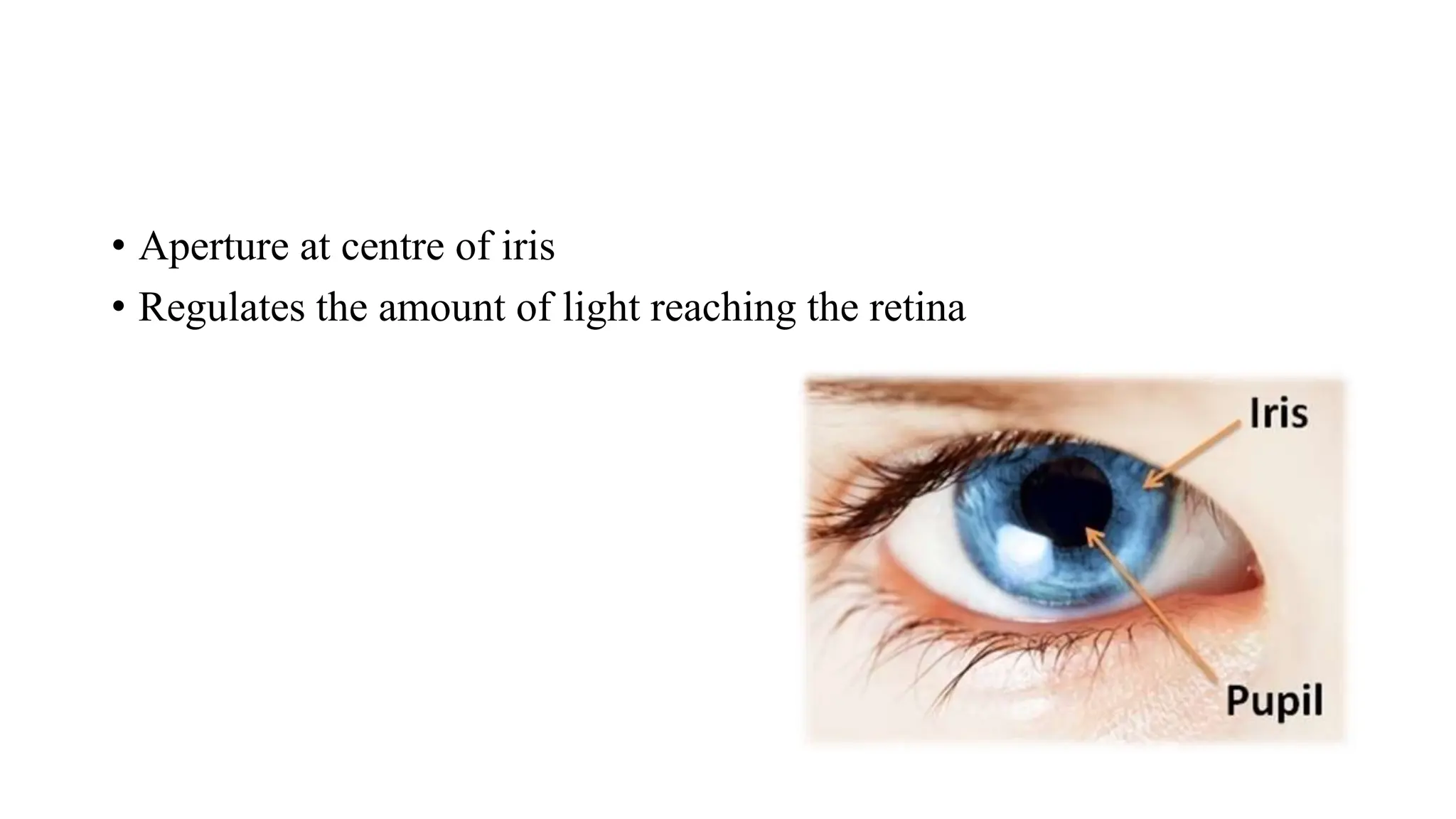
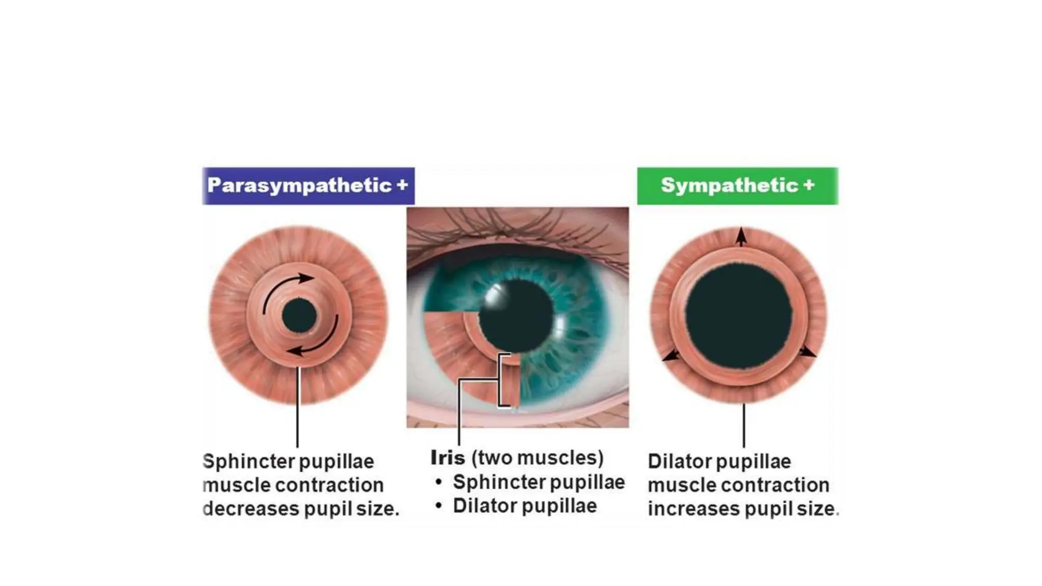
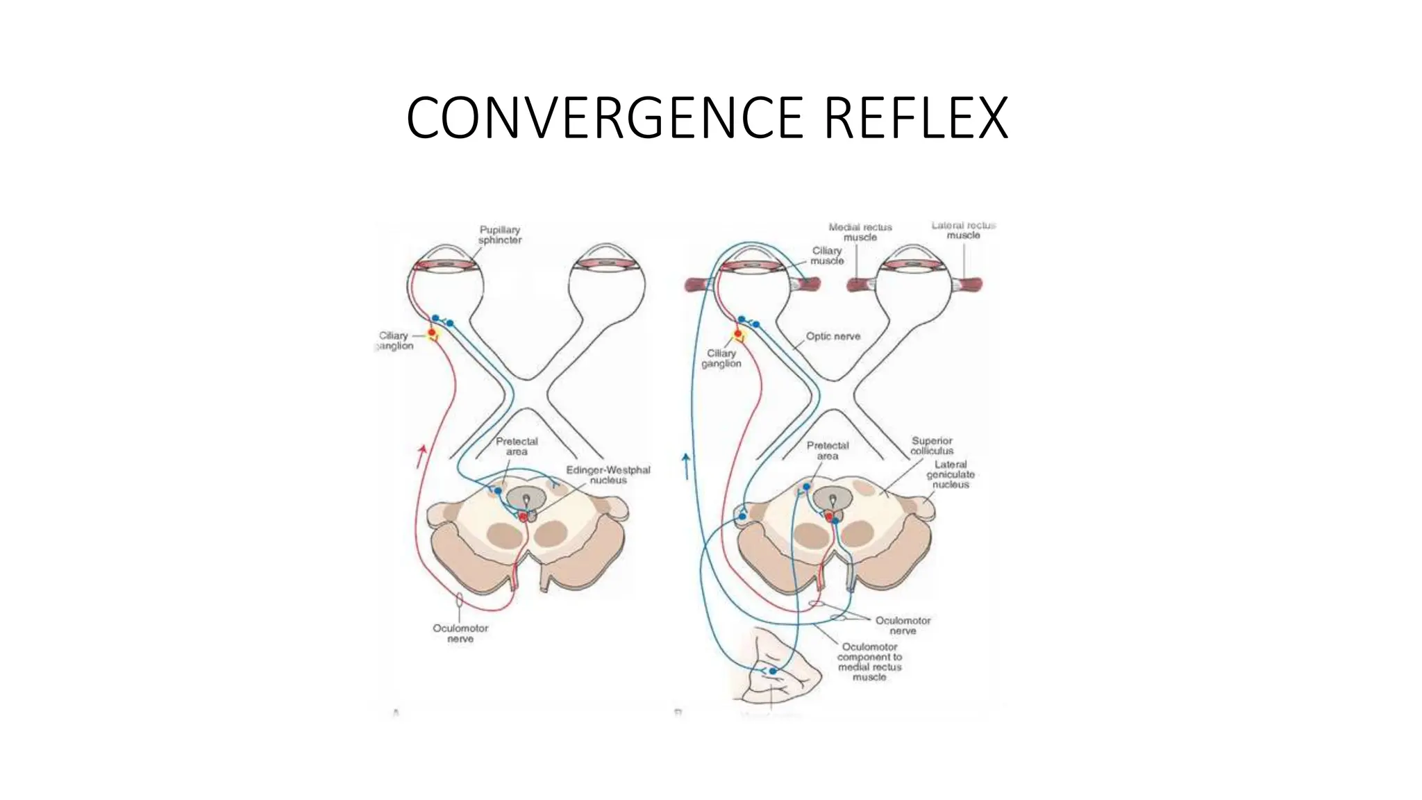
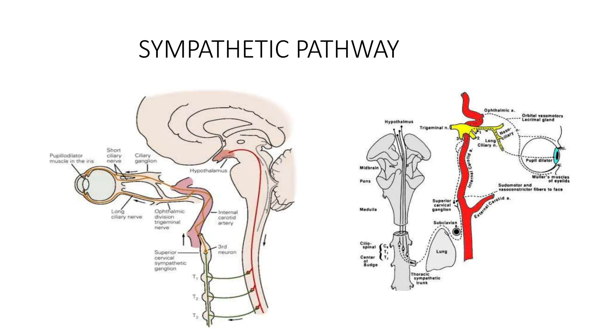
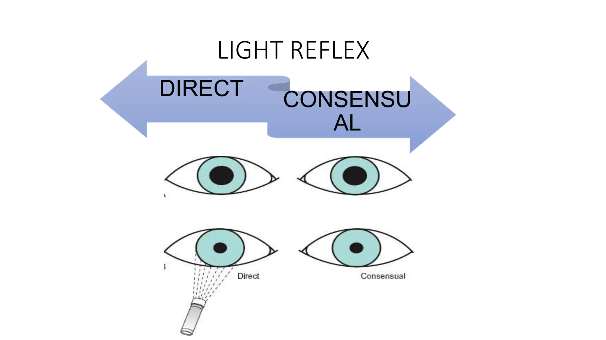
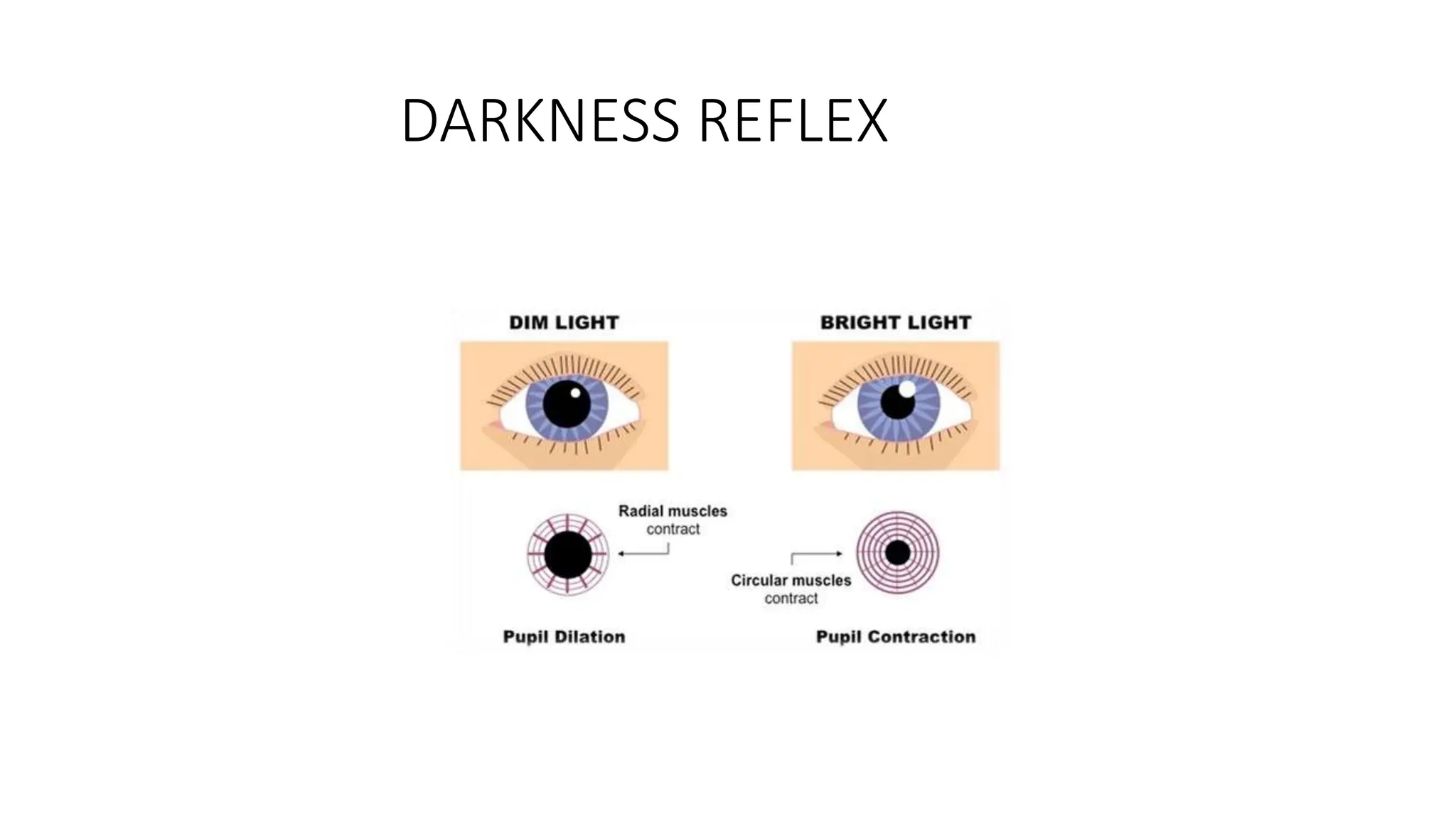
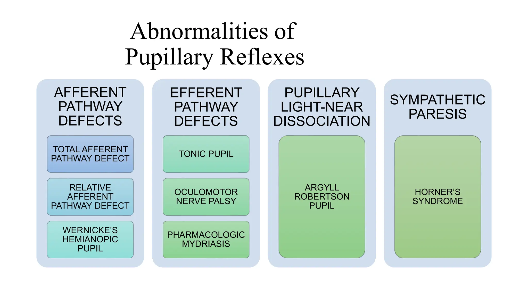
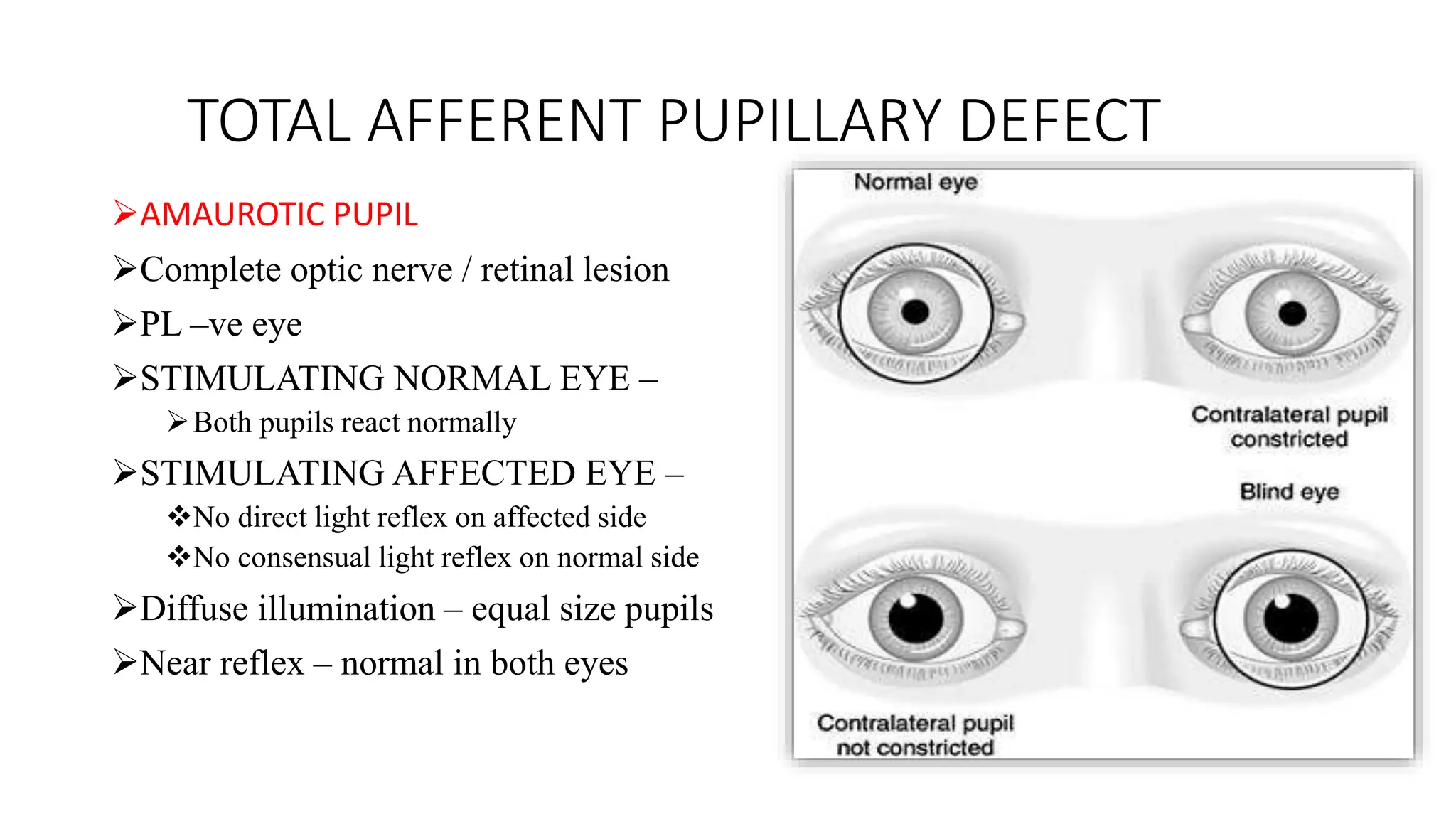
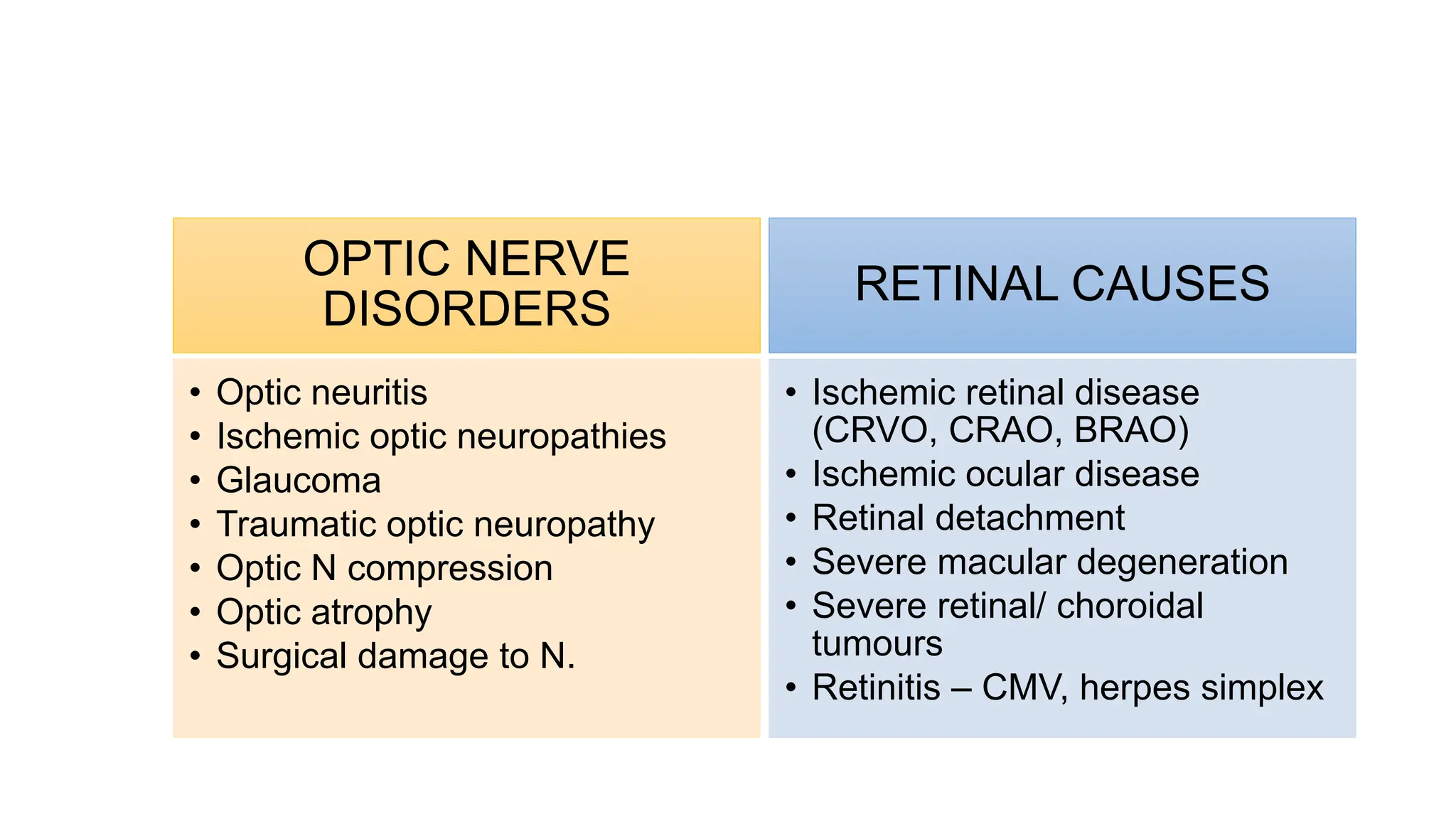
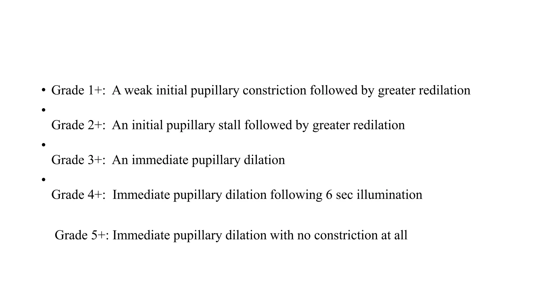
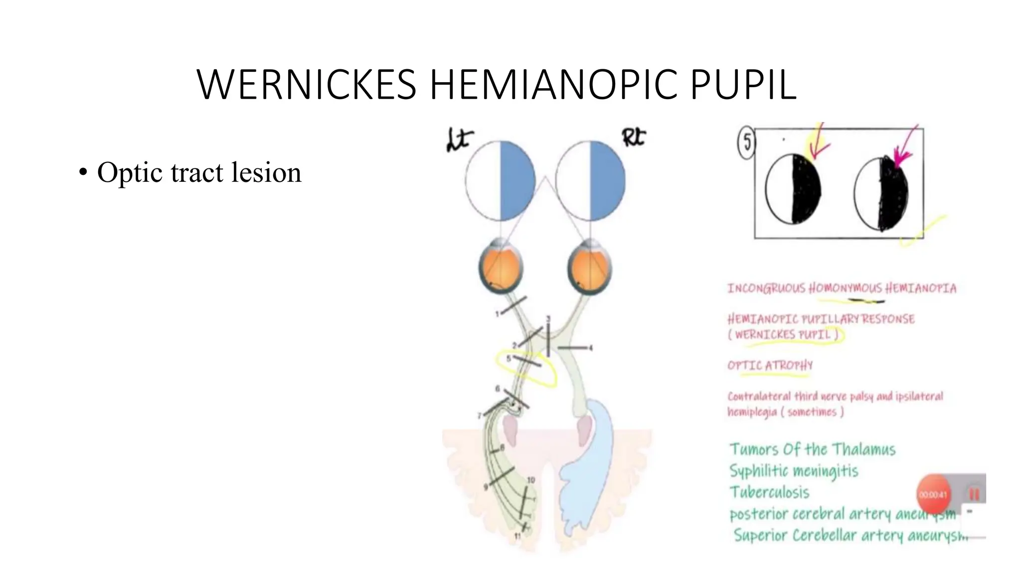
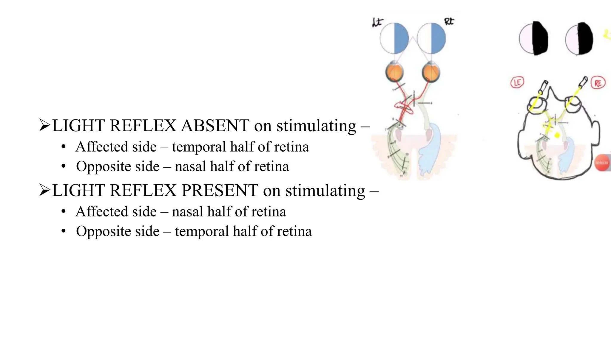
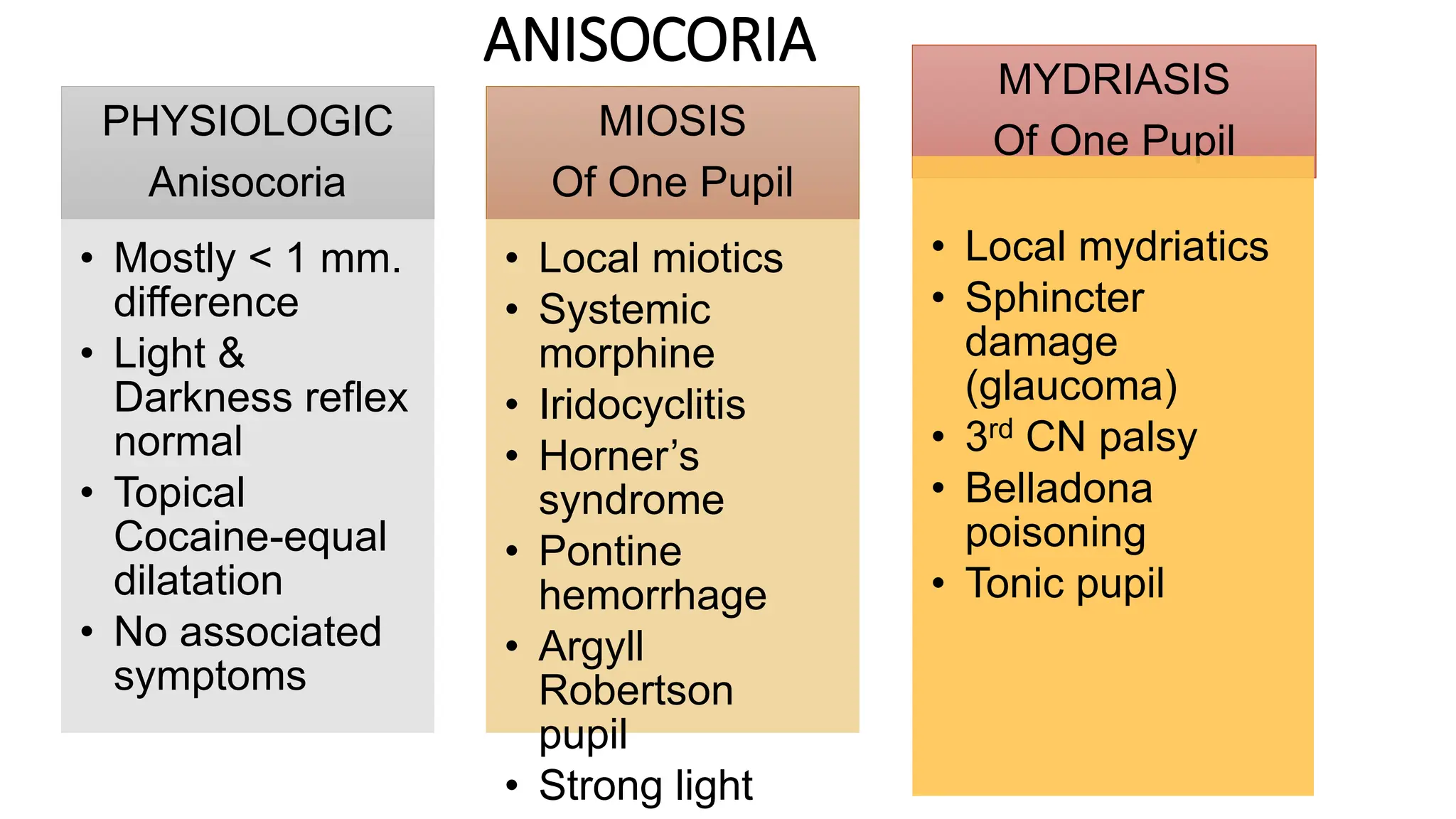
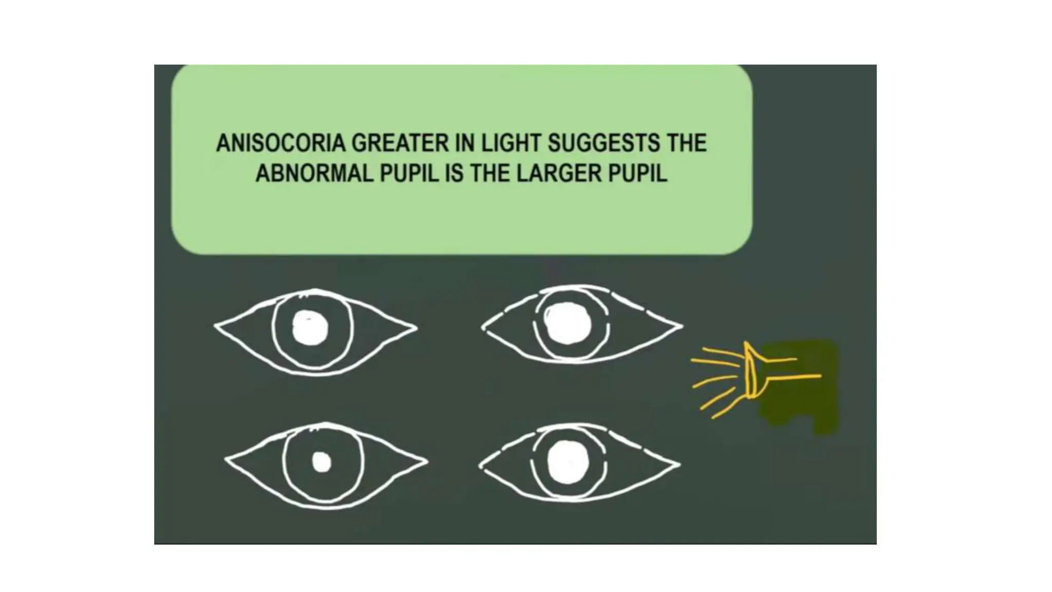
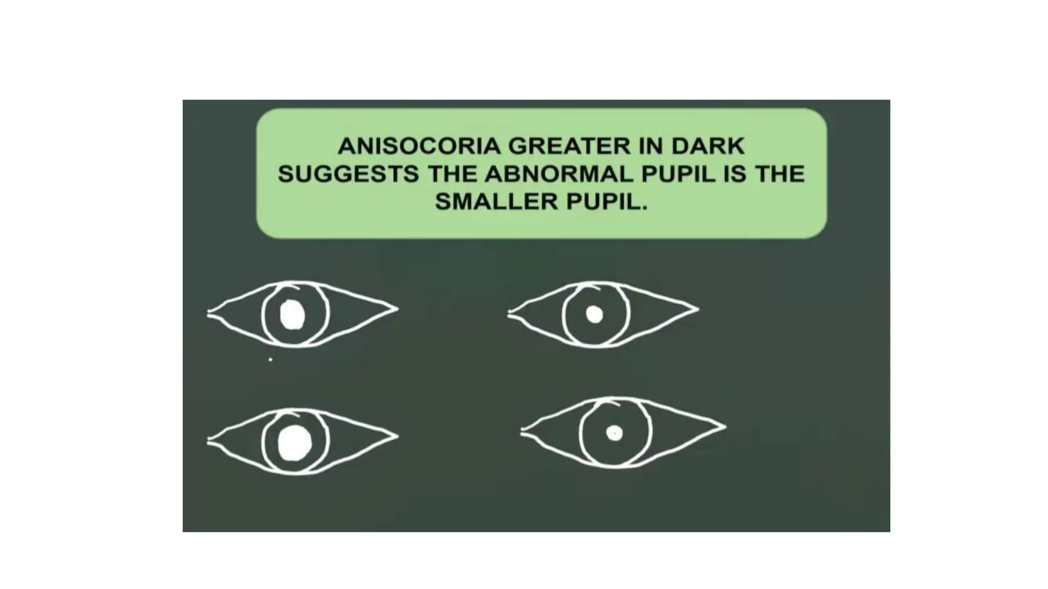
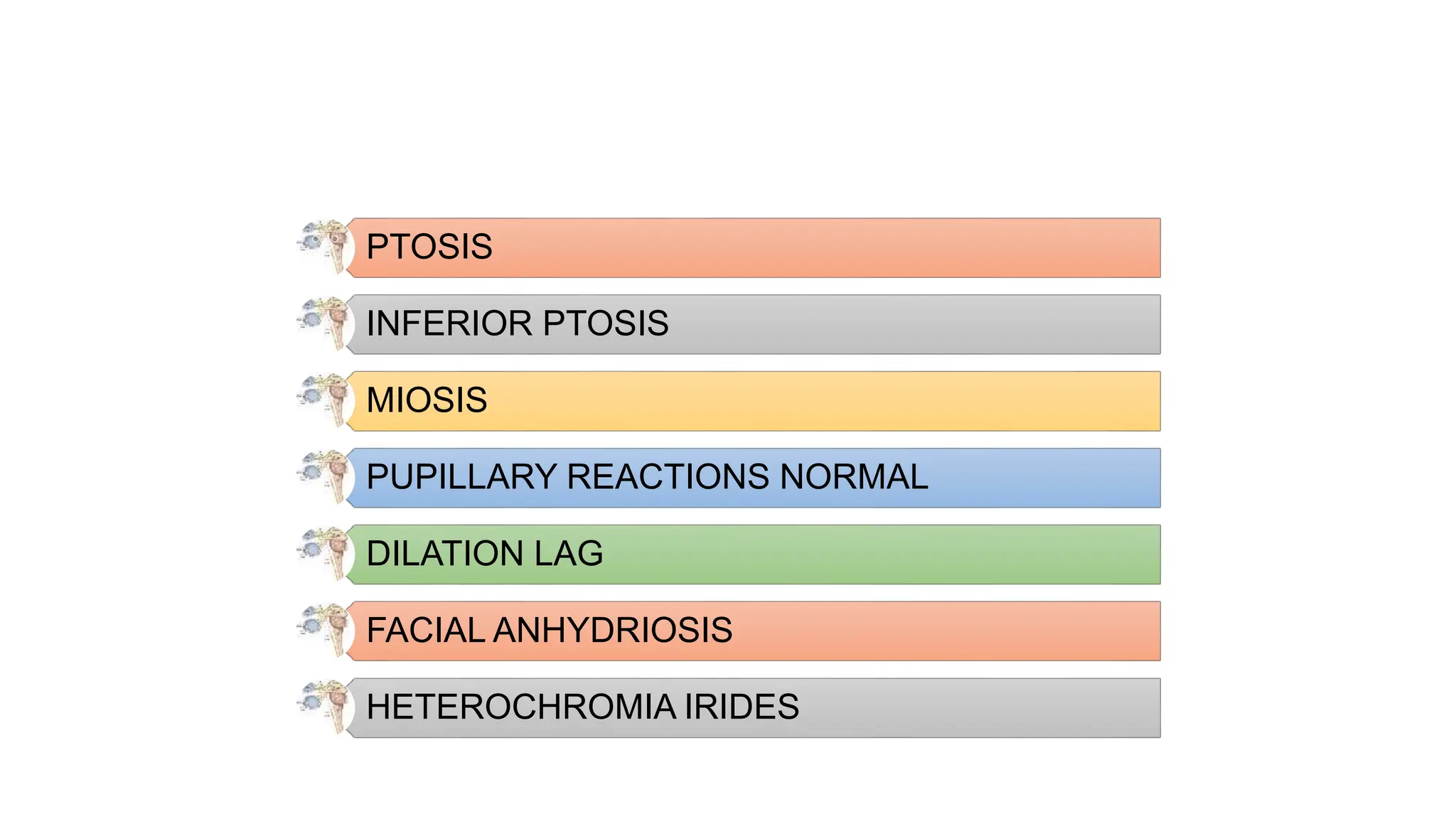
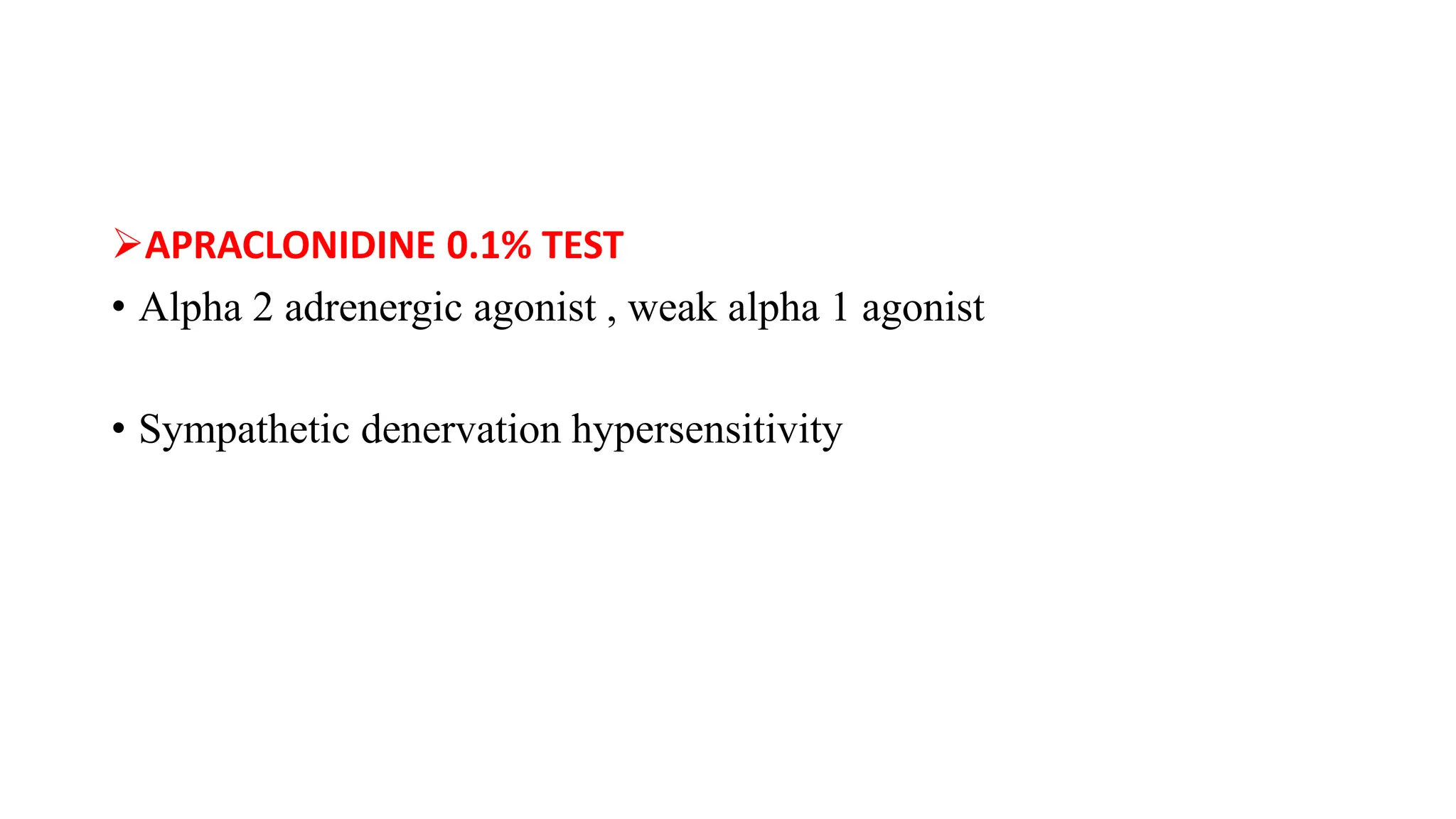
PUPILLARY ABNORMALITIES
• Aperture at centre of iris
• Regulates the amount of light reaching the retina
PUPILLARY PATHWAYS
• LIGHT REFLEX
• CONVERGENCE
• ACCOMODATION
• SYMPATHETIC DISCHARGE
LIGHT REFLEX
CONVERGENCE REFLEX
ACCOMODATION REFLEX
SYMPATHETIC PATHWAY
PUPILLARY REFLEX
• LIGHT RELEX
• NEAR REFLEX
• PSYCHOSENSORY REFLEX
• LID CLOSURE REFLEX
LIGHT REFLEX
DIRECT CONSENSU
AL
NEAR REFLEX
DARKNESS REFLEX
Abnormalities of
Pupillary Reflexes
AFFERENT
PATHWAY
DEFECTS
TOTAL AFFERENT
PATHWAY DEFECT
RELATIVE
AFFERENT
PATHWAY DEFECT
WERNICKE’S
HEMIANOPIC
PUPIL
EFFERENT
PATHWAY
DEFECTS
TONIC PUPIL
OCULOMOTOR
NERVE PALSY
PHARMACOLOGIC
MYDRIASIS
PUPILLARY
LIGHT-NEAR
DISSOCIATION
ARGYLL
ROBERTSON
PUPIL
SYMPATHETIC
PARESIS
HORNER’S
SYNDROME
TOTAL AFFERENT PUPILLARY DEFECT
AMAUROTIC PUPIL
Complete optic nerve / retinal lesion
PL –ve eye
STIMULATING NORMAL EYE –
Both pupils react normally
STIMULATING AFFECTED EYE –
No direct light reflex on affected side
No consensual light reflex on normal side
Diffuse illumination – equal size pupils
Near reflex – normal in both eyes
RELATIVE AFFERENT PUPILLARY DEFECT
Marcus-Gunn Pupil
Incomplete optic nerve lesion/ severe retinal disease
• RAPD cause a reduction in pupil contraction when one eye is stimulated by light
compared with when the opposite eye is stimulated by light
• Detected by swinging flash light
OPTIC NERVE
DISORDERS
• Optic neuritis
• Ischemic optic neuropathies
• Glaucoma
• Traumatic optic neuropathy
• Optic N compression
• Optic atrophy
• Surgical damage to N.
RETINAL CAUSES
• Ischemic retinal disease
(CRVO, CRAO, BRAO)
• Ischemic ocular disease
• Retinal detachment
• Severe macular degeneration
• Severe retinal/ choroidal
tumours
• Retinitis – CMV, herpes simplex
• Grade 1+: A weak initial pupillary constriction followed by greater redilation
•
Grade 2+: An initial pupillary stall followed by greater redilation
•
Grade 3+: An immediate pupillary dilation
•
Grade 4+: Immediate pupillary dilation following 6 sec illumination
Grade 5+: Immediate pupillary dilation with no constriction at all
NEUTRAL DENSITY FILTER
• Estimation of the amount of RAPD in log units
WERNICKES HEMIANOPIC PUPIL
• Optic tract lesion
LIGHT REFLEX ABSENT on stimulating –
• Affected side – temporal half of retina
• Opposite side – nasal half of retina
LIGHT REFLEX PRESENT on stimulating –
• Affected side – nasal half of retina
• Opposite side – temporal half of retina
ANISOCORIA
• Asymmetry of efferent signals to the iris muscles produces inequality
in the diameters of 2 pupil
ANISOCORIA
PHYSIOLOGIC
Anisocoria
• Mostly < 1 mm.
difference
• Light &
Darkness reflex
normal
• Topical
Cocaine-equal
dilatation
• No associated
symptoms
MIOSIS
Of One Pupil
• Local miotics
• Systemic
morphine
• Iridocyclitis
• Horner’s
syndrome
• Pontine
hemorrhage
• Argyll
Robertson
pupil
• Strong light
MYDRIASIS
Of One Pupil
• Local mydriatics
• Sphincter
damage
(glaucoma)
• 3rd CN palsy
• Belladona
poisoning
• Tonic pupil
OCULAR EXAMINATION
• Determine which pupil is abnormal by comparing pupil sizes in light
and in dark
• Test pupillary reaction – light & near
• Look for ptosis , ocular motility
• Examine the pupillary margin with a slit lamp
EFFERENT PATHWAY DEFECTS
STIMULATING AFFECTED EYE –
 Direct light reflex & near reflex absent
 Consensual reflex present
 STIMULATING NORMAL EYE –
 Direct light reflex & near reflex present
 Consensual reflex absent
Brainstem lesions
Fascicular Third Cranial Nerve lesions – compressive third nerve lesions
Lesions of Ciliary Ganglion / Short Ciliary Nerves
Iris damage
Mydriatic Drug
TONIC PUPIL
Lesion of Ciliary Ganglion /
Short Ciliary Nerves
Upregulation of postsynaptic receptor to allow re innervation
Aberrant innervation / Regeneration
Tonic miosis on near reflex along with accomodation
AFFECTED PUPIL
 Is larger
 Reaction to light – absent
 Near Reflex – very slow & tonic
 Accommodative paresis
 Cholinergic supersensitivity of denervated muscle (constricts with 0.125%
pilocarpine
CAUSES
LOCAL TONIC PUPIL
• Viral ciliary ganglionitis (herpes zoster)
• Orbital or choroidal tumours or trauma
NEUROPATHIC TONIC PUPIL
• Diabetes ,Alcoholism
IDIOPATHIC TONIC PUPIL WITH BENIGN AREFLEXIA(ADIE’S TONIC
PUPIL)
ADIE’S TONIC PUPIL
Denervation of postganglionic supply of sphincter pupillae & ciliary muscles of
unknown etiology
 80% U/L
 Affects healthy young women > men
 Absent knee jerk
 Affected pupil is large & irregular
 Light reflex is absent 0r slow
 Near reflex is slow & tonic
 Accomodative paresis
OCULOMOTOR NERVE PALSY
• Pupil mid dialated
• Light ,consensual , near reflexes – affected
Pupillary Light-Near Dissociation
• NEAR RELEX PRESENT , LIGHT REFLEX ABSENT
CAUSES
B/L TAPD
Lesions in midbrain
TNP with aberrant regeneration of medial rectus innervation into sphincter
innervation pathway- PSEUDO –ARGYLL ROBERTSON PUPIL
Ciliary ganglion/ short ciliary nerve lesions with aberrant regeneration of
accomodation
Argyll Robertson Pupil
Dorsal midbrain lesion
Cause – Neurosyphilis
Usually B/L & assymetrical involvement
Vision is good
Pupils – small & irregular
Light Reflex absent
Near Reflex present
Cocaine Test- mydriasis
HORNERS SYNDROME
• Oculosympathetic paresis
CENTRAL
Horner’s Synd
• Lesion located
b/w
hypothalamus
to Ciliospinal
centre of
Budge (C8-T2)
PREGANGLION
IC
Horner’s Synd
• Lesion located
b/w C8 - T2 to
superior
cervical
ganglion
POSTGANGLION
IC
Horner’s Synd
• Lesion located
b/w superior
cervical ganglion
to innervation of
dilator pupillae
PTOSIS
INFERIOR PTOSIS
MIOSIS
PUPILLARY REACTIONS NORMAL
DILATION LAG
FACIAL ANHYDRIOSIS
HETEROCHROMIA IRIDES
PHARMACOLOGICAL TESTS
COCAINE 4% or 10% TEST
• Blocks the reuptake of norepinephrine – increased sympathetic
activity – dialatation of pupil
APRACLONIDINE 0.1% TEST
• Alpha 2 adrenergic agonist , weak alpha 1 agonist
• Sympathetic denervation hypersensitivity
LOCALIZING THE LESION
HYDROXYAMPHETAMINE 1% TEST
• Release of stored epinephrine from the 3rd order neuron junction
with the iris
• IN 3RD Order neuron lesion – no stored NE
No dilatation – post-ganglionic Horner’s syndrome
Miotic pupil dilatation – central & preganglionic
Horner’s syndrome
• PHENYLEPHRINE 1% TEST
Apraclonidine (0.5%) Test
• Dilatation to apraclonidine – Horner’s syndrome
Cocaine (4%) Test
• No dilatation to 4% cocaine – Horner’s syndrome
Phenylephrine (1%) Test
• Dilatation to phenylephrine – only post-ganglionic
Horner’s syndrome
Hydroxyamphetamine (1%) Test
• Miotic pupil dilatation – central & preganglionic
Horner’s syndrome
No dilatation – post-ganglionic Horner’s syndrome
PILOCARPINE TEST
Pupil constriction
to 0.125% pilo
• Adie’s tonic
pupil
Pupil constriction
to 1 – 2% pilo
• 3rd CN palsy
No constriction
with pilo
• Mydriatic drugs
• Traumatic
mydriasis
• Fixed pupils in
Iritis
ABNORMAL
PUPIL
MYDRIASIS
NORMAL LIGHT
REACTION
ESSENTIAL
ANISOCORIA
ABNORMAL
LIGHT REACTION
LIGHT-NEAR
DISSOCIATION
NO LIGHT-NEAR
DISSOCIATION
MIOSIS
LIGHT-NEAR
DISSOCIATION
BILATER
AL
-CONVERGENCE-
RETRACTION
NYSTAGMUS
-VERTICAL GAZE DEFICIT
-CONVERGENCE DEFICIT
-LID RETRACTION
PARINAU
D
SYNDRO
ME
UNILATER
AL
-SEGMENTAL PUPILLARY
CONSTRICTION
-IMPAIRED DARK
ADAPTATION
–IMPAIRED NEAR REFLEX
–TONIC REDILATION
BILATER
AL
ADIE’S
PUPIL
UNILATE
RAL
ADIE’S
PUPIL
NO LIGHT-NEAR
DISSOCIATION
0.125%
PILOCARPIN
E
CONSTRICT
ION
RECHECK
FOR TONIC
PUPILS
NO
RESPONSE
1%
PILOCARPINE
CONSTRICTI
ON
3RD CN
PALSY
NO
RESPONSE
DRUG-
INDUCED
MIOSI
S
NORMAL LIGHT
REACTION
4%
COCAIN
E
DILATION
ESSENTIAL
ANISOCORIA
NO
RESPONS
E
HORNER
SYMPATHETIC
PARESIS
-ABNORMAL LIGHT
REACTION
-IRRREGULAR PUPIL
-LIGHT-NEAR
DISSOCIATION
ARGYLL
ROBERTSO
N PUPILS
HORNER
SYMPATHETIC
PARESIS
1%
HYDROXYAMPHETA
MINE
DILATION
CENTRAL /
PREGANGLI
ONIC LESION
NO
RESPONS
E
POSTGANGLI
ONIC LESION
HETEROCH
ROMIA
IRIDIS
CONGENITAL
HORNER’S
SYNDROME
PUPILLARY ABNORMALITIES and pupillary reflexes

PUPILLARY ABNORMALITIES and pupillary reflexes