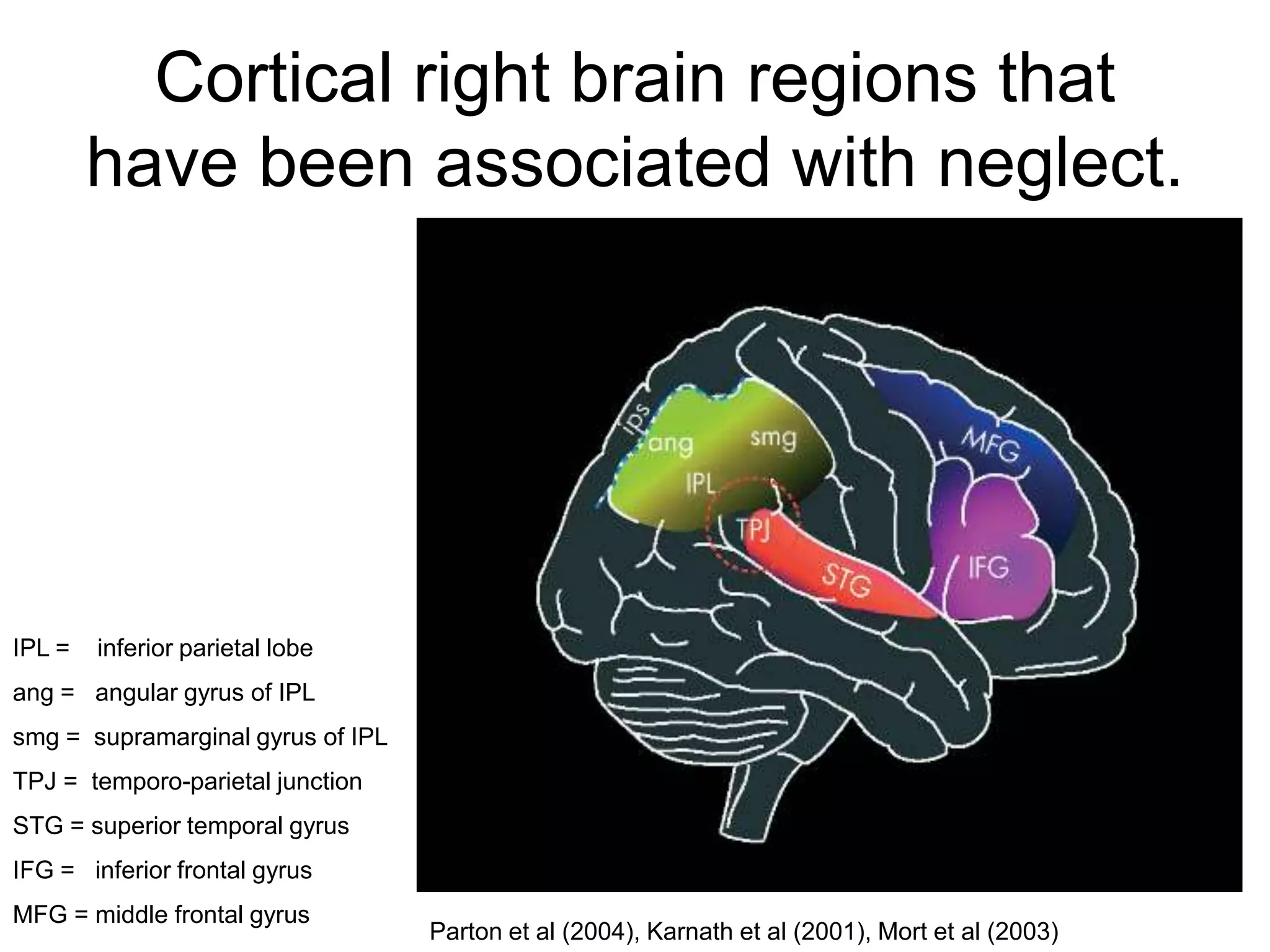
HEMISPATIAL
  NEGLECT
HEMISPATIAL
  NEGLECT
       egocentric
HEMISPATIAL
  NEGLECT
       allocentric
Extinction
               One circle


             One circle!!!
Hemianopsia
 Loss of half the
 vision in both eyes.


Binasal hemianopsia   homonymous hemianopsia
Somatoparaphrenia


 RIGHT


                             LEFT

It’s not
                        RIGHT
mine!!!
Cortical right brain regions that
        have been associated with neglect.


IPL =   inferior parietal lobe
ang = angular gyrus of IPL
smg = supramarginal gyrus of IPL
TPJ = temporo-parietal junction
STG = superior temporal gyrus
IFG = inferior frontal gyrus
MFG = middle frontal gyrus
                                   Parton et al (2004), Karnath et al (2001), Mort et al (2003)
Marotta et al (2002)
What does the Parietal Lobe do?
Integration of sensory information

                         


                         


                         
Enables mapping of visually perceived objects to
                               body coordinates
What can neglect tell us about
   cognitive functioning?
Q1: Do elementary features combine to form
    objects pre-attentively or attentively?


             This is your target!
                            just say if it’s there or not
Kahnaman (1973), Neisser (1967), Treisman (1988)
Neglect is an attention problem, so
attentive tasks like the latter one (colour &
shape variation) should be more difficult
for neglect sufferers, while the earlier
tasks, which required only pre-attentive
processing, should be just as easy for
neglect patients
Q2: Does attentional selection operate early or late
    in the sequence of processing stages?
intact left hemisphere
                           777ms
                               Berti & Rizzolatti (1992)
damaged right hemisphere
intact left hemisphere
                           795ms
                               Berti & Rizzolatti (1992)
damaged right hemisphere
intact left hemisphere
                           890ms
                               Berti & Rizzolatti (1992)
damaged right hemisphere
Summary
• Usually right lobe  left neglect
• PERCEPTION & ATTENTION problem,
  not sense problem
• damage to the ANGULAR GYRUS in the
  PARIETAL lobe
• Enlightening in the study of attention & in
  trying to find out how much of our visual
  processing is pre-attentional
References
•   ArguinM., Joanette Y. & Cavanagh P. (1993) Visual search for feature and conjunction targets
    with an attention deficit. Journal of Cognitive Neuroscience, 54, 436-452.
•   Berti A. & Rizzolatti G. (1992) Visual Processing without awareness: Evidence from unilateral
    neglect. Journal of Cognitive Neuroscience, 4, 345-351.
•   Bird C. M., Malhotra P., Parton A., Coulthard E., Rushworth M. F. S. & Husain M. (2006) Visual
    neglect after right posterior cerebral artery infarction. Journal of Neurology, Neurosurgery, and
    Psychiatry. 77: 1008-1012.
•   Brefczynski J. A. & DeYoe E. A. (1999) A physiological correlate of the ‘spotlight’ of visual
    attention. Nature Neuroscience, 2, 370-374.
•   Driver J., Baylis G. C. & Rafal R. D. (1992) Preserved figure-ground segregation and symmetry
    perception in visual neglect. Nature; 360(6399):73–5.
•   Driver J., Mattingley J.B., Rorden C. & Davis G. (1997). Extinction as a paradigm measure of
    attentional bias and restricted capacity following brain injury. In. P. Thier & H.-O. Karnath
    (Eds.), Parietal Lobe Contributions to Orientation in 3D Space (pp. 401-430). Heidelberg:
    Springer-Verlag.
•   Heinze H., Mangun G. R., Burchert W., Hinrichs M., Scholz M., Muente T. F., Goes A., Scherg
    M., Johannes S., Hundeshagen H., Gazzaniga M. S. & Hillyard S. A. (1994) Combined spatial and
    tomporal imaging of brain activity during visual selective attention in humans. Nature, 172, 543-
    546.
•   Joseph J. S., Chun M. M. & Nakayama K. (1997) Attentional requirements in a ‘preattentive’
    feature search task. Nature, 387, 805-807.
•   Kahneman D. (1973) Attention and Effort. Eglewood Cliffs, NJ: Prentice Hall.
•   Karnath H., Ferber S. & Himmelbach M. (2001) Spatial awareness is a function of the temporal
    not the posterior parietal lobe. Nature, 411:950–3.
•   Mangun G. R. & Hillyard S. A. (1988) Spatial gradients of visual attention: Behavioral and
    electrophysiological evidence. Electroencephalography and Clinical Neurophysiology, 70, 417-
    428.
•   Marotta J. J., McKeeff T. J. & Behrmann M. (2002) Hemispatial neglect: its effects on visual
    preception and visually guided grasping. Neuropsychologia, 41: 1262-1271.
•   Martinez A., Anllo-Vento L., Sereno M. I., Frank L. R., Buxton R. B., Dubowitz D. J., Wong E. C.,
    Hinrichs H., Heinze H. J. & Hillyard S. A. (1999) Involvement of striate and extrastriate visual
    cortical areas in spatial attention. Nature Neuroscience, 4, 364-369.
•   Mesulam M. M. (1999) Spatial attention and neglect: parietal, frontal and cingulate contributions to
    the mental representation and attentional targeting of salient extrapersonal events. Philosophical
    Transactions of The Royal Society London, 354, 1325-1346.
•   Mort D., Malhotra P., Mannan S., et al. (2003) The anatomy of visual neglect. Brain, 126:1986–97.
•   Neisser U. (1967) Cognitive Psychology. New York: Appleton-Century-Crofts.
•   Parton A., Malhotra P. & Husain M. (2004) Review: Hemispatial neglect. J Neurol Neurosurg
    Psychiatry, 75: 13-21.
•   Treisman A. (1988) Features and objects. Quarterly Journal of Experimental Psychology, 40A,
    201-237.
•   Umilta C. (2001) Mechanisms of Attention. In Rapp B. (Ed.), The Handbook of Cognitive
    Neuropsychology. (pp. 135-158). Taylor & Francis.

Hemispatial neglect2007

  • 1.
  • 2.
  • 3.
  • 8.
    Extinction One circle One circle!!!
  • 9.
    Hemianopsia Loss ofhalf the vision in both eyes. Binasal hemianopsia homonymous hemianopsia
  • 10.
    Somatoparaphrenia RIGHT LEFT It’s not RIGHT mine!!!
  • 13.
    Cortical right brainregions that have been associated with neglect. IPL = inferior parietal lobe ang = angular gyrus of IPL smg = supramarginal gyrus of IPL TPJ = temporo-parietal junction STG = superior temporal gyrus IFG = inferior frontal gyrus MFG = middle frontal gyrus Parton et al (2004), Karnath et al (2001), Mort et al (2003)
  • 15.
  • 16.
    What does theParietal Lobe do?
  • 17.
    Integration of sensoryinformation   
  • 18.
    Enables mapping ofvisually perceived objects to body coordinates
  • 19.
    What can neglecttell us about cognitive functioning?
  • 20.
    Q1: Do elementaryfeatures combine to form objects pre-attentively or attentively?  This is your target! just say if it’s there or not
  • 31.
    Kahnaman (1973), Neisser(1967), Treisman (1988)
  • 33.
    Neglect is anattention problem, so attentive tasks like the latter one (colour & shape variation) should be more difficult for neglect sufferers, while the earlier tasks, which required only pre-attentive processing, should be just as easy for neglect patients
  • 34.
    Q2: Does attentionalselection operate early or late in the sequence of processing stages?
  • 35.
    intact left hemisphere 777ms Berti & Rizzolatti (1992) damaged right hemisphere
  • 36.
    intact left hemisphere 795ms Berti & Rizzolatti (1992) damaged right hemisphere
  • 37.
    intact left hemisphere 890ms Berti & Rizzolatti (1992) damaged right hemisphere
  • 38.
    Summary • Usually rightlobe  left neglect • PERCEPTION & ATTENTION problem, not sense problem • damage to the ANGULAR GYRUS in the PARIETAL lobe • Enlightening in the study of attention & in trying to find out how much of our visual processing is pre-attentional
  • 39.
    References • ArguinM., Joanette Y. & Cavanagh P. (1993) Visual search for feature and conjunction targets with an attention deficit. Journal of Cognitive Neuroscience, 54, 436-452. • Berti A. & Rizzolatti G. (1992) Visual Processing without awareness: Evidence from unilateral neglect. Journal of Cognitive Neuroscience, 4, 345-351. • Bird C. M., Malhotra P., Parton A., Coulthard E., Rushworth M. F. S. & Husain M. (2006) Visual neglect after right posterior cerebral artery infarction. Journal of Neurology, Neurosurgery, and Psychiatry. 77: 1008-1012. • Brefczynski J. A. & DeYoe E. A. (1999) A physiological correlate of the ‘spotlight’ of visual attention. Nature Neuroscience, 2, 370-374. • Driver J., Baylis G. C. & Rafal R. D. (1992) Preserved figure-ground segregation and symmetry perception in visual neglect. Nature; 360(6399):73–5. • Driver J., Mattingley J.B., Rorden C. & Davis G. (1997). Extinction as a paradigm measure of attentional bias and restricted capacity following brain injury. In. P. Thier & H.-O. Karnath (Eds.), Parietal Lobe Contributions to Orientation in 3D Space (pp. 401-430). Heidelberg: Springer-Verlag. • Heinze H., Mangun G. R., Burchert W., Hinrichs M., Scholz M., Muente T. F., Goes A., Scherg M., Johannes S., Hundeshagen H., Gazzaniga M. S. & Hillyard S. A. (1994) Combined spatial and tomporal imaging of brain activity during visual selective attention in humans. Nature, 172, 543- 546. • Joseph J. S., Chun M. M. & Nakayama K. (1997) Attentional requirements in a ‘preattentive’ feature search task. Nature, 387, 805-807. • Kahneman D. (1973) Attention and Effort. Eglewood Cliffs, NJ: Prentice Hall.
  • 40.
    Karnath H., Ferber S. & Himmelbach M. (2001) Spatial awareness is a function of the temporal not the posterior parietal lobe. Nature, 411:950–3. • Mangun G. R. & Hillyard S. A. (1988) Spatial gradients of visual attention: Behavioral and electrophysiological evidence. Electroencephalography and Clinical Neurophysiology, 70, 417- 428. • Marotta J. J., McKeeff T. J. & Behrmann M. (2002) Hemispatial neglect: its effects on visual preception and visually guided grasping. Neuropsychologia, 41: 1262-1271. • Martinez A., Anllo-Vento L., Sereno M. I., Frank L. R., Buxton R. B., Dubowitz D. J., Wong E. C., Hinrichs H., Heinze H. J. & Hillyard S. A. (1999) Involvement of striate and extrastriate visual cortical areas in spatial attention. Nature Neuroscience, 4, 364-369. • Mesulam M. M. (1999) Spatial attention and neglect: parietal, frontal and cingulate contributions to the mental representation and attentional targeting of salient extrapersonal events. Philosophical Transactions of The Royal Society London, 354, 1325-1346. • Mort D., Malhotra P., Mannan S., et al. (2003) The anatomy of visual neglect. Brain, 126:1986–97. • Neisser U. (1967) Cognitive Psychology. New York: Appleton-Century-Crofts. • Parton A., Malhotra P. & Husain M. (2004) Review: Hemispatial neglect. J Neurol Neurosurg Psychiatry, 75: 13-21. • Treisman A. (1988) Features and objects. Quarterly Journal of Experimental Psychology, 40A, 201-237. • Umilta C. (2001) Mechanisms of Attention. In Rapp B. (Ed.), The Handbook of Cognitive Neuropsychology. (pp. 135-158). Taylor & Francis.

Editor's Notes

  • #2 Hemispatial neglect = Loss of attention to stimuli coming from the space contra-lateral to a lesionCan be sights, sounds, smells, tactile stimuli, and even imagined stuff (memories, dreams & hallucinations)!!
  • #3 Egocentric neglect = neglectof stuff to the left (or occasionally right) of their own midline.
  • #4 Allocentric neglect = neglect of half of each individual object in the visual field.Neglect can also occur to upper or lower fields, though this is rarer.
  • #5 Which side of this face would a patient with neglect due to damage to the right lobe not perceive?
  • #6 … the left side.Right hemisphere of brain is responsible for left side of visual field, so the part of the picture I’ve deleted above is your left visual field, and is processed in the right hemisphere of your brain.
  • #7 What about this face?
  • #8 The surprising answer is that it may be the right side as we see it cos they manipulate the internal representation before it becomes available to consciousness. 1) Sense it upside-down  2) Flip it the right way up  2) Neglect the left side of the flipped image 4) Flip it back
  • #11 The left side of the brain contains a somatosensory map of the right side of the body.However, the right side of the brain contains a somatosensory map for both sides of the body.Consequently , a lesion on the left can be compensated for by the right, while one on the right cannot.This also explains Somatoparephrenia (rejecting a body part as not one’s own), since the right hemisphere can lay claim to bits on both sides,while the left hemisphere, which also controls language, speaks the truth when saying ‘that arm (on the left side) is not mine!Info. from parietal lobe video:
  • #12 People with Hemispatial neglect tend to ignore parts of pictures, both when copying and drawing spontaneously.Why do they draw the whole circle for a clock?  circle drawing is apparently an automatic thing Image from http://www.undergrad.ahs.uwaterloo.ca/~aktse/assessment.html
  • #13 Look back at the copy of the house on the left that a patient made.If shown the house on the right, the patient is aware that there’s only half a house, but she still doesn’t realise that the copy of the house she made herself is only half complete!She doesn’t report having only seen half a house originally.Why? Does she have the percept, but then ignore half of it?video:http://www.youtube.com/watch?v=ADchGO-0kGo
  • #16 Right: parietal, frontal. 110cm3Right: parietal, frontal. 175cm3Right: parietal, frontal, temopro-occipital. 89cm3Right: frontal, temporal. 61cm3Right: parietal. 1.5cm massFrom Marotta et al 2002.
  • #20 Both Neglectand Extinction are considered to be deficits of Spatial Attention, so problems like neglect can help in the study of attention. Particularly, it has been enlightening in the study of how much of our visual processing is attentional and how much is pre-attentional.(the following slides are based on Umilta, 1993)
  • #21 Tasks that can be performed on large multi-element displays in less than 200 to 250 msec are considered preattentive. Eye movements take at least 200 msec to initiate, and random locations of the elements in the display ensure that attention cannot be prefocused on any particular location, yet viewers report that these tasks can be completed with very little effort. This suggests that certain information in the display is processed in parallel by the low-level visual system. (eg, red circle in a group of blue distractors)http://www.csc.ncsu.edu/faculty/healey/PP/
  • #22 Present
  • #24 Absent
  • #26 Present
  • #28 Absent
  • #30 Absent
  • #32 Presentthe first four slides were examples of properties that are detected pre-attentively – the target just jumps out at you.The last two slides were examples of properties that are detected attentively – they are made up of a conjunction of non-unique features and a time consuming serial search is required to find the target.Increasing the number of distractors in the simple tasks does NOT lead to significant slowing in detection.Increasing the number of distractors in the conjunctive tasks does!2 theories of when objects are parsed from visual information:Kahnaman (1973) & Neisser (1967) propose objects parsed preattentively by Gestalt principles.Treisman (1988) proposes objects parsed attentively.Treisman’s proposal predicts greater imparement for neglect sufferers on the conjunct tasks than simple tasks, when compared with participants without neglect.Some results (Arguin, Joannette & Cavanagh, 1993) support this, others (Joseph, Chun & Nakayama, 1997) suggest such a simple division cannot be made between attentive and preattentive processing.
  • #34 Driver et al (1992) showed that neglect patients could use symmetry cues in their impaired field to parse the visual display into objects, without being able to consciously recognise whether the cues were symmetrical or not.So, symmetry was acting on the impaired field, despite the patient not perceiving it.Driver,Mattingley, Rorden & Davis (1997)  extinction patients. If presented with a circle in each field, the one in the impaired field is extinguished.However, if the circles are connected by a line, to make a barbell like object, the whole is conceived.(more proof for pre-attentive object segmentation)
  • #35 Stroop test – interference due to competition at a late stage between attended and unattended information.Both the colours (physical properties) and the words (semantic properties) are processed preattentionally to quite a late stage, which accounts for the interference when trying to focus attention on just the colours. If attentional selection were early, we could recite the colours without interference from the words.
  • #36 Berti and Rizzolatti (1992) showed line drawings of animals and vegetables to damaged and intact hemispheres.Could be the same; different, but of the same category; or of different categoriesAverage recognition times were respectively; 777ms, 795ms, 890msSo, categorization of the drawing in the right field was affected by the drawing in the left field.This all supports the late selection view (unattended info undergoes semantic analysis), but the neurophysiological evidence (ERPs, fMRIs) supports the early selection view (discarded after simple features are coded and prior to stumulus identification).Scans of what’s happening when during and visual tasks, ERP and fMRI suggest that attention asserts its influence during early stages of processing in the extrastriate cortex (V2 ~ V5) but not in the primary visual cortex. (Heinz et al, 1994; Mangun & Hillyard, 1988; Martinez et al, 1999; Brefczynski & DeYoe, 1999)
  • #37 Berti and Rizzolatti (1992) showed line drawings of animals and vegetables to damaged and intact hemispheres.Could be the same; different, but of the same category; or of different categoriesAverage recognition times were respectively; 777ms, 795ms, 890msSo, categorization of the drawing in the right field was affected by the drawing in the left field.This all supports the late selection view (unattended info undergoes semantic analysis), but the neurophysiological evidence (ERPs, fMRIs) supports the early selection view (discarded after simple features are coded and prior to stumulus identification).Scans of what’s happening when during and visual tasks, ERP and fMRI suggest that attention asserts its influence during early stages of processing in the extrastriate cortex (V2 ~ V5) but not in the primary visual cortex. (Heinz et al, 1994; Mangun & Hillyard, 1988; Martinez et al, 1999; Brefczynski & DeYoe, 1999)
  • #38 Berti and Rizzolatti (1992) showed line drawings of animals and vegetables to damaged and intact hemispheres.Could be the same; different, but of the same category; or of different categoriesAverage recognition times were respectively; 777ms, 795ms, 890msSo, categorization of the drawing in the right field was affected by the drawing in the left field.This all supports the late selection view (unattended info undergoes semantic analysis), but the neurophysiological evidence (ERPs, fMRIs) supports the early selection view (discarded after simple features are coded and prior to stumulus identification).Scans of what’s happening when during and visual tasks, ERP and fMRI suggest that attention asserts its influence during early stages of processing in the extrastriate cortex (V2 ~ V5) but not in the primary visual cortex. (Heinz et al, 1994; Mangun & Hillyard, 1988; Martinez et al, 1999; Brefczynski & DeYoe, 1999)