Definition:
Heart failure (HF)is a complex clinical
syndrome that can result from any
structural or functional cardiac
disorder that impairs the ability of the
ventricle to fill with or eject blood.
3.
Prevalence
Prevalence 0.4-2%overall, 3-5 % in over
65s, 10% of over 80s
Commonest medical reason for admission
Annual mortality of 60% over 80s
> 10% also have AF
Progressive condition - median survival 5
years after diagnosis
4.
REMEMBER LEFTVENTRICULAR
FAILURE IS A TRUE LIFE
THREATENING EMERGENCY
5.
Etiology
It isa common end point for many
diseases of cardiovascular system
It can be caused by :
-Inappropriate work load (volume or pressure
overload)
-Restricted filling
-Myocyte loss
6.
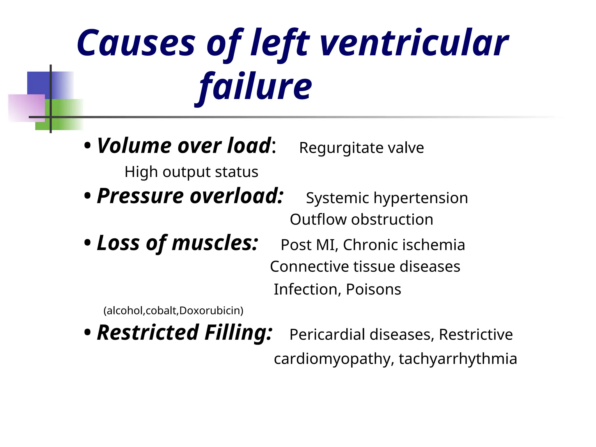
Causes of leftventricular
failure
• Volume over load: Regurgitate valve
High output status
• Pressure overload: Systemic hypertension
Outflow obstruction
• Loss of muscles: Post MI, Chronic ischemia
Connective tissue diseases
Infection, Poisons
(alcohol,cobalt,Doxorubicin)
• Restricted Filling: Pericardial diseases, Restrictive
cardiomyopathy, tachyarrhythmia
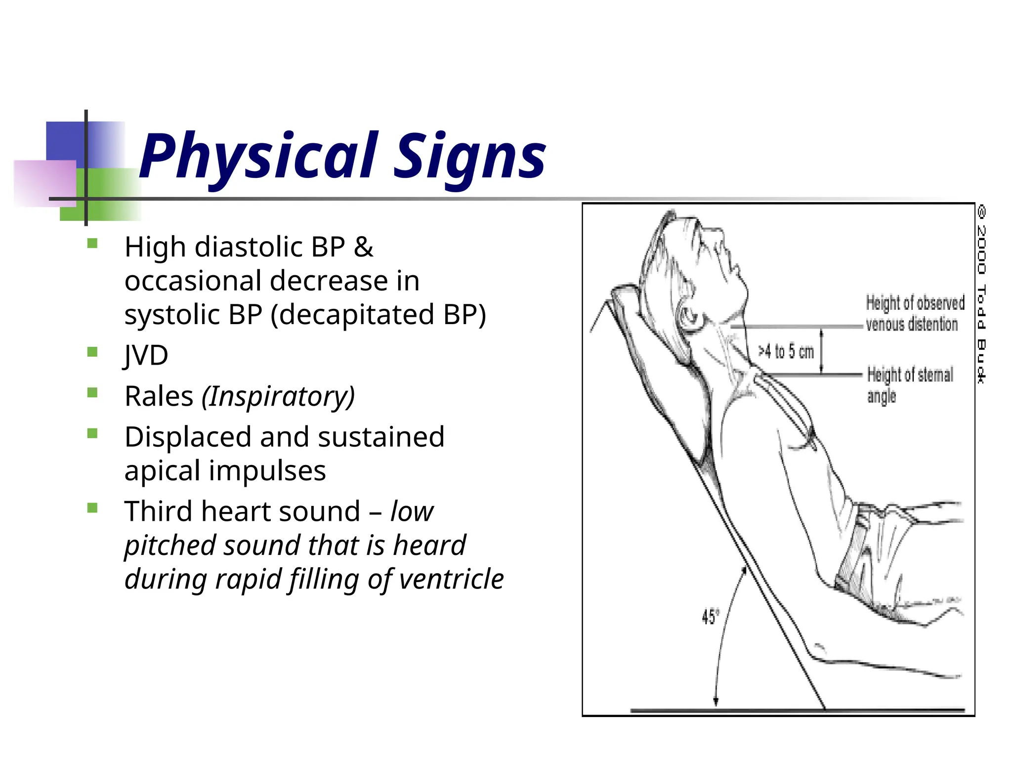
Physical Signs
Highdiastolic BP &
occasional decrease in
systolic BP (decapitated BP)
JVD
Rales (Inspiratory)
Displaced and sustained
apical impulses
Third heart sound – low
pitched sound that is heard
during rapid filling of ventricle
15.
Physical signs (cont.)
Mechanism of S3 sudden deceleration of blood
as elastic limits of the ventricles are
reached
Vibration of the ventricular wall by blood
filling
Common in children
16.
Physical signs (cont.)
Fourth heart Sound (S4)
- Usually at the end of diastole
- Exact mechanism is not known
Could be due to contraction of
atrium against stiff ventricle
Pale, cold sweaty skin
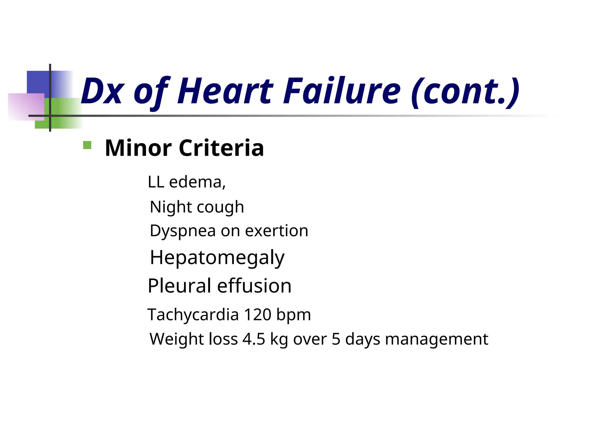
Dx of HeartFailure (cont.)
Minor Criteria
LL edema,
Night cough
Dyspnea on exertion
Hepatomegaly
Pleural effusion
Tachycardia 120 bpm
Weight loss 4.5 kg over 5 days management
19.
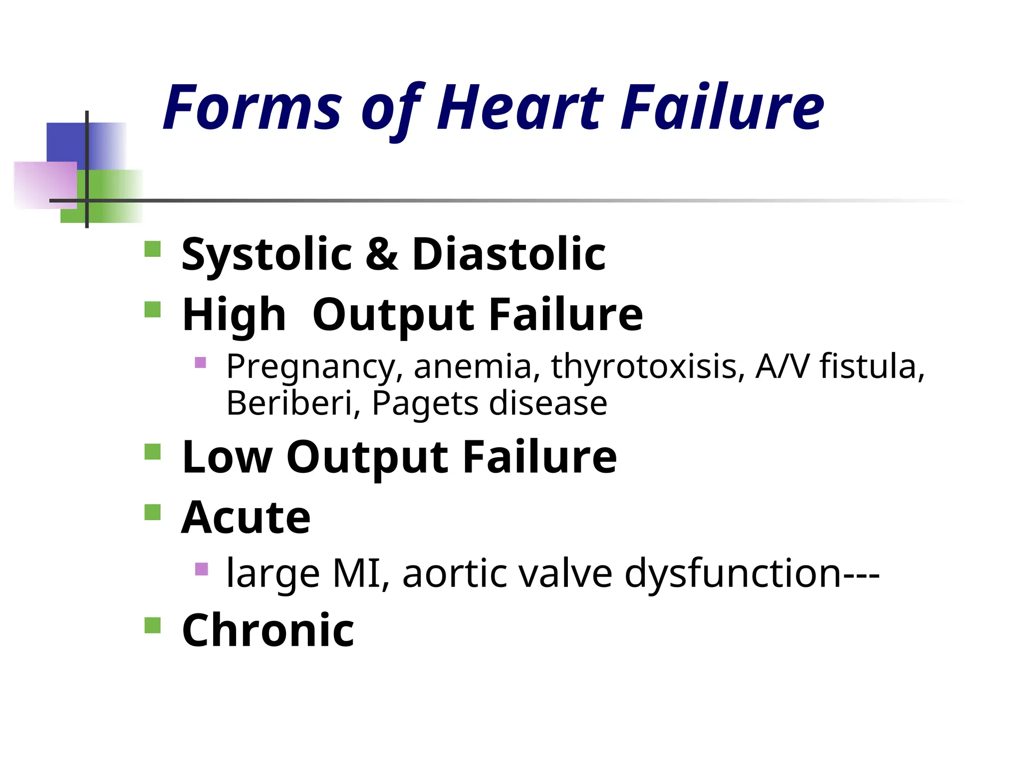
Forms of HeartFailure
Systolic & Diastolic
High Output Failure
Pregnancy, anemia, thyrotoxisis, A/V fistula,
Beriberi, Pagets disease
Low Output Failure
Acute
large MI, aortic valve dysfunction---
Chronic
20.
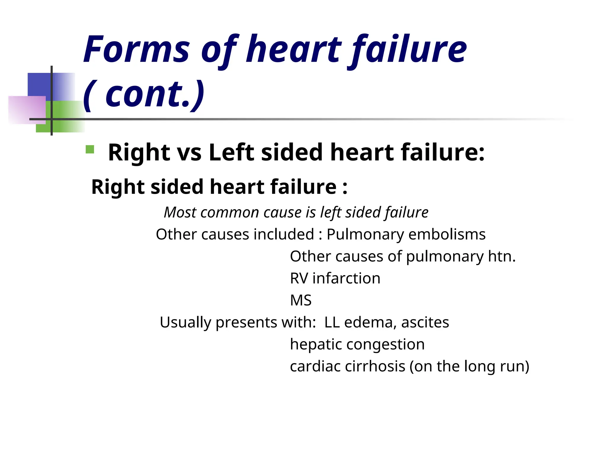
Forms of heartfailure
( cont.)
Right vs Left sided heart failure:
Right sided heart failure :
Most common cause is left sided failure
Other causes included : Pulmonary embolisms
Other causes of pulmonary htn.
RV infarction
MS
Usually presents with: LL edema, ascites
hepatic congestion
cardiac cirrhosis (on the long run)
In conclusion, congestive
heartfailure is often
assumed to be a disease
when in fact it is a syndrome
caused by multiple
disorders.
28.
TREATMENT
Correction ofreversible causes
Ischemia
Valvular heart disease
Thyrotoxicosis and other high output status
Shunts
Arrhythmia
A fib, flutter, PJRT
Medications
Ca channel blockers, some antiarrhythmics
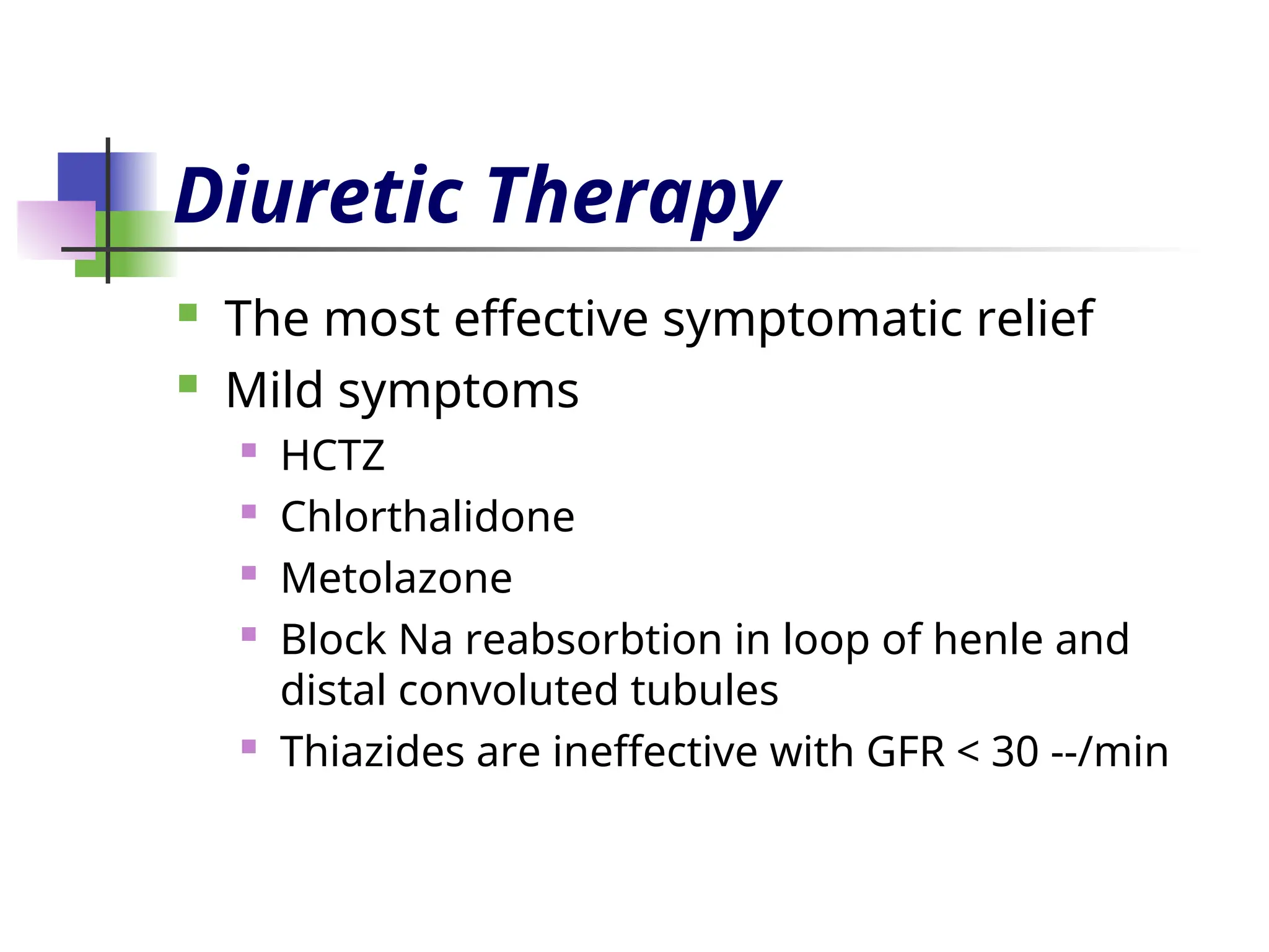
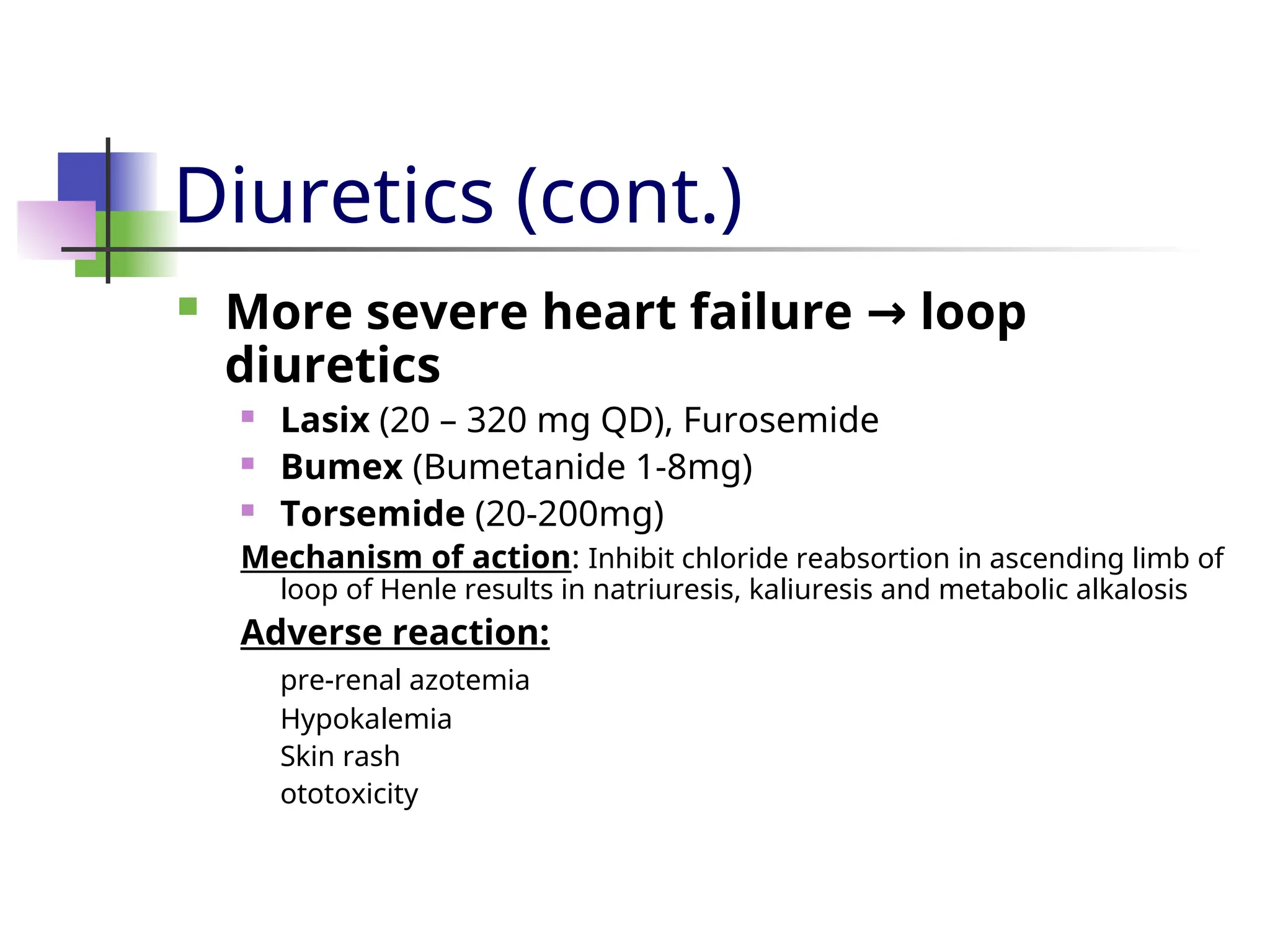
Diuretics (cont.)
Moresevere heart failure loop
→
diuretics
Lasix (20 – 320 mg QD), Furosemide
Bumex (Bumetanide 1-8mg)
Torsemide (20-200mg)
Mechanism of action: Inhibit chloride reabsortion in ascending limb of
loop of Henle results in natriuresis, kaliuresis and metabolic alkalosis
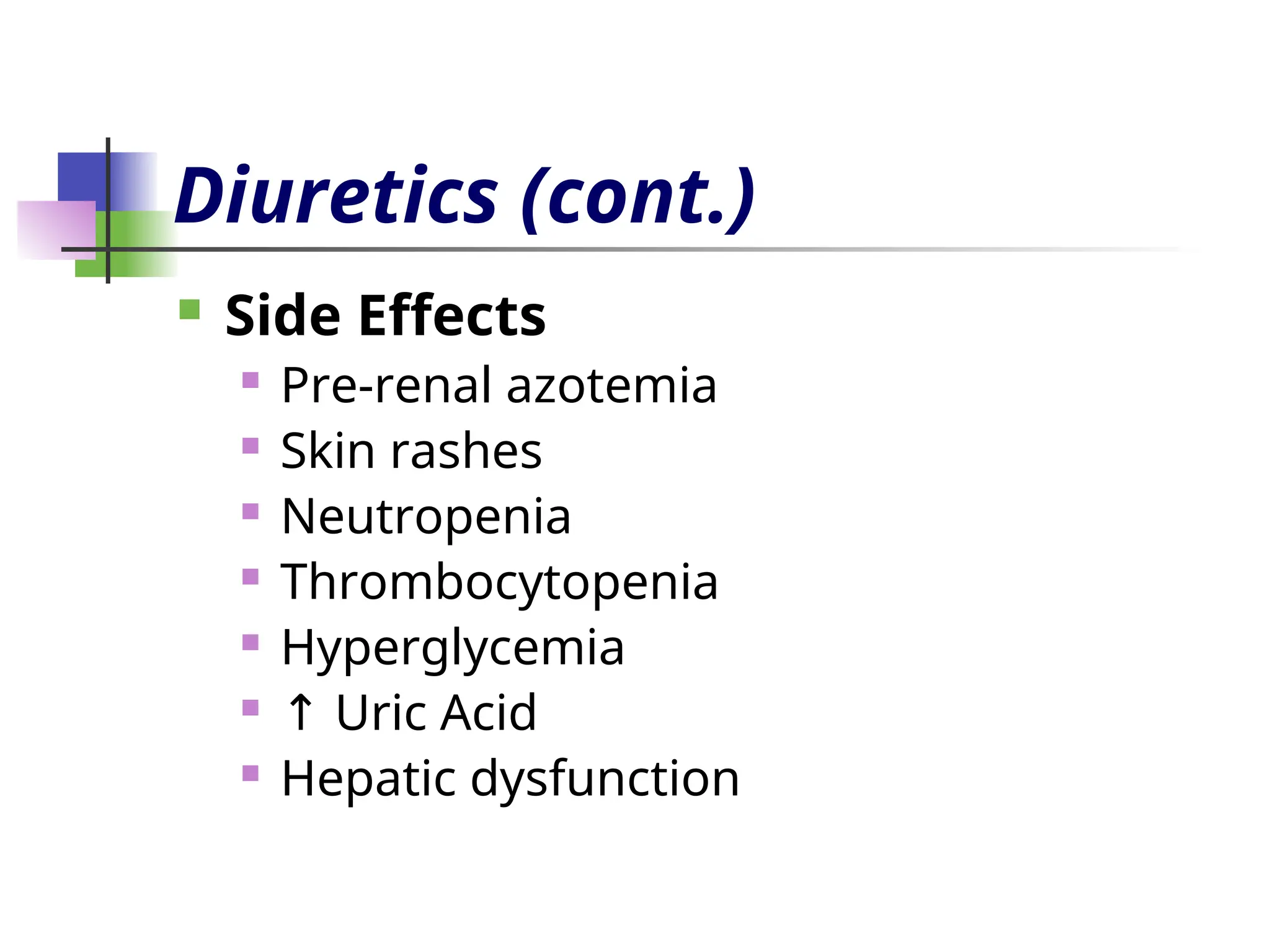
Adverse reaction:
pre-renal azotemia
Hypokalemia
Skin rash
ototoxicity
33.
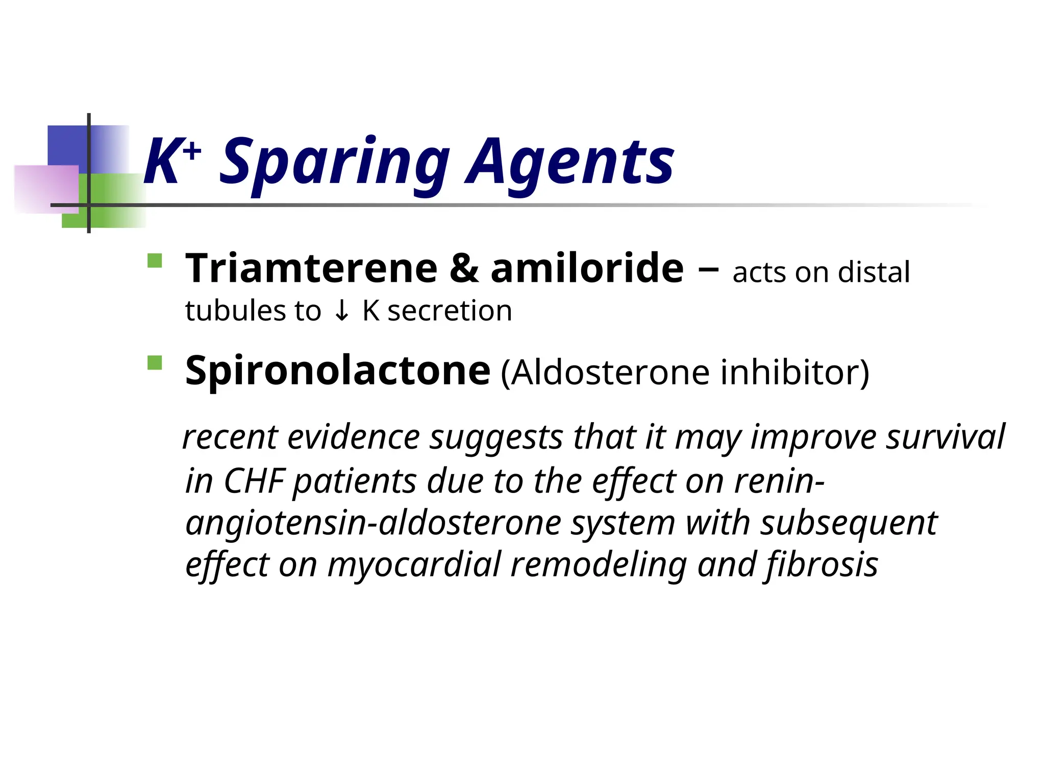
K+
Sparing Agents
Triamterene &amiloride – acts on distal
tubules to K secretion
↓
Spironolactone (Aldosterone inhibitor)
recent evidence suggests that it may improve survival
in CHF patients due to the effect on renin-
angiotensin-aldosterone system with subsequent
effect on myocardial remodeling and fibrosis
34.
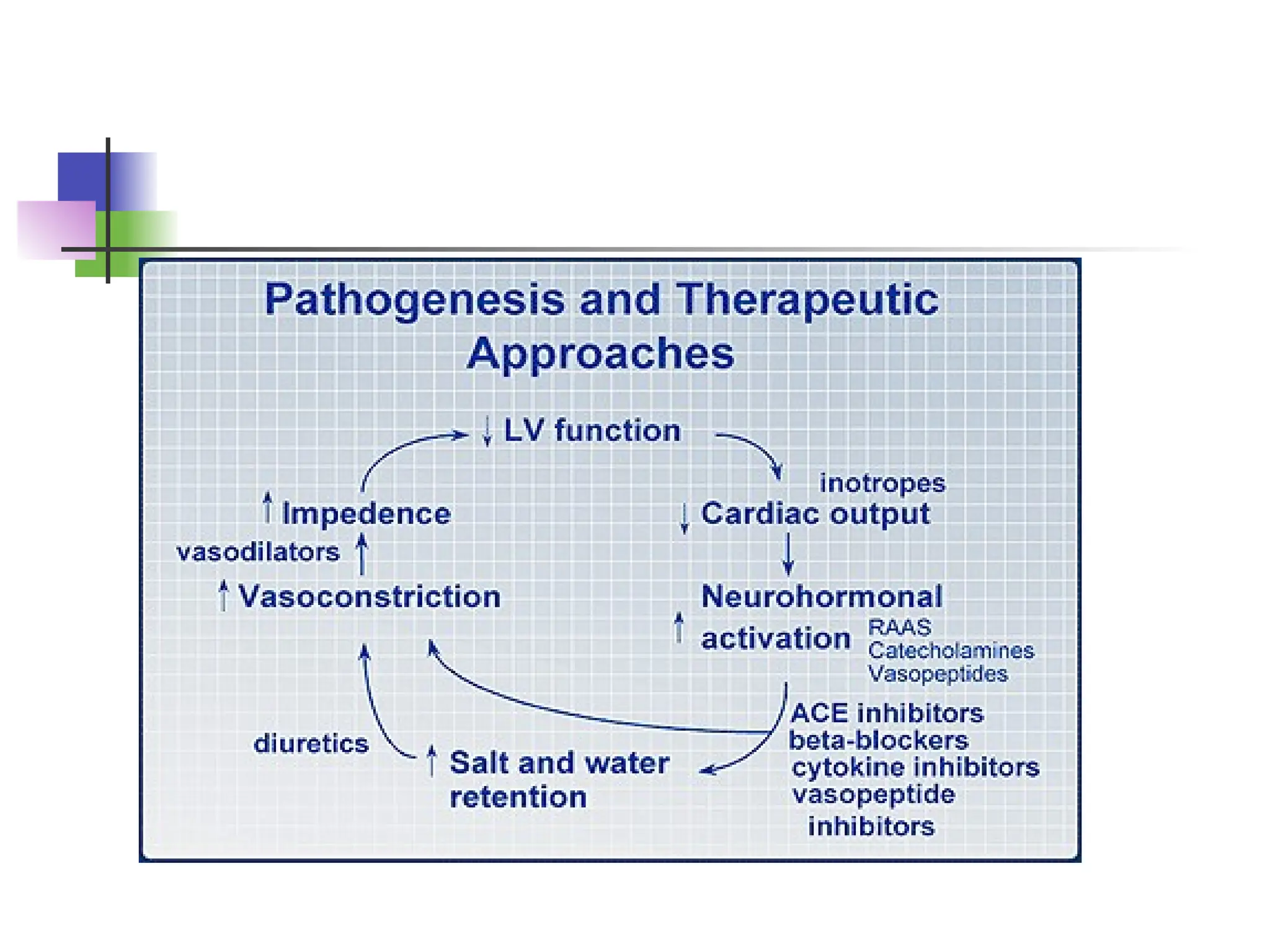
Inhibitors of renin-angiotensin-
aldosteronesystem
Renin-angiotensin-aldosterone system is
activation early in the course of heart failure and
plays an important role in the progression of the
syndrome
Angiotensin converting enzyme
inhibitors
Angiotensin receptors blockers
Spironolactone
35.
Angiotensin Converting
Enzyme Inhibitors
They block the R-A-A system by inhibiting the
conversion of angiotensin I to angiotensin II
vasodilation and Na retention
→ ↓
↓ Bradykinin degradation its
↑ level PG
→ ↑
secretion & nitric oxide
Ace Inhibitors were found to improve
survival in CHF patients
Delay onset & progression of HF in pts with
asymptomatic LV dysfunction
↓ cardiac remodeling
Angiotensin II receptor
blockers
Has comparable effect to ACE I
Can be used in certain conditions when
ACE I are contraindicated (angioneurotic
edema, cough)
38.
Digitalis Glycosides
(Digoxin, Digitoxin)
The role of digitalis has declined somewhat
because of safety concern
Recent studies have shown that digitals
does not affect mortality in CHF patients
but causes significant
Reduction in hospitalization
Reduction in symptoms of HF
39.
Digitalis (cont.)
Mechanism ofAction
+ve inotropic effect by intracellular Ca &
↑
enhancing actin-myosin cross bride
formation (binds to the Na-K ATPase →
inhibits Na pump intracellular Na
→ ↑ → ↑
Na-Ca exchange
Vagotonic effect
Arrhythmogenic effect
40.
Digitalis Toxicity
Narrowtherapeutic to toxic ratio
Non cardiac manifestations
Anorexia,
Nausea, vomiting,
Headache,
Xanthopsia sotoma,
Disorientation
41.
Digitalis Toxicity
Cardiacmanifestations
Sinus bradycardia and arrest
A/V block (usually 2nd
degree)
Atrial tachycardia with A/V Block
Development of junctional rhythm in patients
with a fib
PVC’s, VT/ V fib (bi-directional VT)
42.
Digitalis Toxicity
Treatment
Holdthe medications
Observation
In case of A/V block or severe bradycardia →
atropine followed by temporary PM if
needed
In life threatening arrhythmia digoxin-
→
specific fab antibodies
Lidocaine and phenytoin could be used – try
to avoid D/C cardioversion in non life
threatening arrhythmia
43.
β Blockers
Hasbeen traditionally contraindicated in pts
with CHF
Now they are the main stay in treatment on
CHF & may be the only medication that
shows substantial improvement in LV
function
In addition to improved LV function multiple
studies show improved survival
The only contraindication is severe
decompensated CHF
44.
Vasodilators
Reduction ofafterload by arteriolar
vasodilatation (hydralazin) reduce LVEDP,
O2 consumption,improve myocardial perfusion,
stroke volume and COP
Reduction of preload By venous dilation
( Nitrate) the venous return
↓ the load on
↓
both ventricles.
Usually the maximum benefit is achieved
by using agents with both action.
45.
Positive inotropic agents
These are the drugs that improve myocardial
contractility (β adrenergic agonists, dopaminergic
agents, phosphodiesterase inhibitors),
dopamine, dobutamine, milrinone, amrinone
Several studies showed mortality with oral
↑
inotropic agents
So the only use for them now is in acute
sittings as cardiogenic shock
Antiarrhythmics
Most commoncause of SCD in these
patients is ventricular tachyarrhythmia
Patients with h/o sustained VT or SCD →
ICD implant
48.
Antiarrhythmics (cont.)
Patientswith non sustained ventricular
tachycardia
Correction of electrolytes and acid base
imbalance
In patients with ischemic cardiomyopathy →
ICD implant is the option after r/o acute
ischemia as the cause
In patients wit non ischemic cardiomyopathy
management is ICD implantation
49.
New Methods
Implantableventricular assist devices
Biventricular pacing (only in patient
with LBBB & CHF)
Artificial Heart
50.
Cardiac Transplant
Ithas become more widely used since the
advances in immunosuppressive treatment
Survival rate
1 year 80% - 90%
5 years 70%
51.
Prognosis
Annual mortalityrate depends on patients
symptoms and LV function
5% in patients with mild symptoms and
mild in LV function
↓
30% to 50% in patient with advances LV
dysfunction and severe symptoms
40% – 50% of death is due to SCD


![Apporach to lung biopsy [Auto-saved].pptx latest](https://cdn.slidesharecdn.com/ss_thumbnails/apporachtolungbiopsyauto-saved-251211225655-93258539-thumbnail.jpg?width=640&height=640&fit=bounds)


