2. * The outer ear includes an outer projection and a canal ending at a
membrane.
* The middle ear is an air space containing three small bones.
* The inner ear is the most complex and contains the sensory receptors
for hearing and equilibrium.
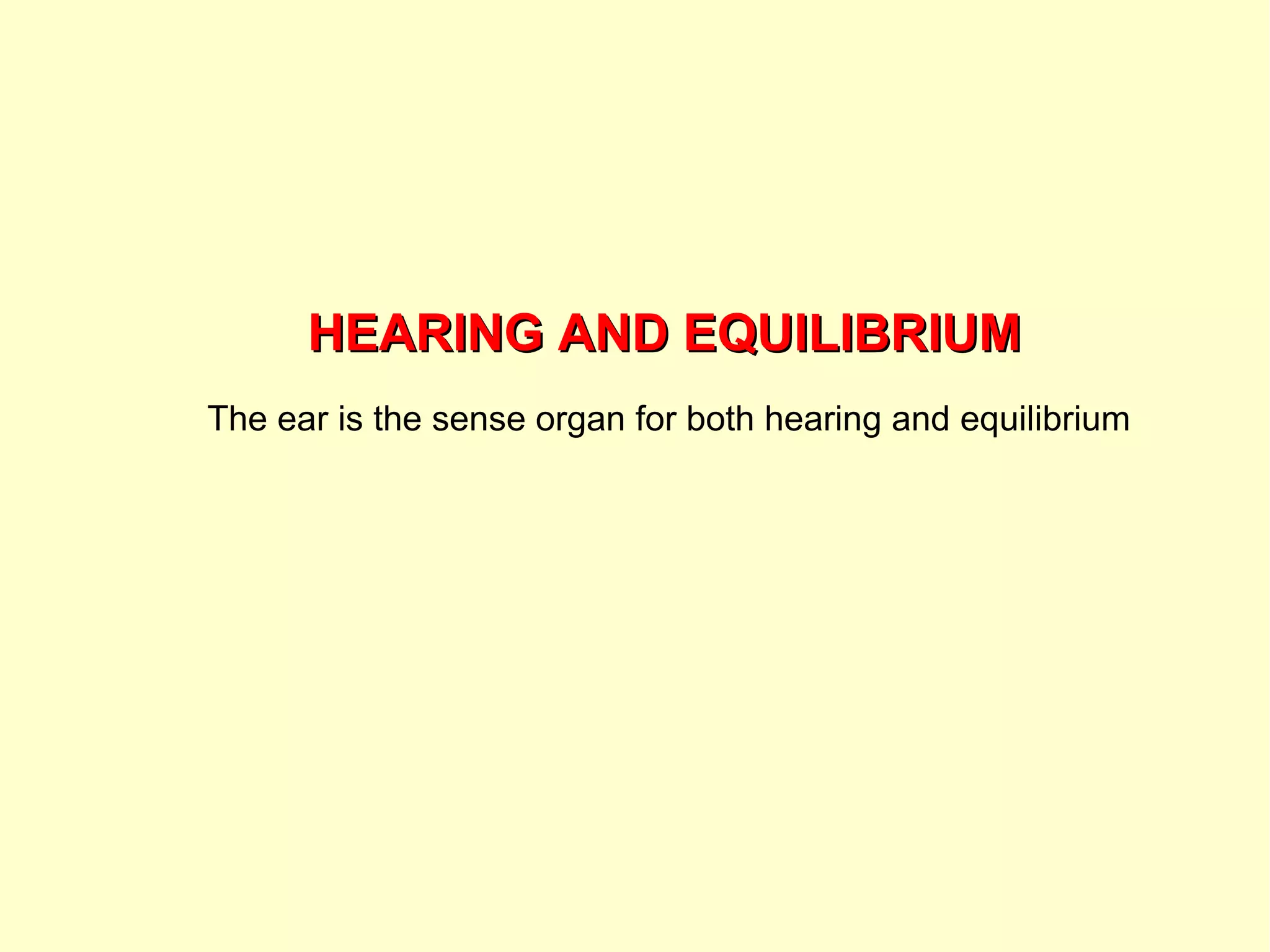
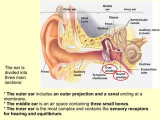
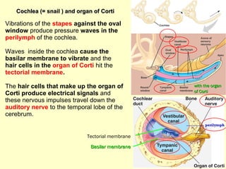
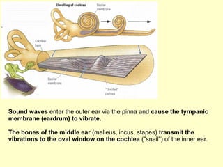
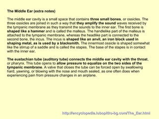
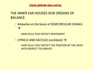
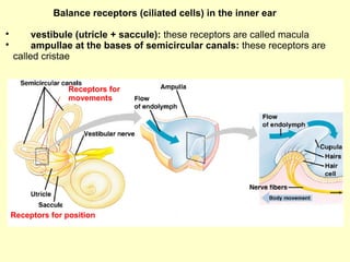
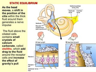
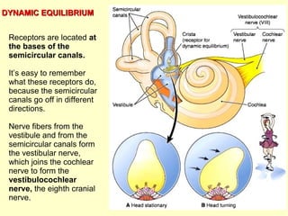
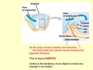
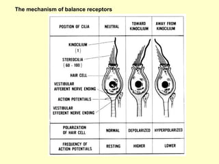
The ear is
divided into
three main
sections:
3. THE EAR CONVERTS AIR PRESSURE WAVES
INTO ELECTRICAL SIGNALS THAT BRAIN
PERCEIVES AS SOUND
1.- OUTER EAR collects
soundwaves to eardrum
PINNA “ear”; flap-like
structure
AUDITORY CANAL
2.- EARDRUM sheet of
tissue that separates outer ear
from middle ear
3.- MIDDLE EAR
Eardrum; HAMMER, ANVIL,
STIRRUP, OVAL WINDOW
4.- INNER EAR. Contains
COCHLEA the actual
hearing organ
THE SENSE OFTHE SENSE OF
HEARINGHEARING
4. Cochlea (= snail ) and organ of CortiCochlea (= snail ) and organ of Corti
Vibrations of the stapes against the oval
window produce pressure waves in the
perilymph of the cochlea.
Waves inside the cochlea cause the
basilar membrane to vibrate and the
hair cells in the organ of Corti hit the
tectorial membrane.
The hair cells that make up the organ of
Corti produce electrical signals and
these nervous impulses travel down the
auditory nerve to the temporal lobe of the
cerebrum.
Basilar membraneBasilar membrane
with the organwith the organ
of Cortiof Corti
Tectorial membrane
5. Sound waves enter the outer ear via the pinna and cause the tympanic
membrane (eardrum) to vibrate.
The bones of the middle ear (malleus, incus, stapes) transmit the
vibrations to the oval window on the cochlea ("snail") of the inner ear.
6.
7. Lower frequency waves
make the distal basilar
membrane vibrate
Higher frequency waves
make the proximal basilar
membrane vibrate
How can we distinguish between different sounds?
8. http://encyclopedia.lubopitko-bg.com/The_Ear.htmlhttp://encyclopedia.lubopitko-bg.com/The_Ear.html
The Middle Ear (extra notes)
The middle ear cavity is a small space that contains three small bones, or ossicles. The
three ossicles are joined in such a way that they amplify the sound waves received by
the tympanic membrane as they transmit the sounds to the inner ear. The first bone is
shaped like a hammer and is called the malleus. The handlelike part of the malleus is
attached to the tympanic membrane, whereas the headlike part is connected to the
second bone, the incus. The incus is shaped like an anvil, an iron block used in
shaping metal, as is used by a blacksmith. The innermost ossicle is shaped somewhat
like the stirrup of a saddle and is called the stapes. The base of the stapes is in contact
with the inner ear.
The eustachian tube (auditory tube) connects the middle ear cavity with the throat,
or pharynx. This tube opens to allow pressure to equalize on the two sides of the
tympanic membrane. A valve that closes the tube can be forced open by swallowing
hard, yawning, or blowing with the nose and mouth sealed, as one often does when
experiencing pain from pressure changes in an airplane.
9. THE INNER EAR HOUSES OUR ORGANS OF
BALANCE
– Ampullae at the bases of SEMICIRCULAR CANALS
•
HAIR CELLS THAT DETECT MOVEMENT
– UTRICLE AND SACCULE (vestibule)
•
HAIR CELLS THAT DETECT THE POSITION OF THE HEAD
WITH RESPECT TO GRAVITY
EQUILIBRIUM (BALANCE)EQUILIBRIUM (BALANCE)
10. Balance receptors (ciliated cells) in the inner ear
vestibule (utricle + saccule): these receptors are called macula
ampullae at the bases of semicircular canals: these receptors are
called cristae
Receptors for position
Receptors for
movements
11. As the headAs the head
movesmoves, a shift in
the position of the
cilia within the thick
fluid around them
generates a nerve
impulse
The fluid above the
ciliated cells
contains small
crystals of
calcium
carbonate, called
otoliths, which add
drag to the fluid
around the receptor
cells and increase
the effect of
gravity’s pull
STATIC EQUILIBRIUMSTATIC EQUILIBRIUM
12. Receptors are located at
the bases of the
semicircular canals.
It’s easy to remember
what these receptors do,
because the semicircular
canals go off in different
directions.
Nerve fibers from the
vestibule and from the
semicircular canals form
the vestibular nerve,
which joins the cochlear
nerve to form the
vestibulocochlear
nerve, the eighth cranial
nerve.
DYNAMIC EQUILIBRIUMDYNAMIC EQUILIBRIUM
13. As the body moves towards one direction..
… the fluid inside the canals moves towards the
opposite direction.
This is due to INERTIA
Inertia is the tendency of an object to resist any
change in its motion.
15. WHAT CAUSES MOTION SICKNESS?
– CONFLICTING SIGNALS FROM THE EYES AND
INNER EAR’S BALANCE MECHANISM
– SPIN AROUND OVER AND OVER, THEN SUDDENLY
STOP…WHAT HAPPENS?!?!?!?