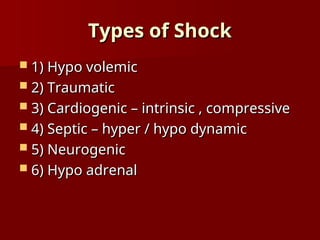
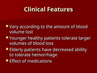
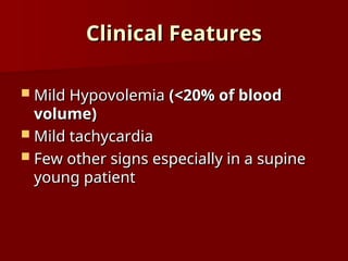
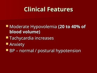
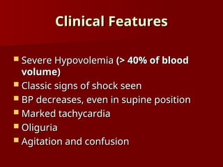
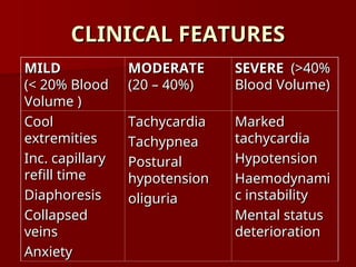
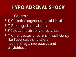
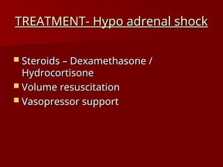
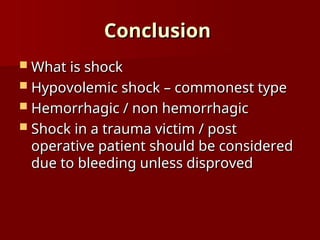
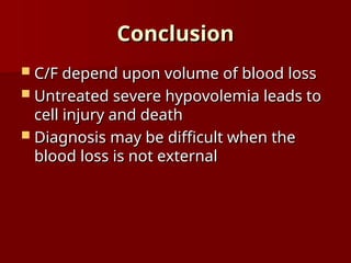
SHOCK
SHOCK
SHOCK
SHOCK
A clinical syndrome that results from
inadequate tissue perfusion
 Hypo perfusion tissue hypoxia
Hypo perfusion tissue hypoxia
 Cellular dysfunction and injury
Cellular dysfunction and injury
 Release of mediators of inflammation
Release of mediators of inflammation
 Further aggravation of hypo perfusion
Further aggravation of hypo perfusion
 Vicious cycle
Vicious cycle
SHOCK
SHOCK
 Clinical manifestations attributable to
Clinical manifestations attributable to
1) sympathetic neuro endocrine responses
2) impaired organ function due to cellular
injury
injury
 Clinical hallmark of shock –
Clinical hallmark of shock –
HYPOTENSION
HYPOTENSION i.e. a
i.e. a MAP < 60 mmhg
MAP < 60 mmhg in
in
previously normotensive patient
Types of Shock
Types of Shock
 1) Hypo volemic
1) Hypo volemic
 2) Traumatic
2) Traumatic
 3) Cardiogenic – intrinsic , compressive
3) Cardiogenic – intrinsic , compressive
 4) Septic – hyper / hypo dynamic
4) Septic – hyper / hypo dynamic
 5) Neurogenic
5) Neurogenic
 6) Hypo adrenal
6) Hypo adrenal
HYPOVOLEMIC SHOCK
HYPOVOLEMIC SHOCK
Introduction
Introduction
 Most common form of shock
Most common form of shock
Caused by:-
Caused by:-
1. Loss of RBC mass and plasma
(hemorrhagic)
(hemorrhagic)
Introduction
Introduction
2. Loss of plasma alone
2. Loss of plasma alone (non
(non
hemorrhagic)
hemorrhagic)
 Extra vascular fluid sequestration
Extra vascular fluid sequestration
 GIT losses
GIT losses
 Urinary losses
Urinary losses
 Insensible losses
Insensible losses
Physiologic response
AIMS:-
AIMS:-
1. Restore effective circulating blood volume
2. Maintain perfusion to the brain and heart
 Increased sympathetic activity
Increased sympathetic activity
- Tachycardia
- Tachycardia
- Tachypnoea
- Tachypnoea
- Collapse of venous capacitance vessels
Physiologic response
 Release of stress hormones
Release of stress hormones
 Expansion of intravascular volume
Expansion of intravascular volume
- Recruitment of interstitial and
intracellular fluid
intracellular fluid
- Reduction in urine output
Clinical Features
Clinical Features
 Vary according to the amount of blood
Vary according to the amount of blood
volume lost
volume lost
 Younger healthy patients tolerate larger
Younger healthy patients tolerate larger
volumes of blood loss
 Elderly patients have decreased ability
Elderly patients have decreased ability
to tolerate hemorrhage
 Effect of medications
Effect of medications
Clinical Features
Clinical Features
 Mild Hypovolemia
Mild Hypovolemia (<20% of blood
(<20% of blood
volume)
volume)
 Mild tachycardia
Mild tachycardia
 Few other signs especially in a supine
Few other signs especially in a supine
young patient
young patient
Clinical Features
Clinical Features
 Moderate Hypovolemia
Moderate Hypovolemia (20 to 40% of
(20 to 40% of
blood volume)
blood volume)
 Tachycardia increases
Tachycardia increases
 Anxiety
Anxiety
 BP – normal / postural hypotension
BP – normal / postural hypotension
Clinical Features
Clinical Features
 Severe Hypovolemia
Severe Hypovolemia (> 40% of blood
(> 40% of blood
volume)
volume)
 Classic signs of shock seen
Classic signs of shock seen
 BP decreases, even in supine position
BP decreases, even in supine position
 Marked tachycardia
Marked tachycardia
 Oliguria
Oliguria
 Agitation and confusion
Agitation and confusion
Clinical Features
Clinical Features
 Perfusion of the CNS is maintained till
Perfusion of the CNS is maintained till
shock becomes severe. Mental
obtundation is an ominous sign
CLINICAL FEATURES
CLINICAL FEATURES
MILD
MILD
(< 20% Blood
(< 20% Blood
Volume )
Volume )
MODERATE
MODERATE
(20 – 40%)
(20 – 40%)
SEVERE
SEVERE (>40%
(>40%
Blood Volume)
Blood Volume)
Cool
Cool
extremities
extremities
Inc. capillary
Inc. capillary
refill time
refill time
Diaphoresis
Diaphoresis
Collapsed
Collapsed
veins
veins
Anxiety
Anxiety
Tachycardia
Tachycardia
Tachypnea
Tachypnea
Postural
Postural
hypotension
hypotension
oliguria
oliguria
Marked
Marked
tachycardia
tachycardia
Hypotension
Hypotension
Haemodynami
Haemodynami
c instability
c instability
Mental status
Mental status
deterioration
deterioration
DIAGNOSIS
DIAGNOSIS
 Easy when source of volume loss is
Easy when source of volume loss is
obvious and there is haemodynamic
instability
instability
 May be difficult when source of volume
May be difficult when source of volume
loss is occult or when plasma volume
alone is depleted
alone is depleted
DIAGNOSIS
DIAGNOSIS
 Shock in a trauma victim or post operative
Shock in a trauma victim or post operative
patient should be presumed to be due to
hemorrhage until proved otherwise
 In Trauma – understanding the patterns of
In Trauma – understanding the patterns of
injury helps to direct the evaluation and
management
management
DIAGNOSIS
DIAGNOSIS
 Penetrating trauma
Penetrating trauma – source of
– source of
bleeding is usually located along the
path of the wounding object.
 Operative intervention is required if
Operative intervention is required if
there are features of shock
DIAGNOSIS
DIAGNOSIS
 Blunt trauma –
Blunt trauma – may have multiple
may have multiple
sources of potential hemorrhage
 Blood loss sufficient to cause shock
Blood loss sufficient to cause shock
usually occurs in the following sites:-
- External
- External
- Intrabdominal – commonest
contd…
contd…
DIAGNOSIS
DIAGNOSIS
- Intrathoracic
- Intrathoracic
- Retroperitoneal
- Retroperitoneal
- extremities
- extremities
DIAGNOSIS
DIAGNOSIS
 The site of bleeding should be looked
The site of bleeding should be looked
for aggressively and treated
 Severe hypovolemia, if untreated, leads
Severe hypovolemia, if untreated, leads
to irreversible cell injury and death
 Time frame available is very narrow
Time frame available is very narrow
DIAGNOSIS
DIAGNOSIS
 Non trauma
Non trauma – usually the site of loss is
– usually the site of loss is
the GIT
the GIT
DIAGNOSIS
DIAGNOSIS
Hypovolemic v/s cardiogenic shock
 Both forms have a similar picture
Both forms have a similar picture
 Presence of raised JVP, rales, S3 gallop
Presence of raised JVP, rales, S3 gallop
rhythm in cardiogenic shock
 Definitive therapy differs significantly
Definitive therapy differs significantly
 Volume expansion is undesirable in
Volume expansion is undesirable in
cardiogenic shock
cardiogenic shock
Treatment
Treatment
 Initial treatment is empiric
Initial treatment is empiric
 Instituted concurrently along with
Instituted concurrently along with
diagnostic evaluation
 Time is of essence
Time is of essence
 Aims:-
Aims:-
Rapid re-expansion
Rapid re-expansion
Control ongoing losses
Treatment
Treatment
1.
1. Volume Resuscitation
Volume Resuscitation
 Place large bore IV lines
Place large bore IV lines
 Rapid infusion of Normal Saline /
Rapid infusion of Normal Saline /
Ringer’s lactate solution
 No distinct benefit demonstrated from
No distinct benefit demonstrated from
use of colloids
use of colloids
Treatment
Treatment
 Infusion of 2 to 3 liters should restore
Infusion of 2 to 3 liters should restore
normal haemodynamic parameters, if the
bleeding has stopped
 If haemodynamic instability persists
If haemodynamic instability persists
? Ongoing losses / inadequate replacement
Treatment
Treatment
Inadequate replacement
 Blood transfusion – if Hb < 10gm% with
Blood transfusion – if Hb < 10gm% with
continuing blood loss
 Fully cross matched blood preferred
Fully cross matched blood preferred
 In extreme emergencies, type specific
In extreme emergencies, type specific
or ‘O’ negative packed red cells may be
given
given
Treatment
Treatment
Ongoing losses
Ongoing losses
–
– prompt surgical intervention required
prompt surgical intervention required
to control persistent bleeding, usually
from a large vessel.
Treatment
Treatment
 Patients who respond to initial
Patients who respond to initial
resuscitation but then deteriorate also
have injuries that require control by
surgery or interventional radiology
Treatment
Treatment
 Persistent haemodynamic instability
Persistent haemodynamic instability
despite control of bleeding and
restoration of intravascular volume
 Presence of the trauma triad of
Presence of the trauma triad of
hypothermia, acidosis and
coagulopathy
coagulopathy
 Mortality inevitable
Mortality inevitable
Treatment
Treatment
2. Ionotropic support
 Dopamine, vasopressin and
Dopamine, vasopressin and
dobutamine
dobutamine
 To maintain adequate ventricular
To maintain adequate ventricular
performance, after blood volume has
been replaced
been replaced
Treatment
Treatment
3. Support of respiratory function
 Also required for successful
Also required for successful
resuscitation
resuscitation
 Supplemental oxygen
Supplemental oxygen
 Endotracheal intubation, if required
Endotracheal intubation, if required
Treatment
Treatment
 4)
4) Adjunctive measures
Adjunctive measures
Positioning
Positioning
PASG and MAST
PASG and MAST
Re warming
Re warming
Treatment
Treatment
 Recovery after resuscitation from
Recovery after resuscitation from
hemorrhagic shock is usually better
than following Septic or traumatic shock
– because of less severe activation of
the inflammatory mediator response
system
system
TRAUMATIC SHOCK
TRAUMATIC SHOCK
TRAUMATIC SHOCK
TRAUMATIC SHOCK
 Caused by bleeding and loss of plasma
Caused by bleeding and loss of plasma
in the injured tissues
 In addition –
In addition – injury induced
injury induced
inflammatory responses
inflammatory responses contribute to
contribute to
secondary micro circulatory injury
 Mal distribution of blood flow
Mal distribution of blood flow
 Tissue hypoxia and MOSF
Tissue hypoxia and MOSF
Traumatic Shock - Treatment
 A – ensure airway
A – ensure airway
 B – breathing and adequate ventilation
B – breathing and adequate ventilation
 C – restore blood volume, support
C – restore blood volume, support
circulation
circulation
 Control haemorrhage
Control haemorrhage
 Reduce inflammatory response by
Reduce inflammatory response by
fracture stabilization, debridement and
haematoma evacuation
HYPO ADRENAL SHOCK
HYPO ADRENAL SHOCK
HYPO ADRENAL SHOCK
HYPO ADRENAL SHOCK
Causes
Causes :-
:-
 1) Chronic exogenous steroid intake
1) Chronic exogenous steroid intake
 2) Prolonged critical state
2) Prolonged critical state
 3) Idiopathic atrophy of adrenals
3) Idiopathic atrophy of adrenals
 4) other causes of adrenal insufficiency
4) other causes of adrenal insufficiency
like Tuberculosis , bilateral
haemorrhage, metastases and
amyloidosis
amyloidosis
TREATMENT- Hypo adrenal shock
 Steroids – Dexamethasone /
Steroids – Dexamethasone /
Hydrocortisone
Hydrocortisone
 Volume resuscitation
Volume resuscitation
 Vasopressor support
Vasopressor support
Conclusion
Conclusion
 What is shock
What is shock
 Hypovolemic shock – commonest type
Hypovolemic shock – commonest type
 Hemorrhagic / non hemorrhagic
Hemorrhagic / non hemorrhagic
 Shock in a trauma victim / post
Shock in a trauma victim / post
operative patient should be considered
due to bleeding unless disproved
Conclusion
Conclusion
 C/F depend upon volume of blood loss
C/F depend upon volume of blood loss
 Untreated severe hypovolemia leads to
Untreated severe hypovolemia leads to
cell injury and death
 Diagnosis may be difficult when the
Diagnosis may be difficult when the
blood loss is not external
Conclusion
Conclusion
 The pattern of injury may help to
The pattern of injury may help to
discover site of blood loss
 Therapy aims at re-expansion of
Therapy aims at re-expansion of
intravascular volume and control of
ongoing losses, along with supportive
measures
measures

SHOCK PRESENTATION PPT EASY WAY TO UNDERSTAND

  • 1.
  • 2.
    SHOCK SHOCK A clinical syndromethat results from A clinical syndrome that results from inadequate tissue perfusion inadequate tissue perfusion  Hypo perfusion tissue hypoxia Hypo perfusion tissue hypoxia  Cellular dysfunction and injury Cellular dysfunction and injury  Release of mediators of inflammation Release of mediators of inflammation  Further aggravation of hypo perfusion Further aggravation of hypo perfusion  Vicious cycle Vicious cycle
  • 3.
    SHOCK SHOCK  Clinical manifestationsattributable to Clinical manifestations attributable to 1) sympathetic neuro endocrine responses 1) sympathetic neuro endocrine responses 2) impaired organ function due to cellular 2) impaired organ function due to cellular injury injury  Clinical hallmark of shock – Clinical hallmark of shock – HYPOTENSION HYPOTENSION i.e. a i.e. a MAP < 60 mmhg MAP < 60 mmhg in in previously normotensive patient previously normotensive patient
  • 4.
    Types of Shock Typesof Shock  1) Hypo volemic 1) Hypo volemic  2) Traumatic 2) Traumatic  3) Cardiogenic – intrinsic , compressive 3) Cardiogenic – intrinsic , compressive  4) Septic – hyper / hypo dynamic 4) Septic – hyper / hypo dynamic  5) Neurogenic 5) Neurogenic  6) Hypo adrenal 6) Hypo adrenal
  • 5.
  • 6.
    Introduction Introduction  Most commonform of shock Most common form of shock Caused by:- Caused by:- 1. Loss of RBC mass and plasma 1. Loss of RBC mass and plasma (hemorrhagic) (hemorrhagic)
  • 7.
    Introduction Introduction 2. Loss ofplasma alone 2. Loss of plasma alone (non (non hemorrhagic) hemorrhagic)  Extra vascular fluid sequestration Extra vascular fluid sequestration  GIT losses GIT losses  Urinary losses Urinary losses  Insensible losses Insensible losses
  • 8.
    Physiologic response Physiologic response AIMS:- AIMS:- 1.Restore effective circulating blood volume 1. Restore effective circulating blood volume 2. Maintain perfusion to the brain and heart 2. Maintain perfusion to the brain and heart  Increased sympathetic activity Increased sympathetic activity - Tachycardia - Tachycardia - Tachypnoea - Tachypnoea - Collapse of venous capacitance vessels - Collapse of venous capacitance vessels
  • 9.
    Physiologic response Physiologic response Release of stress hormones Release of stress hormones  Expansion of intravascular volume Expansion of intravascular volume - Recruitment of interstitial and - Recruitment of interstitial and intracellular fluid intracellular fluid - Reduction in urine output - Reduction in urine output
  • 10.
    Clinical Features Clinical Features Vary according to the amount of blood Vary according to the amount of blood volume lost volume lost  Younger healthy patients tolerate larger Younger healthy patients tolerate larger volumes of blood loss volumes of blood loss  Elderly patients have decreased ability Elderly patients have decreased ability to tolerate hemorrhage to tolerate hemorrhage  Effect of medications Effect of medications
  • 11.
    Clinical Features Clinical Features Mild Hypovolemia Mild Hypovolemia (<20% of blood (<20% of blood volume) volume)  Mild tachycardia Mild tachycardia  Few other signs especially in a supine Few other signs especially in a supine young patient young patient
  • 12.
    Clinical Features Clinical Features Moderate Hypovolemia Moderate Hypovolemia (20 to 40% of (20 to 40% of blood volume) blood volume)  Tachycardia increases Tachycardia increases  Anxiety Anxiety  BP – normal / postural hypotension BP – normal / postural hypotension
  • 13.
    Clinical Features Clinical Features Severe Hypovolemia Severe Hypovolemia (> 40% of blood (> 40% of blood volume) volume)  Classic signs of shock seen Classic signs of shock seen  BP decreases, even in supine position BP decreases, even in supine position  Marked tachycardia Marked tachycardia  Oliguria Oliguria  Agitation and confusion Agitation and confusion
  • 14.
    Clinical Features Clinical Features Perfusion of the CNS is maintained till Perfusion of the CNS is maintained till shock becomes severe. Mental shock becomes severe. Mental obtundation is an ominous sign obtundation is an ominous sign
  • 15.
    CLINICAL FEATURES CLINICAL FEATURES MILD MILD (<20% Blood (< 20% Blood Volume ) Volume ) MODERATE MODERATE (20 – 40%) (20 – 40%) SEVERE SEVERE (>40% (>40% Blood Volume) Blood Volume) Cool Cool extremities extremities Inc. capillary Inc. capillary refill time refill time Diaphoresis Diaphoresis Collapsed Collapsed veins veins Anxiety Anxiety Tachycardia Tachycardia Tachypnea Tachypnea Postural Postural hypotension hypotension oliguria oliguria Marked Marked tachycardia tachycardia Hypotension Hypotension Haemodynami Haemodynami c instability c instability Mental status Mental status deterioration deterioration
  • 16.
    DIAGNOSIS DIAGNOSIS  Easy whensource of volume loss is Easy when source of volume loss is obvious and there is haemodynamic obvious and there is haemodynamic instability instability  May be difficult when source of volume May be difficult when source of volume loss is occult or when plasma volume loss is occult or when plasma volume alone is depleted alone is depleted
  • 17.
    DIAGNOSIS DIAGNOSIS  Shock ina trauma victim or post operative Shock in a trauma victim or post operative patient should be presumed to be due to patient should be presumed to be due to hemorrhage until proved otherwise hemorrhage until proved otherwise  In Trauma – understanding the patterns of In Trauma – understanding the patterns of injury helps to direct the evaluation and injury helps to direct the evaluation and management management
  • 18.
    DIAGNOSIS DIAGNOSIS  Penetrating trauma Penetratingtrauma – source of – source of bleeding is usually located along the bleeding is usually located along the path of the wounding object. path of the wounding object.  Operative intervention is required if Operative intervention is required if there are features of shock there are features of shock
  • 19.
    DIAGNOSIS DIAGNOSIS  Blunt trauma– Blunt trauma – may have multiple may have multiple sources of potential hemorrhage sources of potential hemorrhage  Blood loss sufficient to cause shock Blood loss sufficient to cause shock usually occurs in the following sites:- usually occurs in the following sites:- - External - External - Intrabdominal – commonest - Intrabdominal – commonest contd… contd…
  • 20.
    DIAGNOSIS DIAGNOSIS - Intrathoracic - Intrathoracic -Retroperitoneal - Retroperitoneal - extremities - extremities
  • 21.
    DIAGNOSIS DIAGNOSIS  The siteof bleeding should be looked The site of bleeding should be looked for aggressively and treated for aggressively and treated  Severe hypovolemia, if untreated, leads Severe hypovolemia, if untreated, leads to irreversible cell injury and death to irreversible cell injury and death  Time frame available is very narrow Time frame available is very narrow
  • 22.
    DIAGNOSIS DIAGNOSIS  Non trauma Nontrauma – usually the site of loss is – usually the site of loss is the GIT the GIT
  • 23.
    DIAGNOSIS DIAGNOSIS Hypovolemic v/s cardiogenicshock Hypovolemic v/s cardiogenic shock  Both forms have a similar picture Both forms have a similar picture  Presence of raised JVP, rales, S3 gallop Presence of raised JVP, rales, S3 gallop rhythm in cardiogenic shock rhythm in cardiogenic shock  Definitive therapy differs significantly Definitive therapy differs significantly  Volume expansion is undesirable in Volume expansion is undesirable in cardiogenic shock cardiogenic shock
  • 24.
    Treatment Treatment  Initial treatmentis empiric Initial treatment is empiric  Instituted concurrently along with Instituted concurrently along with diagnostic evaluation diagnostic evaluation  Time is of essence Time is of essence  Aims:- Aims:- Rapid re-expansion Rapid re-expansion Control ongoing losses Control ongoing losses
  • 25.
    Treatment Treatment 1. 1. Volume Resuscitation VolumeResuscitation  Place large bore IV lines Place large bore IV lines  Rapid infusion of Normal Saline / Rapid infusion of Normal Saline / Ringer’s lactate solution Ringer’s lactate solution  No distinct benefit demonstrated from No distinct benefit demonstrated from use of colloids use of colloids
  • 26.
    Treatment Treatment  Infusion of2 to 3 liters should restore Infusion of 2 to 3 liters should restore normal haemodynamic parameters, if the normal haemodynamic parameters, if the bleeding has stopped bleeding has stopped  If haemodynamic instability persists If haemodynamic instability persists ? Ongoing losses / inadequate replacement ? Ongoing losses / inadequate replacement
  • 27.
    Treatment Treatment Inadequate replacement Inadequate replacement Blood transfusion – if Hb < 10gm% with Blood transfusion – if Hb < 10gm% with continuing blood loss continuing blood loss  Fully cross matched blood preferred Fully cross matched blood preferred  In extreme emergencies, type specific In extreme emergencies, type specific or ‘O’ negative packed red cells may be or ‘O’ negative packed red cells may be given given
  • 28.
    Treatment Treatment Ongoing losses Ongoing losses – –prompt surgical intervention required prompt surgical intervention required to control persistent bleeding, usually to control persistent bleeding, usually from a large vessel. from a large vessel.
  • 29.
    Treatment Treatment  Patients whorespond to initial Patients who respond to initial resuscitation but then deteriorate also resuscitation but then deteriorate also have injuries that require control by have injuries that require control by surgery or interventional radiology surgery or interventional radiology
  • 30.
    Treatment Treatment  Persistent haemodynamicinstability Persistent haemodynamic instability despite control of bleeding and despite control of bleeding and restoration of intravascular volume restoration of intravascular volume  Presence of the trauma triad of Presence of the trauma triad of hypothermia, acidosis and hypothermia, acidosis and coagulopathy coagulopathy  Mortality inevitable Mortality inevitable
  • 31.
    Treatment Treatment 2. Ionotropic support 2.Ionotropic support  Dopamine, vasopressin and Dopamine, vasopressin and dobutamine dobutamine  To maintain adequate ventricular To maintain adequate ventricular performance, after blood volume has performance, after blood volume has been replaced been replaced
  • 32.
    Treatment Treatment 3. Support ofrespiratory function 3. Support of respiratory function  Also required for successful Also required for successful resuscitation resuscitation  Supplemental oxygen Supplemental oxygen  Endotracheal intubation, if required Endotracheal intubation, if required
  • 33.
    Treatment Treatment  4) 4) Adjunctivemeasures Adjunctive measures Positioning Positioning PASG and MAST PASG and MAST Re warming Re warming
  • 34.
    Treatment Treatment  Recovery afterresuscitation from Recovery after resuscitation from hemorrhagic shock is usually better hemorrhagic shock is usually better than following Septic or traumatic shock than following Septic or traumatic shock – because of less severe activation of – because of less severe activation of the inflammatory mediator response the inflammatory mediator response system system
  • 35.
  • 36.
    TRAUMATIC SHOCK TRAUMATIC SHOCK Caused by bleeding and loss of plasma Caused by bleeding and loss of plasma in the injured tissues in the injured tissues  In addition – In addition – injury induced injury induced inflammatory responses inflammatory responses contribute to contribute to secondary micro circulatory injury secondary micro circulatory injury  Mal distribution of blood flow Mal distribution of blood flow  Tissue hypoxia and MOSF Tissue hypoxia and MOSF
  • 37.
    Traumatic Shock -Treatment Traumatic Shock - Treatment  A – ensure airway A – ensure airway  B – breathing and adequate ventilation B – breathing and adequate ventilation  C – restore blood volume, support C – restore blood volume, support circulation circulation  Control haemorrhage Control haemorrhage  Reduce inflammatory response by Reduce inflammatory response by fracture stabilization, debridement and fracture stabilization, debridement and haematoma evacuation haematoma evacuation
  • 38.
  • 39.
    HYPO ADRENAL SHOCK HYPOADRENAL SHOCK Causes Causes :- :-  1) Chronic exogenous steroid intake 1) Chronic exogenous steroid intake  2) Prolonged critical state 2) Prolonged critical state  3) Idiopathic atrophy of adrenals 3) Idiopathic atrophy of adrenals  4) other causes of adrenal insufficiency 4) other causes of adrenal insufficiency like Tuberculosis , bilateral like Tuberculosis , bilateral haemorrhage, metastases and haemorrhage, metastases and amyloidosis amyloidosis
  • 40.
    TREATMENT- Hypo adrenalshock TREATMENT- Hypo adrenal shock  Steroids – Dexamethasone / Steroids – Dexamethasone / Hydrocortisone Hydrocortisone  Volume resuscitation Volume resuscitation  Vasopressor support Vasopressor support
  • 41.
    Conclusion Conclusion  What isshock What is shock  Hypovolemic shock – commonest type Hypovolemic shock – commonest type  Hemorrhagic / non hemorrhagic Hemorrhagic / non hemorrhagic  Shock in a trauma victim / post Shock in a trauma victim / post operative patient should be considered operative patient should be considered due to bleeding unless disproved due to bleeding unless disproved
  • 42.
    Conclusion Conclusion  C/F dependupon volume of blood loss C/F depend upon volume of blood loss  Untreated severe hypovolemia leads to Untreated severe hypovolemia leads to cell injury and death cell injury and death  Diagnosis may be difficult when the Diagnosis may be difficult when the blood loss is not external blood loss is not external
  • 43.
    Conclusion Conclusion  The patternof injury may help to The pattern of injury may help to discover site of blood loss discover site of blood loss  Therapy aims at re-expansion of Therapy aims at re-expansion of intravascular volume and control of intravascular volume and control of ongoing losses, along with supportive ongoing losses, along with supportive measures measures

Editor's Notes

  • #3 MAP – 1/3 of pulse pressure plus diastolic BP