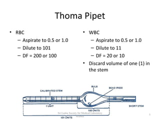
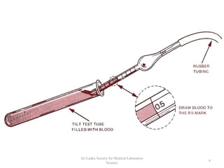
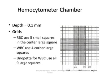
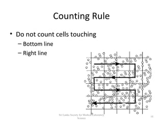
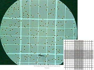
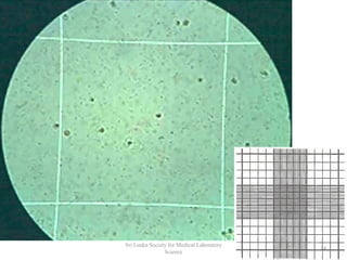
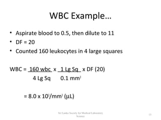
Cell Counting Using a Haemocytometer Equipment & Reagents Haemocytometer plus a supply of cover-slips 0.4% Trypan Blue stain (fresh & filtered) in phosphate buffered saline Tally Counter Cell Suspension Gilson pipettes or similar Inverted microscope (preferably phase contrast) Procedure 1. Ensure the cover-slip and haemocytometer are clean and grease-free (use alcohol to clean). 2. Moisten (with water or exhaled breath) and affix cover-slip to the haemocytometer. 3. Look for "Newton's Rings" which indicate that the cover slip has adhered via suction to the haemocytometer. Newton's refraction rings are seen as rainbow-like rings under the cover-slip. 4. Mix equal volumes of 0.4% trypan blue stain and a well mixed cell suspension (not too vigorous) e.g. mix 100µl trypan blue stain with 100 µl cell suspension. 5. Pipette trypan blue/cell mix (approximately 10µl) at the edge of the cover-slip and allow to run under the cover slip. 6. Visualise the haemocytometer grid under the microscope, refer to figure 1 for layout of grid. Please note: 7. Trypan Blue is a "vital stain"; it is excluded from live cells. 8. Live cells appear colourless and bright (refractile) under phase contrast. 9. Dead cells stain blue and are non-refractile. 10. To aid accuracy and consistency of cell counts use counting system illustrated in figure 2. 11. Count viable (live) and dead cells in one or more large corner squares and record cell counts. 12. It is advisable to count around 40 to 70 cells to obtain an accurate cell count - therefore it may be necessary to count more than one large corner square. 13. To calculate cell concentration per ml: Average number of cells in one large square x dilution factor* x 104 *dilution factor is usually 2 (1:1 dilution with trypan blue), but may need to further dilute (or concentrate) cell suspensions. 104 = conversion factor to convert 10-4ml to 1ml Calculation of Cell Viability No. of Viable Cells Counted / Total Cells Counted (viable and dead) x 100 = % viable cells Ravi Kumudesh / slsmls.org