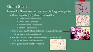
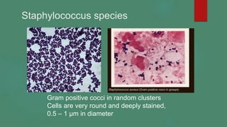
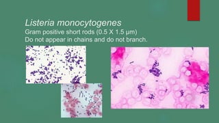
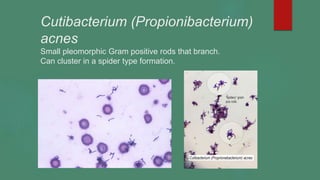
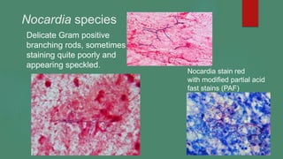
Gram stains 2023
Margie A. Morgan, PhD. D(ABMM)
Gram Stain
Assess for Gram reaction and morphology of organism
 Gram negative (red) /Gram positive (blue)
 Crystal violet – primary stain
 Gram’s iodine – mordant
 Acetone/Alcohol – decolorizer
 Safranin – counterstain
 How to judge quality of gram staining – is everything blue?
 If so, slide is under-decolorized.
 Are the polys and other cells washed out in appearance?
 If so, the slide is over-decolorized
 Poor quality stains must be repeated
Gram Stain: Prepare a thin smear of material onto a glass slide.
Air dry and then heat fix specimen on slide prior to staining.
Stain approximately one minute per step and rinse with sterile
water between each step. Air dry before scope reading.
Presence of large amount of peptidoglycan in the Gram positive cell wall,
traps the crystal violet making for Gram positive staining with the primary
stain crystal violet and the organism appearing blue in color.
Cellular Material –
Gram stains are not able to
differentiate many cell types.
Best able to identify
polymorphonuclear
leukocytes (PMNs),
mononuclear cells, and
epithelial cells. For other cell
types, differential stains must
be used, such as Wright-
Giemsa.
Artifacts in Gram stains
Safranin
crystals
Crystal Violet crystals
When reading a Gram stain, check for the uniformity of the cell borders of
suspected organisms, there should also be repeating/consistent size and
shape of the cells to support identification as a micro-organism. Artifacts
such as stain crystals are usually not consistent in shape or size.
Most common
Gram Positive Bacteria
Staphylococcus species
Gram positive cocci in random clusters
Cells are very round and deeply stained,
0.5 – 1 µm in diameter
Streptococcus / Beta hemolytic
 Gram positive cocci in chains of variable length
 Cocci are 0.5 wide by 2.0 µm in length (oval)
 Beta hemolytic Streptococci tend to form longer
chains than related groups
Streptococcus pneumoniae
 Gram positive cocci in pairs, 0.5 - 1.5 µm in diameter
 Cocci are lancet or bullet shaped with flattened ends together
 Sometimes can visualize a capsule on gram stain – which appears as
clearing around the stained organism
Viridans Streptococcus
 Gram positive cocci in short chains and pairs
 Chains usually shorter than the Beta hemolytic
Streptococcus, oval in shape, 0.5 µm -1.5 µm diameter
Enterococcus species
 Gram positive cocci in short chains and pairs
 Can be difficult to differentiate from the viridans Streptococcus, size
for both organisms is similar 0.5 µm - 1.5 µm diameter
Bacillus species
Gram positive rods – large and rectangular(1.0 X 4.0 µm)
Described as box car shaped. Spores will not stain with gentian violet and
appear as vacant spots in the cell.
Even when stained properly can appear over-decolorized and Gram
negative (reddish)
Gram negative appearing
Spores
Listeria monocytogenes
Gram positive short rods (0.5 X 1.5 µm)
Do not appear in chains and do not branch.
Corynebacterium species
Gram positive rods that cluster in Chinese
letter forms and palisade cell arrangement (0.5 X 4 µm)
Palisade cell
arrangement
Cutibacterium (Propionibacterium)
acnes
Small pleomorphic Gram positive rods that branch.
Can cluster in a spider type formation.
Nocardia species
Delicate Gram positive
branching rods, sometimes
staining quite poorly and
appearing speckled.
Nocardia stain red
with modified partial acid
fast stains (PAF)
Actinomyces species
Gram positive rods with antler like branching, can be seen within
sulfur granules in tissue
Sulfur Granule
Formation
Gardnerella vaginalis
Gram variable (not quite red or blue) rods
Associated with “clue cells” in bacterial vaginosis
Lactobacillus species
Gram positive rods, thin, appear sometimes in short chains
Clostridium species
Anaerobic Gram positive rods, large, with spores sometimes visible.
Spores will not stain with the Gram stain and appear as colorless areas in
the cell.
Spores are located throughout the length of the rod-shaped organism
depending on the species.
Mycobacterium species
Do not stain well with Gram stain due to high amount of
lipid in the cell wall, appear beaded and barely visible.
Acid fast stains are superior for the staining of
Mycobacteria
Gram negative bacteria
Enteric Gram negative rods
(Enterobacterales)
Plump rectangular in shape(1 x 3 µm), stain vividly red with
Gram stain.
Pseudomonas aeruginosa
thinner and a bit longer than the Enterobacterales
0.5-0.8 X 1.5-3µm
Haemophilus influenza
pleomorphic in size and shape, thin and faintly staining
Vibrio species
Curved or comma shaped Gram negative rods
0.5-0.8um X 1-3 um
Fusobacterium species – faintly staining
unusually shaped Gram negative rods
F. nucleatum F. necrophorum
Spindle shaped, with pointed Chaining pleomorphic rods
ends, 0.5-1um X 8-10um
Campylobacter species
Sea gull wing shaped, faintly staining Gram negative rods
0.2-0.5um X 0.5 -5.0um
Capnocytophyga spp –
Pleomorphic Gram negative bacilli, variable in size and shape
Bacteroiodes spp
Pleomorphic Gram negative bacilli, variable in length and
sometimes variable in staining intensity
Yersinia pestis
Safety pin like with bipolar staining
---
(0.5 X 1.5 µm)
Neisseria species (meningitidis and gonorrhoeae)
Gram negative diplococci, 0.6-0.8 X 1.0–3.0 um
Kidney bean shaped and always in pairs, often times intracellular
Yeast – Candida species
 Large Gram positive oval globules (@ 8 um) that produce
a smaller attached cell known as a bud
 Sometimes pseudo-hyphae can be seen which are
extensions of the yeast cells into long hyphae like forms
Cryptococcus neoformans and C. gatti
Due to the polysaccharide capsule of
C. neoformans and C. gatti the yeast
appear somewhat like a round blob
when Gram stained
An India ink prep can be used
to visualize the polysaccharide capsule
Mycelial Fungi
Variable staining of fungal hyphae on Gram stain, not
usually considered a sensitive way to search out fungi.
Calcofluor white is a more sensitive staining method

Gram Stain 2023

  • 1.
    Gram stains 2023 MargieA. Morgan, PhD. D(ABMM)
  • 2.
    Gram Stain Assess forGram reaction and morphology of organism  Gram negative (red) /Gram positive (blue)  Crystal violet – primary stain  Gram’s iodine – mordant  Acetone/Alcohol – decolorizer  Safranin – counterstain  How to judge quality of gram staining – is everything blue?  If so, slide is under-decolorized.  Are the polys and other cells washed out in appearance?  If so, the slide is over-decolorized  Poor quality stains must be repeated
  • 3.
    Gram Stain: Preparea thin smear of material onto a glass slide. Air dry and then heat fix specimen on slide prior to staining. Stain approximately one minute per step and rinse with sterile water between each step. Air dry before scope reading.
  • 4.
    Presence of largeamount of peptidoglycan in the Gram positive cell wall, traps the crystal violet making for Gram positive staining with the primary stain crystal violet and the organism appearing blue in color.
  • 5.
    Cellular Material – Gramstains are not able to differentiate many cell types. Best able to identify polymorphonuclear leukocytes (PMNs), mononuclear cells, and epithelial cells. For other cell types, differential stains must be used, such as Wright- Giemsa.
  • 6.
    Artifacts in Gramstains Safranin crystals Crystal Violet crystals When reading a Gram stain, check for the uniformity of the cell borders of suspected organisms, there should also be repeating/consistent size and shape of the cells to support identification as a micro-organism. Artifacts such as stain crystals are usually not consistent in shape or size.
  • 7.
  • 8.
    Staphylococcus species Gram positivecocci in random clusters Cells are very round and deeply stained, 0.5 – 1 µm in diameter
  • 9.
    Streptococcus / Betahemolytic  Gram positive cocci in chains of variable length  Cocci are 0.5 wide by 2.0 µm in length (oval)  Beta hemolytic Streptococci tend to form longer chains than related groups
  • 10.
    Streptococcus pneumoniae  Grampositive cocci in pairs, 0.5 - 1.5 µm in diameter  Cocci are lancet or bullet shaped with flattened ends together  Sometimes can visualize a capsule on gram stain – which appears as clearing around the stained organism
  • 11.
    Viridans Streptococcus  Grampositive cocci in short chains and pairs  Chains usually shorter than the Beta hemolytic Streptococcus, oval in shape, 0.5 µm -1.5 µm diameter
  • 12.
    Enterococcus species  Grampositive cocci in short chains and pairs  Can be difficult to differentiate from the viridans Streptococcus, size for both organisms is similar 0.5 µm - 1.5 µm diameter
  • 13.
    Bacillus species Gram positiverods – large and rectangular(1.0 X 4.0 µm) Described as box car shaped. Spores will not stain with gentian violet and appear as vacant spots in the cell. Even when stained properly can appear over-decolorized and Gram negative (reddish) Gram negative appearing Spores
  • 14.
    Listeria monocytogenes Gram positiveshort rods (0.5 X 1.5 µm) Do not appear in chains and do not branch.
  • 15.
    Corynebacterium species Gram positiverods that cluster in Chinese letter forms and palisade cell arrangement (0.5 X 4 µm) Palisade cell arrangement
  • 16.
    Cutibacterium (Propionibacterium) acnes Small pleomorphicGram positive rods that branch. Can cluster in a spider type formation.
  • 17.
    Nocardia species Delicate Grampositive branching rods, sometimes staining quite poorly and appearing speckled. Nocardia stain red with modified partial acid fast stains (PAF)
  • 18.
    Actinomyces species Gram positiverods with antler like branching, can be seen within sulfur granules in tissue Sulfur Granule Formation
  • 19.
    Gardnerella vaginalis Gram variable(not quite red or blue) rods Associated with “clue cells” in bacterial vaginosis
  • 20.
    Lactobacillus species Gram positiverods, thin, appear sometimes in short chains
  • 21.
    Clostridium species Anaerobic Grampositive rods, large, with spores sometimes visible. Spores will not stain with the Gram stain and appear as colorless areas in the cell. Spores are located throughout the length of the rod-shaped organism depending on the species.
  • 22.
    Mycobacterium species Do notstain well with Gram stain due to high amount of lipid in the cell wall, appear beaded and barely visible. Acid fast stains are superior for the staining of Mycobacteria
  • 23.
  • 24.
    Enteric Gram negativerods (Enterobacterales) Plump rectangular in shape(1 x 3 µm), stain vividly red with Gram stain.
  • 25.
    Pseudomonas aeruginosa thinner anda bit longer than the Enterobacterales 0.5-0.8 X 1.5-3µm
  • 26.
    Haemophilus influenza pleomorphic insize and shape, thin and faintly staining
  • 27.
    Vibrio species Curved orcomma shaped Gram negative rods 0.5-0.8um X 1-3 um
  • 28.
    Fusobacterium species –faintly staining unusually shaped Gram negative rods F. nucleatum F. necrophorum Spindle shaped, with pointed Chaining pleomorphic rods ends, 0.5-1um X 8-10um
  • 29.
    Campylobacter species Sea gullwing shaped, faintly staining Gram negative rods 0.2-0.5um X 0.5 -5.0um
  • 30.
    Capnocytophyga spp – PleomorphicGram negative bacilli, variable in size and shape
  • 31.
    Bacteroiodes spp Pleomorphic Gramnegative bacilli, variable in length and sometimes variable in staining intensity
  • 32.
    Yersinia pestis Safety pinlike with bipolar staining ---
  • 33.
  • 34.
    Neisseria species (meningitidisand gonorrhoeae) Gram negative diplococci, 0.6-0.8 X 1.0–3.0 um Kidney bean shaped and always in pairs, often times intracellular
  • 35.
    Yeast – Candidaspecies  Large Gram positive oval globules (@ 8 um) that produce a smaller attached cell known as a bud  Sometimes pseudo-hyphae can be seen which are extensions of the yeast cells into long hyphae like forms
  • 36.
    Cryptococcus neoformans andC. gatti Due to the polysaccharide capsule of C. neoformans and C. gatti the yeast appear somewhat like a round blob when Gram stained An India ink prep can be used to visualize the polysaccharide capsule
  • 37.
    Mycelial Fungi Variable stainingof fungal hyphae on Gram stain, not usually considered a sensitive way to search out fungi. Calcofluor white is a more sensitive staining method