Downloaded 119 times


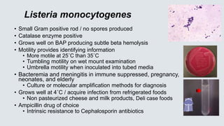
![• Escherichia coli
• Normal flora in human intestine
• #1 cause of UTI [@80% of cases]
• Bacteremia, neonatal meningitis,
and abdominal infections
• MacConkey agar / Lactose fermenter
• Spot indole reaction = positive / turns robin’s egg blue
• Detects breakdown of tryptophan from growth on BAP
• Eosin methylene blue agar (EMB) green sheen produced
• Pathogen of diarrhea:
• Enterotoxigenic (ETEC) E. coli cause of traveler’s diarrhea
• Enterohemorrhagic E. coli (EHEC) (such as 0157:H7)
• Bloody diarrhea acquired from eating undercooked beef
• Pathogenicity from Shiga toxin production
• Hemolytic uremic syndrome (HUS) can result with hemolytic anemia,
thrombocytopenia, and renal failure] particularly in young children
• Culture on MacConkey agar with sorbitol (not lactose) / does NOT ferment
sorbitol/ most all E. coli except EHEC ferment sorbitol
Green sheen on
EMB agar
Indole positive
Lactose
fermentor](https://image.slidesharecdn.com/bacteriologypart2-210219005911/85/Bacteriology-Update-2021-Part-2-11-320.jpg)
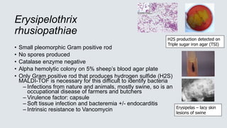
![1. Glu/lac/suc
fermented
with gas
2. Glucose
fermented
3. Glucose
fermented
with H2S
4. No CHO
fermentation
Non fermenter
Triple Sugar Iron Agar (TSI)– Used to detect fermentation of glucose,
lactose and/or sucrose and production of hydrogen sulfide [H2S] in GNRs
CHO Fermentation= yellow medium
Gas production= Disruption of the agar
H2S
No CHO
fermentation =
Red medium
1 2 3 4](https://image.slidesharecdn.com/bacteriologypart2-210219005911/85/Bacteriology-Update-2021-Part-2-14-320.jpg)
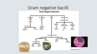
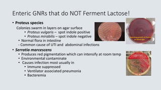
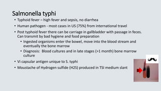
![• Salmonella species
• Diarrhea with +/- fever and PMNs in the stool
• Infection from eating contaminated food (raw eggs, poultry, ground beef
or dairy) or direct contact with a sick animal
– Must ingest large #’s of organisms to make you ill (1,000,000
bacteria), normal levels of stomach acid is protective
• MacConkey agar - does not ferment lactose
• Produces hydrogen sulfide on selective agars
• Motile
• Identification based on biochemical reactions and serologic typing
• Kaufman White serologic typing for speciation of Salmonella
• O Somatic (cell wall) antigen – Salmonella group “B”
• H flagellar antigens – 2 phases [h1 & h2]
• Vi capsular antigen – Salmonella typhi only](https://image.slidesharecdn.com/bacteriologypart2-210219005911/85/Bacteriology-Update-2021-Part-2-15-320.jpg)
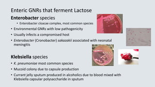
![Shigella
• Diarrhea, +/-vomiting, fluid loss, PMNs and blood in stool
• Infection: Human to human transmission /control with good hand
hygiene
• Ingestion of low #’s of organisms make you ill [10 – 100 bacteria]
• No lactose fermentation on agar
• Non motile
• No Hydrogen sulfide (H2S) produced
• 4 species based on somatic (cell wall) antigen
• S. boydii Group C
• S. dysenteriae Group A
• S. flexneri Group B
• S. sonnei Group D](https://image.slidesharecdn.com/bacteriologypart2-210219005911/85/Bacteriology-Update-2021-Part-2-17-320.jpg)
![Salmonella Shigella Agar (SS agar)
Shigella are colorless due to lactose
not being fermented. Salmonella does
not ferment lactose, but H2S produced
by Salmonella turning the colony black
Hektoen agar –
Salmonella produces H2S [Hydrogen sulfide]
producing black colonies
Shigella – green colonies
Normal flora – orange colored due to
fermentation of lactose (E. coli)
Non-Lactose fermenter
Shigella
Salmonella
Normal Flora
Lactose fermented
H2S
Salmonella
Shigella
Salmonella](https://image.slidesharecdn.com/bacteriologypart2-210219005911/85/Bacteriology-Update-2021-Part-2-18-320.jpg)
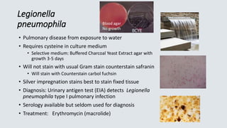
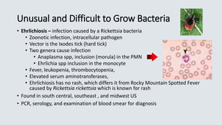
![Haemophilus influenzae
• Transmission – close contact/secretions
• Virulence factor – capsular polysaccharide
• Small pleomorphic Gram negative rod
• Requires 2 nutritional factors for growth:
• X factor = hemin
• V factor = NAD (nicotinamide adenine dinucleotide)
• Grows on chocolate agar (contains X and V factor)
• Will not grow on 5% sheep’s blood agar
• Requires 5-8% C0₂ for growth
• Effective vaccine targets invasive infections with H. influenzae type
B (HIB) effectively eliminating most childhood invasive infections
• Ampicillin resistance from beta lactamase enzyme productions [25
%], 3rd generation Cephalosporin becomes the antibiotic of choice
(Ceftriaxone) for invasive infections
Satellite phenomenon
Staph aureus supplies
the X and V factors
required](https://image.slidesharecdn.com/bacteriologypart2-210219005911/85/Bacteriology-Update-2021-Part-2-26-320.jpg)

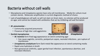
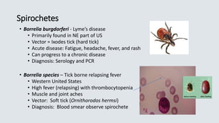


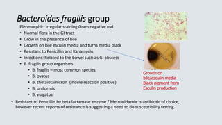
![• C. septicum –
• Bacteremia or gas gangrene in patient with underlying
malignancy
• Hematogenous spread from GI tract – no trauma necessary
• Clostridioides (Clostridium) difficile –
• Disease: antibiotic associated colitis, pseudomembranous
colitis from toxin production
• Toxin A – enterotoxin causing fluid accumulation
• Toxin B – potent cell cytotoxin, primary virulence factor (TcdB)
• Binary toxin – Nap1 strain produces large amount of toxin
• Diagnosis of infection:
• EIA methods [toxin A/B] are insensitive but detect presence of active
toxin
• Molecular methods are sensitive but only detect toxin genes
• Selective medium Cycloserine, Cefoxitin, Fructose Agar [CCFA] –
culture usually only for research purposes](https://image.slidesharecdn.com/bacteriologypart2-210219005911/85/Bacteriology-Update-2021-Part-2-50-320.jpg)

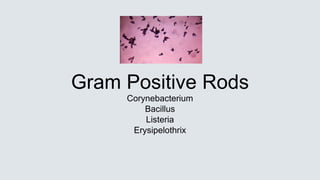
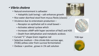
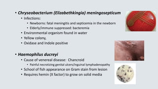
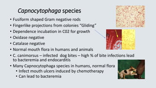
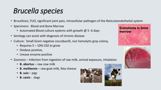
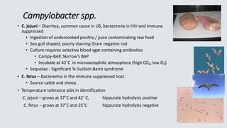
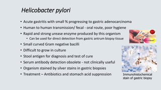
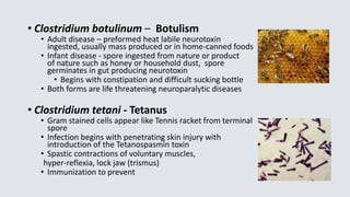
This document summarizes several Gram positive and Gram negative bacterial species. It describes their morphology, biochemical characteristics, pathogenicity and role in causing diseases like diphtheria, anthrax, listeriosis, plague and more. Identification of these bacteria involves examining their growth patterns, hemolytic properties, enzyme production and reactions on selective media.


