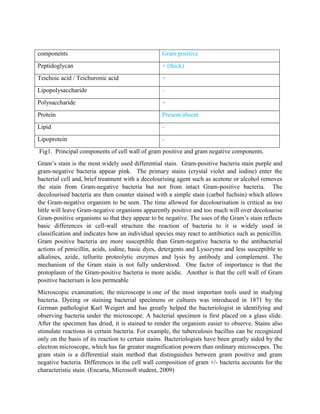
SlideShare a Scribd company logo
1 of 6
Title: culture identification through culture characteristics, appearance, grams staining reactions
and catalase tests of cultures K & J.

Aim: To identify the bacteria in culture K & J through culture appearance, staining
characteristics and microscopic examination by the gram staining method.

Introduction
A fundamental method of studying bacteria is by culturing them in liquid media or on the surface
of media that have been solidified by agar. Media contain nutrients, varying from simple sugars
to complex substances such as meat broth. To purify or isolate a single bacterial species from a
mixture of different bacteria, solidified media generally are used. Individual cells dividing on the
surface of solidified media do not move away from each other as they do in liquid, and after
many rounds of replication they form visible colonies composed of tens of millions of cells all
derived by binary fission from a single cell. If a portion of a colony is then transferred to a liquid
medium, it will grow as a pure culture free of all other bacteria except the single species that was
found in the colony.
Many different species of bacteria so closely resemble one another in appearance that they
cannot be differentiated from one another under the microscope. Various culture techniques have
been developed to aid in species identification. Some media contain substances to inhibit the
growth of many bacteria, but not the species of interest. Others contain sugars that some but not
all bacteria can utilize for growth. Some media contain pH indicators that change color to
indicate that a constituent of the media has been fermented, yielding acid end products. Gas
production as an end product of fermentation can be detected by inoculating bacteria in solidified
media in tubes rather than on plates. Sufficient gas production will result in the formation in the
agar of bubbles that can easily be seen. Still other media are formulated to identify bacteria that
produce certain enzymes that can break down constituents in the media; for example, blood agar
plates, which can detect whether bacteria produce an enzyme to lyse, that is, dissolve, red blood
cells. The various culture media and culture techniques are essential to the hospital laboratory,
whose job it is to identify the cause of various infectious diseases.Sterilization is maintained
during this experiment: Drying or freezing kills many species of bacteria and causes others to
become inactive. Heat or moist heat above a certain temperature kills all bacteria. Sterilization of
many different objects, such as spacecraft and surgical instruments, are important facets of
bacteriological work.
There are several number of bacteria that is gram positive and these include;
STAPHYLOCOCCUS, STREPTOCOCCUS, CORYNRBACTERIUM, LISTERIA, ERYSIPELOTHRIX, CLOSTRIDIUM,
BACILLUS, ACTINOMYCES, NOCARDIA, MYCOBACTERIUM and DERMATOPHILUS

Culture appearances refer to the physical or grossly viewed features such as colonies on the
plate: size, shape, elevation, texture and colonies. Hemolysis in blood agar and lactose
fermentation in MacConkey agar were also taken note of by simple gross observation. Besides
being differential, MacConkey is used to detect bacteria that produces hemolysin, a substance
that is capable of lysing red blood cells. The lysis appears around the colony on the agar plate
and it is used to identify the characteristics.
Streptococcus exhibits a kind of hemolysis called alpha hemolysis, characterized by a greenish
zone around the colonies (Nester, E, L et al, 2004).
Some bacteria are covered with a layer of firm gelatinous material in direct contact with the cell
wall and visible by light microscopy. This material is the capsule. The capsule is normally
composed of complex polysaccharides. Streptococcus pneumoniae (Pneumococcus) is an
organism with prominent capsule composed of polysaccharide. Capsules may also take the form
of a very thin microcapsule or a loose layer of slime. The form of capsule is influenced by the
culture conditions. The bacterial genera streptococcus and staphylococcus belong to the family
Enterobacteriaciae together with several other bacteria such as Escherichia, Salmonella,
Brucellaceae, Brucella, Bacteroidaceae, Sphaerophorus, Micrococcaceae, Lactobacillaceae,
Corynebacteriaceae and Corynebacterium.
The structure of the cell wall in bacterial are more complex than those of eukaryotic cells and
they contain substances unique to bacteria. Peptidoglycan, formally called mucopeptide is the
most important component of the bacterial cell wall; being common to both Gram positive and
Gram-negative cells. It surrounds the cell external to the cytoplasmic membrane as a single baglike molecule. It is composed of linear glycan chains of alternating residues of N-acetyl
glucosamine and N-acetyl muramic acid. These are linked together by short peptide bridges to
form a cross-linked insoluble polymer. Thus, peptidoglycan forms the basic structure of the
bacterial cell wall.
In addition to peptidoglycan, other accessory polymers are also found in most bacteria. Gram positive organisms such as staphylococci, contain teichoic acids composed of either polyglycerol
phosphate or polyribitol phosphate. These occur both within and on the surface of the cell wall
and may account for 20-50% of the cell wall mass.
In Gram - negative cells, the peptidoglycan is much thinner than in Gram - positive organisms.
External to the peptidoglycan lies a membrane (the outer membrane) which contains different
proteins and a unique component, lipopolysaccharide (endotoxin). Endotoxin is important in
pathogenicity of some disease and is also known as somatic O antigen. The LPS is responsible
for many of the biologic activities associated with gram negative bacteria. A major protein of
outer membrane is called porin which forms transmembrane pores or diffusion channels.
components

Gram positive

Peptidoglycan

+ (thick)

Teichoic acid / Teichuronic acid

+

Lipopolysaccharide

-

Polysaccharide

+

Protein

Present/absent

Lipid

-

Lipoprotein

-

Fig1. Principal components of cell wall of gram positive and gram negative components.
Gram’s stain is the most widely used differential stain. Gram-positive bacteria stain purple and
gram-negative bacteria appear pink. The primary stains (crystal violet and iodine) enter the
bacterial cell and, brief treatment with a decolourising agent such as acetone or alcohol removes
the stain from Gram-negative bacteria but not from intact Gram-positive bacteria. The
decolourised bacteria are then counter stained with a simple stain (carbol fuchsin) which allows
the Gram-negative organism to be seen. The time allowed for decolourisation is critical as too
little will leave Gram-negative organisms apparently positive and too much will over decolourise
Gram-positive organisms so that they appear to be negative. The uses of the Gram’s stain reflects
basic differences in cell-wall structure the reaction of bacteria to it is widely used in
classification and indicates how an individual species may react to antibiotics such as penicillin.
Gram positive bacteria are more susceptible than Gram-negative bacteria to the antibacterial
actions of penicillin, acids, iodine, basic dyes, detergents and Lysozyme and less susceptible to
alkalines, azide, tellurite proteolytic enzymes and lysis by antibody and complement. The
mechanism of the Gram stain is not fully understood. One factor of importance is that the
protoplasm of the Gram-positive bacteria is more acidic. Another is that the cell wall of Gram
positive bacterium is less permeable
Microscopic examination; the microscope is one of the most important tools used in studying
bacteria. Dyeing or staining bacterial specimens or cultures was introduced in 1871 by the
German pathologist Karl Weigert and has greatly helped the bacteriologist in identifying and
observing bacteria under the microscope. A bacterial specimen is first placed on a glass slide.
After the specimen has dried, it is stained to render the organism easier to observe. Stains also
stimulate reactions in certain bacteria. For example, the tuberculosis bacillus can be recognized
only on the basis of its reaction to certain stains. Bacteriologists have been greatly aided by the
electron microscope, which has far greater magnification powers than ordinary microscopes. The
gram stain is a differential stain method that distinguishes between gram positive and gram
negative bacteria. Differences in the cell wall composition of gram +/- bacteria accounts for the
characteristic stain. (Encarta, Microsoft student, 2009)
Material













Prepared cultures of K & J on blood agar and MacConkey
Microscopic glass slide
Light Microscope
Loop
Normal saline
Bunsen burner
Staining rack
Oil
crystal violet dye
70% alcohol
Safranin dye
Lugols iodine solution

Procedure
Slide preparation & fixing
1.
2.
3.
4.
5.
6.

The slide was cleaned in order to remove all traces of grease
A loop full of normal saline or distilled water sterile was transferred onto the glass slide
Using a sterile loop, the surface of an independent colony was touched gently
The loop full was emulsified/mixed with a drop of normal saline and it was spread
The slide was then left to dry on the bench at room temperature
The slide was then fixed over a Bunsen burner flame by passing it over the flame 3 times
–noting that the surface with bacteria should be on top so as to kill the bacterial thereby
making it less viable and also to make bacteria to adhere to the slide.
7. The slide was left to cool and the staining procedure followed.
Staining: Gram Stain procedure
1. The slide was flooded with crystal violet solution for 1 minute
2. The stain was washed off under running tap water
3. The slide was Counter stained with lugols iodine; a mordant that does not take part in the
actual staining but only enhances the performance of crystal violet
4. The slide was Washed off again under running tap water
5. The slide was decolorized using 70% alcohol /acetone in brief
6. The slide was washed off under running tap water
7. The slide was counter stained with dilute safranin/ carbofushin
8. The slide was washed off under running tap water
9. The slide was then left to dry
10. An oil drop was added on the slide to enhance visibility under the light microscope
11. The slide was examined under a light microscope at x100 oil power
Results
Fig2: showing results of different physical/microscopic bacterial parameters
Parameter

Shape
Size
Hemolysis
color
Picture

Blood agar
K
Entire
0.5-1.0mm
positive
Greyish

J
Entire
2mm
Positive
Greyish

MacConkey
K/J
No growth in
both media

No growth

Discussion
In this laboratory practical several methods of culture identification were employed and it was
observed that some evidence streptococcus and staphylococcus bacteria were noted. On blood
agar plate the observations that were made were the same for culture K & J: entire shaped,
positive hemolysis and greyish in color. The exceptions were in terms ofsize: where k was 0.51.0mm and J was 2mm. Another difference is in the arrangement of the cocci: k was long
chained, while J was clustered like grapes. Most bacteria come in one of three shapes: rod,
sphere, or spiral. Rod-shaped bacteria are called bacilli. Spherical bacteria are called cocci, and
spiral or corkscrew-shaped bacteria are called spirilla. Some bacteria come in more complex
shapes. A hairlike form of spiral bacteria is called spirochete. Streptococci and staphylococci are
well-known disease-causing bacteria among the cocci.
Streptococcus, genus of spherical, gram-positive, aerobic bacteria. The streptococci occur in
pairs or chains, and some species are pathogenic in humans. Streptococcal infections include
strep throat, scarlet fever, erysipelas, puerperal fever, and some pneumonias. The drugs of choice
for treating such infections are penicillin and erythromycin. Cultures of nonpathogenic lactic
streptococci are used in the fermentation of dairy products such as cheese and buttermilk
(Microsoft ® Encarta ® 2009).
Staphylococcus, genus of round, parasitic bacteria, commonly found in air and water and on the
skin and upper part of the human pharynx. These bacteria are known to cause pneumonia and
septicemia as well as boils and kidney and wound infections.
The antibiotic drug penicillin was once effective for the treatment and control of staphylococci,
but the increase of resistant strains has required use of other antibiotic agents such as semisynthetic penicillins, cephalosporins, or vancomycin. Studies announced in 2006 showed that the
antibiotic daptomycin is effective against staphylococcal infections of the bloodstream, heart,
and skin. No vaccine against Staphylococcus is currently available.
Two common species of Staphylococcus include Staphylococcus aureus, which is commonly
responsible for skin infections, and Staphylococcus epidermis, which does not normally cause
infection. However, either of these bacteria can cause serious infections under the right
conditions.
Staphylococcus aureus is found on the skin and in the nostrils of many healthy individuals.
These bacteria often give rise to minor superficial diseases, including the formation of pustules
or boils in hair follicles. Staphylococcus aureus infections are characterized by the presence of
pus and formation of abscesses. In addition to skin pustules, boils, and carbuncles,
Staphylococcus aureus is responsible for impetigo, infections of wounds and burns (particularly
in a hospital environment), breast abscesses, whitlow (inflammation of a finger or toe near the
nail), osteomyelitis, bronchopneumonia, septicemia, bacteremia, acute endocarditis, food
poisoning, and scalded skin syndrome. Scalded skin syndrome occurs in newborns and is due to
infection by toxigenic strains of Staphylococcus aureus. The toxins cause the skin to exfoliate,
which leaves an appearance of having been scalded. In some cases Staphylococcus aureus can
also cause necrotizing fasciitis, commonly called flesh-eating bacteria. This severe infection can
be life threatening and is more.
Conclusion
Based on the cultures K and J appearance, staining characteristics of gram stain and the catalase
biochemical test. Culture K and J from blood agar exhibited (alpha) hemolysis-incomplete
hemolysis. Gram positive bacteria were identifiedin J & K asStaphylococcus & streptococcus
respectively. And in MacConkey agar both cultures K and J showed no growth.
References
Hawkey M.P and Lewis A.D, 1989, medical bacteriology and practical approach, IRC
Press at oxford university press, England, page8.
Nester W.E et al, 2004, microbiology: A human perspective, 4th edition, McGraw Hill,
New York, pages 83, 84, and 94.
Microsoft ® Encarta ® 2009. © 1993-2008 Microsoft Corporation. All rights reserved.

More Related Content

What's hot

Identification of bacteria
Identification of bacteriaIdentification of bacteria
Identification of bacteriaDhanashri Mali
 
Chapter 3 tools of the laboratory e mail
Chapter 3 tools of the laboratory e mailChapter 3 tools of the laboratory e mail
Chapter 3 tools of the laboratory e mailErika Brockmann
 
Diagnosis of bacterial diseases
Diagnosis of bacterial diseasesDiagnosis of bacterial diseases
Diagnosis of bacterial diseasesSeham Fawzy
 
Medical Microbiology Laboratory (bacterial shapes, arrangement and staining t...
Medical Microbiology Laboratory (bacterial shapes, arrangement and staining t...Medical Microbiology Laboratory (bacterial shapes, arrangement and staining t...
Medical Microbiology Laboratory (bacterial shapes, arrangement and staining t...Hussein Al-tameemi
 
Isolation and Characterization of Bacteria from Tropical Soil
Isolation and Characterization of Bacteria from Tropical SoilIsolation and Characterization of Bacteria from Tropical Soil
Isolation and Characterization of Bacteria from Tropical SoilCarolina Ayala
 
Identification of bacteria by staining methods
Identification of bacteria by staining methodsIdentification of bacteria by staining methods
Identification of bacteria by staining methodsNAGALAKSHMI R
 
Identification and Detection of Microorganism
Identification and Detection of Microorganism Identification and Detection of Microorganism
Identification and Detection of Microorganism esraa alaa
 
Gram positive bacteria
Gram positive bacteria Gram positive bacteria
Gram positive bacteria Ramza Rasheed
 
Acid-Fast Bacteria. Spores of Bacteria. Complete methods of staining
Acid-Fast Bacteria. Spores of Bacteria. Complete methods of stainingAcid-Fast Bacteria. Spores of Bacteria. Complete methods of staining
Acid-Fast Bacteria. Spores of Bacteria. Complete methods of stainingEneutron
 
Introduction and Brief about Bacteriology, Bacterial Structure, Difference Be...
Introduction and Brief about Bacteriology, Bacterial Structure, Difference Be...Introduction and Brief about Bacteriology, Bacterial Structure, Difference Be...
Introduction and Brief about Bacteriology, Bacterial Structure, Difference Be...Zunaira Gillani
 
Identificationofbacteria 180120102225
Identificationofbacteria 180120102225Identificationofbacteria 180120102225
Identificationofbacteria 180120102225LaraMelisaROlguera
 
General Bacteriology
General BacteriologyGeneral Bacteriology
General BacteriologyMrudulaBhalke
 
bacterial morphology and classification
bacterial morphology and classificationbacterial morphology and classification
bacterial morphology and classificationAshish Jawarkar
 
Analysis of potential vectors of Serratia marcescens as a Caretta caretta nes...
Analysis of potential vectors of Serratia marcescens as a Caretta caretta nes...Analysis of potential vectors of Serratia marcescens as a Caretta caretta nes...
Analysis of potential vectors of Serratia marcescens as a Caretta caretta nes...BHIConservancy
 
Pure culture techniques
Pure culture techniquesPure culture techniques
Pure culture techniquesAyush Singh
 
Presentation: Isolation and Characterization of Bacteria from Tropical Soils
Presentation: Isolation and Characterization of Bacteria from Tropical SoilsPresentation: Isolation and Characterization of Bacteria from Tropical Soils
Presentation: Isolation and Characterization of Bacteria from Tropical SoilsNicole Rivera
 

What's hot (20)

Identification of bacteria
Identification of bacteriaIdentification of bacteria
Identification of bacteria
 
Chapter 3 tools of the laboratory e mail
Chapter 3 tools of the laboratory e mailChapter 3 tools of the laboratory e mail
Chapter 3 tools of the laboratory e mail
 
Bacterial morphology
Bacterial morphology Bacterial morphology
Bacterial morphology
 
Diagnosis of bacterial diseases
Diagnosis of bacterial diseasesDiagnosis of bacterial diseases
Diagnosis of bacterial diseases
 
Medical Microbiology Laboratory (bacterial shapes, arrangement and staining t...
Medical Microbiology Laboratory (bacterial shapes, arrangement and staining t...Medical Microbiology Laboratory (bacterial shapes, arrangement and staining t...
Medical Microbiology Laboratory (bacterial shapes, arrangement and staining t...
 
Isolation and Characterization of Bacteria from Tropical Soil
Isolation and Characterization of Bacteria from Tropical SoilIsolation and Characterization of Bacteria from Tropical Soil
Isolation and Characterization of Bacteria from Tropical Soil
 
Identification of bacteria by staining methods
Identification of bacteria by staining methodsIdentification of bacteria by staining methods
Identification of bacteria by staining methods
 
Bactriology
BactriologyBactriology
Bactriology
 
Identification and Detection of Microorganism
Identification and Detection of Microorganism Identification and Detection of Microorganism
Identification and Detection of Microorganism
 
Gram positive bacteria
Gram positive bacteria Gram positive bacteria
Gram positive bacteria
 
Acid-Fast Bacteria. Spores of Bacteria. Complete methods of staining
Acid-Fast Bacteria. Spores of Bacteria. Complete methods of stainingAcid-Fast Bacteria. Spores of Bacteria. Complete methods of staining
Acid-Fast Bacteria. Spores of Bacteria. Complete methods of staining
 
Introduction and Brief about Bacteriology, Bacterial Structure, Difference Be...
Introduction and Brief about Bacteriology, Bacterial Structure, Difference Be...Introduction and Brief about Bacteriology, Bacterial Structure, Difference Be...
Introduction and Brief about Bacteriology, Bacterial Structure, Difference Be...
 
Faridchapter4microbiology
Faridchapter4microbiologyFaridchapter4microbiology
Faridchapter4microbiology
 
Clinical Microbiology
Clinical MicrobiologyClinical Microbiology
Clinical Microbiology
 
Identificationofbacteria 180120102225
Identificationofbacteria 180120102225Identificationofbacteria 180120102225
Identificationofbacteria 180120102225
 
General Bacteriology
General BacteriologyGeneral Bacteriology
General Bacteriology
 
bacterial morphology and classification
bacterial morphology and classificationbacterial morphology and classification
bacterial morphology and classification
 
Analysis of potential vectors of Serratia marcescens as a Caretta caretta nes...
Analysis of potential vectors of Serratia marcescens as a Caretta caretta nes...Analysis of potential vectors of Serratia marcescens as a Caretta caretta nes...
Analysis of potential vectors of Serratia marcescens as a Caretta caretta nes...
 
Pure culture techniques
Pure culture techniquesPure culture techniques
Pure culture techniques
 
Presentation: Isolation and Characterization of Bacteria from Tropical Soils
Presentation: Isolation and Characterization of Bacteria from Tropical SoilsPresentation: Isolation and Characterization of Bacteria from Tropical Soils
Presentation: Isolation and Characterization of Bacteria from Tropical Soils
 

Viewers also liked

Medical dissection lab optical microscope
Medical dissection lab  optical microscopeMedical dissection lab  optical microscope
Medical dissection lab optical microscopeDIYYALA CHAITANYA KUMAR
 
Power point mineral notes
Power point mineral notesPower point mineral notes
Power point mineral notesharvey09
 
optical microscope
optical microscopeoptical microscope
optical microscopemastx
 
Microscope lab
Microscope labMicroscope lab
Microscope labarasumed
 
Microbiology (lab report 1 format)
Microbiology (lab report 1 format)Microbiology (lab report 1 format)
Microbiology (lab report 1 format)MBBS IMS MSU
 
Gram stain demonstration
Gram stain demonstrationGram stain demonstration
Gram stain demonstrationsanjay singh
 

Viewers also liked (6)

Medical dissection lab optical microscope
Medical dissection lab  optical microscopeMedical dissection lab  optical microscope
Medical dissection lab optical microscope
 
Power point mineral notes
Power point mineral notesPower point mineral notes
Power point mineral notes
 
optical microscope
optical microscopeoptical microscope
optical microscope
 
Microscope lab
Microscope labMicroscope lab
Microscope lab
 
Microbiology (lab report 1 format)
Microbiology (lab report 1 format)Microbiology (lab report 1 format)
Microbiology (lab report 1 format)
 
Gram stain demonstration
Gram stain demonstrationGram stain demonstration
Gram stain demonstration
 

Similar to Bacteriology&immunology lab2

MICRO FOR nurses introduction to microbiology
MICRO FOR nurses introduction to microbiologyMICRO FOR nurses introduction to microbiology
MICRO FOR nurses introduction to microbiologyajadoon84
 
Bacteria Classification By Gram Staining Essay
Bacteria Classification By Gram Staining EssayBacteria Classification By Gram Staining Essay
Bacteria Classification By Gram Staining EssayChristy Hunt
 
Mici 1100 sept_08_lectures_1-5
Mici 1100 sept_08_lectures_1-5Mici 1100 sept_08_lectures_1-5
Mici 1100 sept_08_lectures_1-5Star Reddy
 
Morphology of bacteria lecture.pptx
Morphology of bacteria lecture.pptxMorphology of bacteria lecture.pptx
Morphology of bacteria lecture.pptxmohammedkhateeb16
 
2.-Cell-structers.pdf
2.-Cell-structers.pdf2.-Cell-structers.pdf
2.-Cell-structers.pdfmetti007
 
Ssusume Patrick- Bacteria culture,Makerere University.pdf
Ssusume Patrick- Bacteria culture,Makerere University.pdfSsusume Patrick- Bacteria culture,Makerere University.pdf
Ssusume Patrick- Bacteria culture,Makerere University.pdfPATRICKSSUSUME
 
Cellular differentiation
Cellular differentiationCellular differentiation
Cellular differentiationsanamTulja1
 
MICROBIOLOGY-1-1.pptx
MICROBIOLOGY-1-1.pptxMICROBIOLOGY-1-1.pptx
MICROBIOLOGY-1-1.pptxJaanuJaanu20
 
Gram-Negative Staining Lab Report
Gram-Negative Staining Lab ReportGram-Negative Staining Lab Report
Gram-Negative Staining Lab ReportTiffany Surratt
 
Lec 01. introduction to microbiology
Lec 01. introduction to microbiologyLec 01. introduction to microbiology
Lec 01. introduction to microbiologySebghatullah Mansoor
 
Salmonell A Common Bacteria
Salmonell A Common BacteriaSalmonell A Common Bacteria
Salmonell A Common BacteriaKaren Thompson
 
1st lec microbiology Dentistry-- .Dr.Olfet.pptx
1st lec microbiology Dentistry-- .Dr.Olfet.pptx1st lec microbiology Dentistry-- .Dr.Olfet.pptx
1st lec microbiology Dentistry-- .Dr.Olfet.pptxraely0885
 
identificationof bacteria.pptx
identificationof bacteria.pptxidentificationof bacteria.pptx
identificationof bacteria.pptxobedcudjoe1
 
lecture 3.pptxhhhjjjiiikjjjjjjjjiuhhjjjjj
lecture 3.pptxhhhjjjiiikjjjjjjjjiuhhjjjjjlecture 3.pptxhhhjjjiiikjjjjjjjjiuhhjjjjj
lecture 3.pptxhhhjjjiiikjjjjjjjjiuhhjjjjjzebarehabeb
 
Bacteria cell structure.pptx
 Bacteria cell structure.pptx Bacteria cell structure.pptx
Bacteria cell structure.pptxShayanAhmad48
 

Similar to Bacteriology&immunology lab2 (20)

MICRO FOR nurses introduction to microbiology
MICRO FOR nurses introduction to microbiologyMICRO FOR nurses introduction to microbiology
MICRO FOR nurses introduction to microbiology
 
Bacteria Classification By Gram Staining Essay
Bacteria Classification By Gram Staining EssayBacteria Classification By Gram Staining Essay
Bacteria Classification By Gram Staining Essay
 
Mici 1100 sept_08_lectures_1-5
Mici 1100 sept_08_lectures_1-5Mici 1100 sept_08_lectures_1-5
Mici 1100 sept_08_lectures_1-5
 
Morphology of bacteria lecture.pptx
Morphology of bacteria lecture.pptxMorphology of bacteria lecture.pptx
Morphology of bacteria lecture.pptx
 
2.-Cell-structers.pdf
2.-Cell-structers.pdf2.-Cell-structers.pdf
2.-Cell-structers.pdf
 
Strep. Agalactiae
Strep. AgalactiaeStrep. Agalactiae
Strep. Agalactiae
 
Ssusume Patrick- Bacteria culture,Makerere University.pdf
Ssusume Patrick- Bacteria culture,Makerere University.pdfSsusume Patrick- Bacteria culture,Makerere University.pdf
Ssusume Patrick- Bacteria culture,Makerere University.pdf
 
Bacteria pdf
Bacteria pdfBacteria pdf
Bacteria pdf
 
Cellular differentiation
Cellular differentiationCellular differentiation
Cellular differentiation
 
MICROBIOLOGY-1-1.pptx
MICROBIOLOGY-1-1.pptxMICROBIOLOGY-1-1.pptx
MICROBIOLOGY-1-1.pptx
 
Gram-Negative Staining Lab Report
Gram-Negative Staining Lab ReportGram-Negative Staining Lab Report
Gram-Negative Staining Lab Report
 
Lec 01. introduction to microbiology
Lec 01. introduction to microbiologyLec 01. introduction to microbiology
Lec 01. introduction to microbiology
 
Discussion for microbial growth
Discussion for microbial growthDiscussion for microbial growth
Discussion for microbial growth
 
Salmonell A Common Bacteria
Salmonell A Common BacteriaSalmonell A Common Bacteria
Salmonell A Common Bacteria
 
Prokaryotes
ProkaryotesProkaryotes
Prokaryotes
 
1st lec microbiology Dentistry-- .Dr.Olfet.pptx
1st lec microbiology Dentistry-- .Dr.Olfet.pptx1st lec microbiology Dentistry-- .Dr.Olfet.pptx
1st lec microbiology Dentistry-- .Dr.Olfet.pptx
 
identificationof bacteria.pptx
identificationof bacteria.pptxidentificationof bacteria.pptx
identificationof bacteria.pptx
 
Microbilogy
MicrobilogyMicrobilogy
Microbilogy
 
lecture 3.pptxhhhjjjiiikjjjjjjjjiuhhjjjjj
lecture 3.pptxhhhjjjiiikjjjjjjjjiuhhjjjjjlecture 3.pptxhhhjjjiiikjjjjjjjjiuhhjjjjj
lecture 3.pptxhhhjjjiiikjjjjjjjjiuhhjjjjj
 
Bacteria cell structure.pptx
 Bacteria cell structure.pptx Bacteria cell structure.pptx
Bacteria cell structure.pptx
 

More from Brian Musalo

Protozoology lab1.
Protozoology lab1.Protozoology lab1.
Protozoology lab1.Brian Musalo
 
Pharmacol report2..
Pharmacol report2..Pharmacol report2..
Pharmacol report2..Brian Musalo
 
Pharmacol lab report
Pharmacol lab reportPharmacol lab report
Pharmacol lab reportBrian Musalo
 
Postmortem report1
Postmortem report1Postmortem report1
Postmortem report1Brian Musalo
 
Post mortem on a dog great dane
Post mortem on a dog great danePost mortem on a dog great dane
Post mortem on a dog great daneBrian Musalo
 
Post mortem on a dog
Post mortem on a dogPost mortem on a dog
Post mortem on a dogBrian Musalo
 
Immunology lab report
Immunology lab reportImmunology lab report
Immunology lab reportBrian Musalo
 
Fecal examination lab report
Fecal examination lab reportFecal examination lab report
Fecal examination lab reportBrian Musalo
 
Effects of drugs on the eye and to demonstrate antagonism
Effects of drugs on the eye and to demonstrate antagonismEffects of drugs on the eye and to demonstrate antagonism
Effects of drugs on the eye and to demonstrate antagonismBrian Musalo
 
Classical&african swine fever
Classical&african swine feverClassical&african swine fever
Classical&african swine feverBrian Musalo
 
Candling & innoculation
Candling & innoculationCandling & innoculation
Candling & innoculationBrian Musalo
 
Cell pathology assignment
Cell pathology  assignmentCell pathology  assignment
Cell pathology assignmentBrian Musalo
 
Identification of insects2
Identification of insects2Identification of insects2
Identification of insects2Brian Musalo
 
Identification of ticks and mites
Identification of ticks and mitesIdentification of ticks and mites
Identification of ticks and mitesBrian Musalo
 
Identification of insects
Identification of insectsIdentification of insects
Identification of insectsBrian Musalo
 

More from Brian Musalo (20)

Protozoology lab1.
Protozoology lab1.Protozoology lab1.
Protozoology lab1.
 
Pharmacol report2..
Pharmacol report2..Pharmacol report2..
Pharmacol report2..
 
Protozology
ProtozologyProtozology
Protozology
 
Pharmacol lab report
Pharmacol lab reportPharmacol lab report
Pharmacol lab report
 
Postmortem report1
Postmortem report1Postmortem report1
Postmortem report1
 
Post mortem
Post mortemPost mortem
Post mortem
 
Post mortem on a dog great dane
Post mortem on a dog great danePost mortem on a dog great dane
Post mortem on a dog great dane
 
Post mortem on a dog
Post mortem on a dogPost mortem on a dog
Post mortem on a dog
 
Immunology lab report
Immunology lab reportImmunology lab report
Immunology lab report
 
Fecal examination lab report
Fecal examination lab reportFecal examination lab report
Fecal examination lab report
 
Effects of drugs on the eye and to demonstrate antagonism
Effects of drugs on the eye and to demonstrate antagonismEffects of drugs on the eye and to demonstrate antagonism
Effects of drugs on the eye and to demonstrate antagonism
 
Classical&african swine fever
Classical&african swine feverClassical&african swine fever
Classical&african swine fever
 
Candling & innoculation
Candling & innoculationCandling & innoculation
Candling & innoculation
 
Cell pathology assignment
Cell pathology  assignmentCell pathology  assignment
Cell pathology assignment
 
Parasitology lab3
Parasitology lab3Parasitology lab3
Parasitology lab3
 
Parasitology lab1
Parasitology lab1Parasitology lab1
Parasitology lab1
 
Parasitology lab2
Parasitology lab2Parasitology lab2
Parasitology lab2
 
Identification of insects2
Identification of insects2Identification of insects2
Identification of insects2
 
Identification of ticks and mites
Identification of ticks and mitesIdentification of ticks and mites
Identification of ticks and mites
 
Identification of insects
Identification of insectsIdentification of insects
Identification of insects
 

Recently uploaded

ECONOMIC CONTEXT - LONG FORM TV DRAMA - PPT
ECONOMIC CONTEXT - LONG FORM TV DRAMA - PPTECONOMIC CONTEXT - LONG FORM TV DRAMA - PPT
ECONOMIC CONTEXT - LONG FORM TV DRAMA - PPTiammrhaywood
 
Science 7 - LAND and SEA BREEZE and its Characteristics
Science 7 - LAND and SEA BREEZE and its CharacteristicsScience 7 - LAND and SEA BREEZE and its Characteristics
Science 7 - LAND and SEA BREEZE and its CharacteristicsKarinaGenton
 
URLs and Routing in the Odoo 17 Website App
URLs and Routing in the Odoo 17 Website AppURLs and Routing in the Odoo 17 Website App
URLs and Routing in the Odoo 17 Website AppCeline George
 
Accessible design: Minimum effort, maximum impact
Accessible design: Minimum effort, maximum impactAccessible design: Minimum effort, maximum impact
Accessible design: Minimum effort, maximum impactdawncurless
 
Contemporary philippine arts from the regions_PPT_Module_12 [Autosaved] (1).pptx
Contemporary philippine arts from the regions_PPT_Module_12 [Autosaved] (1).pptxContemporary philippine arts from the regions_PPT_Module_12 [Autosaved] (1).pptx
Contemporary philippine arts from the regions_PPT_Module_12 [Autosaved] (1).pptxRoyAbrique
 
How to Configure Email Server in Odoo 17
How to Configure Email Server in Odoo 17How to Configure Email Server in Odoo 17
How to Configure Email Server in Odoo 17Celine George
 
Mastering the Unannounced Regulatory Inspection
Mastering the Unannounced Regulatory InspectionMastering the Unannounced Regulatory Inspection
Mastering the Unannounced Regulatory InspectionSafetyChain Software
 
18-04-UA_REPORT_MEDIALITERAСY_INDEX-DM_23-1-final-eng.pdf
18-04-UA_REPORT_MEDIALITERAСY_INDEX-DM_23-1-final-eng.pdf18-04-UA_REPORT_MEDIALITERAСY_INDEX-DM_23-1-final-eng.pdf
18-04-UA_REPORT_MEDIALITERAСY_INDEX-DM_23-1-final-eng.pdfssuser54595a
 
Enzyme, Pharmaceutical Aids, Miscellaneous Last Part of Chapter no 5th.pdf
Enzyme, Pharmaceutical Aids, Miscellaneous Last Part of Chapter no 5th.pdfEnzyme, Pharmaceutical Aids, Miscellaneous Last Part of Chapter no 5th.pdf
Enzyme, Pharmaceutical Aids, Miscellaneous Last Part of Chapter no 5th.pdfSumit Tiwari
 
Hybridoma Technology ( Production , Purification , and Application )
Hybridoma Technology  ( Production , Purification , and Application  ) Hybridoma Technology  ( Production , Purification , and Application  )
Hybridoma Technology ( Production , Purification , and Application ) Sakshi Ghasle
 
The Most Excellent Way | 1 Corinthians 13
The Most Excellent Way | 1 Corinthians 13The Most Excellent Way | 1 Corinthians 13
The Most Excellent Way | 1 Corinthians 13Steve Thomason
 
Introduction to ArtificiaI Intelligence in Higher Education
Introduction to ArtificiaI Intelligence in Higher EducationIntroduction to ArtificiaI Intelligence in Higher Education
Introduction to ArtificiaI Intelligence in Higher Educationpboyjonauth
 
Software Engineering Methodologies (overview)
Software Engineering Methodologies (overview)Software Engineering Methodologies (overview)
Software Engineering Methodologies (overview)eniolaolutunde
 
Interactive Powerpoint_How to Master effective communication
Interactive Powerpoint_How to Master effective communicationInteractive Powerpoint_How to Master effective communication
Interactive Powerpoint_How to Master effective communicationnomboosow
 
“Oh GOSH! Reflecting on Hackteria's Collaborative Practices in a Global Do-It...
“Oh GOSH! Reflecting on Hackteria's Collaborative Practices in a Global Do-It...“Oh GOSH! Reflecting on Hackteria's Collaborative Practices in a Global Do-It...
“Oh GOSH! Reflecting on Hackteria's Collaborative Practices in a Global Do-It...Marc Dusseiller Dusjagr
 

Recently uploaded (20)

ECONOMIC CONTEXT - LONG FORM TV DRAMA - PPT
ECONOMIC CONTEXT - LONG FORM TV DRAMA - PPTECONOMIC CONTEXT - LONG FORM TV DRAMA - PPT
ECONOMIC CONTEXT - LONG FORM TV DRAMA - PPT
 
Science 7 - LAND and SEA BREEZE and its Characteristics
Science 7 - LAND and SEA BREEZE and its CharacteristicsScience 7 - LAND and SEA BREEZE and its Characteristics
Science 7 - LAND and SEA BREEZE and its Characteristics
 
Código Creativo y Arte de Software | Unidad 1
Código Creativo y Arte de Software | Unidad 1Código Creativo y Arte de Software | Unidad 1
Código Creativo y Arte de Software | Unidad 1
 
URLs and Routing in the Odoo 17 Website App
URLs and Routing in the Odoo 17 Website AppURLs and Routing in the Odoo 17 Website App
URLs and Routing in the Odoo 17 Website App
 
Accessible design: Minimum effort, maximum impact
Accessible design: Minimum effort, maximum impactAccessible design: Minimum effort, maximum impact
Accessible design: Minimum effort, maximum impact
 
Contemporary philippine arts from the regions_PPT_Module_12 [Autosaved] (1).pptx
Contemporary philippine arts from the regions_PPT_Module_12 [Autosaved] (1).pptxContemporary philippine arts from the regions_PPT_Module_12 [Autosaved] (1).pptx
Contemporary philippine arts from the regions_PPT_Module_12 [Autosaved] (1).pptx
 
How to Configure Email Server in Odoo 17
How to Configure Email Server in Odoo 17How to Configure Email Server in Odoo 17
How to Configure Email Server in Odoo 17
 
Mastering the Unannounced Regulatory Inspection
Mastering the Unannounced Regulatory InspectionMastering the Unannounced Regulatory Inspection
Mastering the Unannounced Regulatory Inspection
 
18-04-UA_REPORT_MEDIALITERAСY_INDEX-DM_23-1-final-eng.pdf
18-04-UA_REPORT_MEDIALITERAСY_INDEX-DM_23-1-final-eng.pdf18-04-UA_REPORT_MEDIALITERAСY_INDEX-DM_23-1-final-eng.pdf
18-04-UA_REPORT_MEDIALITERAСY_INDEX-DM_23-1-final-eng.pdf
 
Model Call Girl in Bikash Puri Delhi reach out to us at 🔝9953056974🔝
Model Call Girl in Bikash Puri  Delhi reach out to us at 🔝9953056974🔝Model Call Girl in Bikash Puri  Delhi reach out to us at 🔝9953056974🔝
Model Call Girl in Bikash Puri Delhi reach out to us at 🔝9953056974🔝
 
Model Call Girl in Tilak Nagar Delhi reach out to us at 🔝9953056974🔝
Model Call Girl in Tilak Nagar Delhi reach out to us at 🔝9953056974🔝Model Call Girl in Tilak Nagar Delhi reach out to us at 🔝9953056974🔝
Model Call Girl in Tilak Nagar Delhi reach out to us at 🔝9953056974🔝
 
Enzyme, Pharmaceutical Aids, Miscellaneous Last Part of Chapter no 5th.pdf
Enzyme, Pharmaceutical Aids, Miscellaneous Last Part of Chapter no 5th.pdfEnzyme, Pharmaceutical Aids, Miscellaneous Last Part of Chapter no 5th.pdf
Enzyme, Pharmaceutical Aids, Miscellaneous Last Part of Chapter no 5th.pdf
 
Hybridoma Technology ( Production , Purification , and Application )
Hybridoma Technology  ( Production , Purification , and Application  ) Hybridoma Technology  ( Production , Purification , and Application  )
Hybridoma Technology ( Production , Purification , and Application )
 
The Most Excellent Way | 1 Corinthians 13
The Most Excellent Way | 1 Corinthians 13The Most Excellent Way | 1 Corinthians 13
The Most Excellent Way | 1 Corinthians 13
 
Introduction to ArtificiaI Intelligence in Higher Education
Introduction to ArtificiaI Intelligence in Higher EducationIntroduction to ArtificiaI Intelligence in Higher Education
Introduction to ArtificiaI Intelligence in Higher Education
 
9953330565 Low Rate Call Girls In Rohini Delhi NCR
9953330565 Low Rate Call Girls In Rohini  Delhi NCR9953330565 Low Rate Call Girls In Rohini  Delhi NCR
9953330565 Low Rate Call Girls In Rohini Delhi NCR
 
TataKelola dan KamSiber Kecerdasan Buatan v022.pdf
TataKelola dan KamSiber Kecerdasan Buatan v022.pdfTataKelola dan KamSiber Kecerdasan Buatan v022.pdf
TataKelola dan KamSiber Kecerdasan Buatan v022.pdf
 
Software Engineering Methodologies (overview)
Software Engineering Methodologies (overview)Software Engineering Methodologies (overview)
Software Engineering Methodologies (overview)
 
Interactive Powerpoint_How to Master effective communication
Interactive Powerpoint_How to Master effective communicationInteractive Powerpoint_How to Master effective communication
Interactive Powerpoint_How to Master effective communication
 
“Oh GOSH! Reflecting on Hackteria's Collaborative Practices in a Global Do-It...
“Oh GOSH! Reflecting on Hackteria's Collaborative Practices in a Global Do-It...“Oh GOSH! Reflecting on Hackteria's Collaborative Practices in a Global Do-It...
“Oh GOSH! Reflecting on Hackteria's Collaborative Practices in a Global Do-It...
 

Bacteriology&immunology lab2

  • 1. Title: culture identification through culture characteristics, appearance, grams staining reactions and catalase tests of cultures K & J. Aim: To identify the bacteria in culture K & J through culture appearance, staining characteristics and microscopic examination by the gram staining method. Introduction A fundamental method of studying bacteria is by culturing them in liquid media or on the surface of media that have been solidified by agar. Media contain nutrients, varying from simple sugars to complex substances such as meat broth. To purify or isolate a single bacterial species from a mixture of different bacteria, solidified media generally are used. Individual cells dividing on the surface of solidified media do not move away from each other as they do in liquid, and after many rounds of replication they form visible colonies composed of tens of millions of cells all derived by binary fission from a single cell. If a portion of a colony is then transferred to a liquid medium, it will grow as a pure culture free of all other bacteria except the single species that was found in the colony. Many different species of bacteria so closely resemble one another in appearance that they cannot be differentiated from one another under the microscope. Various culture techniques have been developed to aid in species identification. Some media contain substances to inhibit the growth of many bacteria, but not the species of interest. Others contain sugars that some but not all bacteria can utilize for growth. Some media contain pH indicators that change color to indicate that a constituent of the media has been fermented, yielding acid end products. Gas production as an end product of fermentation can be detected by inoculating bacteria in solidified media in tubes rather than on plates. Sufficient gas production will result in the formation in the agar of bubbles that can easily be seen. Still other media are formulated to identify bacteria that produce certain enzymes that can break down constituents in the media; for example, blood agar plates, which can detect whether bacteria produce an enzyme to lyse, that is, dissolve, red blood cells. The various culture media and culture techniques are essential to the hospital laboratory, whose job it is to identify the cause of various infectious diseases.Sterilization is maintained during this experiment: Drying or freezing kills many species of bacteria and causes others to become inactive. Heat or moist heat above a certain temperature kills all bacteria. Sterilization of many different objects, such as spacecraft and surgical instruments, are important facets of bacteriological work.
  • 2. There are several number of bacteria that is gram positive and these include; STAPHYLOCOCCUS, STREPTOCOCCUS, CORYNRBACTERIUM, LISTERIA, ERYSIPELOTHRIX, CLOSTRIDIUM, BACILLUS, ACTINOMYCES, NOCARDIA, MYCOBACTERIUM and DERMATOPHILUS Culture appearances refer to the physical or grossly viewed features such as colonies on the plate: size, shape, elevation, texture and colonies. Hemolysis in blood agar and lactose fermentation in MacConkey agar were also taken note of by simple gross observation. Besides being differential, MacConkey is used to detect bacteria that produces hemolysin, a substance that is capable of lysing red blood cells. The lysis appears around the colony on the agar plate and it is used to identify the characteristics. Streptococcus exhibits a kind of hemolysis called alpha hemolysis, characterized by a greenish zone around the colonies (Nester, E, L et al, 2004). Some bacteria are covered with a layer of firm gelatinous material in direct contact with the cell wall and visible by light microscopy. This material is the capsule. The capsule is normally composed of complex polysaccharides. Streptococcus pneumoniae (Pneumococcus) is an organism with prominent capsule composed of polysaccharide. Capsules may also take the form of a very thin microcapsule or a loose layer of slime. The form of capsule is influenced by the culture conditions. The bacterial genera streptococcus and staphylococcus belong to the family Enterobacteriaciae together with several other bacteria such as Escherichia, Salmonella, Brucellaceae, Brucella, Bacteroidaceae, Sphaerophorus, Micrococcaceae, Lactobacillaceae, Corynebacteriaceae and Corynebacterium. The structure of the cell wall in bacterial are more complex than those of eukaryotic cells and they contain substances unique to bacteria. Peptidoglycan, formally called mucopeptide is the most important component of the bacterial cell wall; being common to both Gram positive and Gram-negative cells. It surrounds the cell external to the cytoplasmic membrane as a single baglike molecule. It is composed of linear glycan chains of alternating residues of N-acetyl glucosamine and N-acetyl muramic acid. These are linked together by short peptide bridges to form a cross-linked insoluble polymer. Thus, peptidoglycan forms the basic structure of the bacterial cell wall. In addition to peptidoglycan, other accessory polymers are also found in most bacteria. Gram positive organisms such as staphylococci, contain teichoic acids composed of either polyglycerol phosphate or polyribitol phosphate. These occur both within and on the surface of the cell wall and may account for 20-50% of the cell wall mass. In Gram - negative cells, the peptidoglycan is much thinner than in Gram - positive organisms. External to the peptidoglycan lies a membrane (the outer membrane) which contains different proteins and a unique component, lipopolysaccharide (endotoxin). Endotoxin is important in pathogenicity of some disease and is also known as somatic O antigen. The LPS is responsible for many of the biologic activities associated with gram negative bacteria. A major protein of outer membrane is called porin which forms transmembrane pores or diffusion channels.
  • 3. components Gram positive Peptidoglycan + (thick) Teichoic acid / Teichuronic acid + Lipopolysaccharide - Polysaccharide + Protein Present/absent Lipid - Lipoprotein - Fig1. Principal components of cell wall of gram positive and gram negative components. Gram’s stain is the most widely used differential stain. Gram-positive bacteria stain purple and gram-negative bacteria appear pink. The primary stains (crystal violet and iodine) enter the bacterial cell and, brief treatment with a decolourising agent such as acetone or alcohol removes the stain from Gram-negative bacteria but not from intact Gram-positive bacteria. The decolourised bacteria are then counter stained with a simple stain (carbol fuchsin) which allows the Gram-negative organism to be seen. The time allowed for decolourisation is critical as too little will leave Gram-negative organisms apparently positive and too much will over decolourise Gram-positive organisms so that they appear to be negative. The uses of the Gram’s stain reflects basic differences in cell-wall structure the reaction of bacteria to it is widely used in classification and indicates how an individual species may react to antibiotics such as penicillin. Gram positive bacteria are more susceptible than Gram-negative bacteria to the antibacterial actions of penicillin, acids, iodine, basic dyes, detergents and Lysozyme and less susceptible to alkalines, azide, tellurite proteolytic enzymes and lysis by antibody and complement. The mechanism of the Gram stain is not fully understood. One factor of importance is that the protoplasm of the Gram-positive bacteria is more acidic. Another is that the cell wall of Gram positive bacterium is less permeable Microscopic examination; the microscope is one of the most important tools used in studying bacteria. Dyeing or staining bacterial specimens or cultures was introduced in 1871 by the German pathologist Karl Weigert and has greatly helped the bacteriologist in identifying and observing bacteria under the microscope. A bacterial specimen is first placed on a glass slide. After the specimen has dried, it is stained to render the organism easier to observe. Stains also stimulate reactions in certain bacteria. For example, the tuberculosis bacillus can be recognized only on the basis of its reaction to certain stains. Bacteriologists have been greatly aided by the electron microscope, which has far greater magnification powers than ordinary microscopes. The gram stain is a differential stain method that distinguishes between gram positive and gram negative bacteria. Differences in the cell wall composition of gram +/- bacteria accounts for the characteristic stain. (Encarta, Microsoft student, 2009)
  • 4. Material             Prepared cultures of K & J on blood agar and MacConkey Microscopic glass slide Light Microscope Loop Normal saline Bunsen burner Staining rack Oil crystal violet dye 70% alcohol Safranin dye Lugols iodine solution Procedure Slide preparation & fixing 1. 2. 3. 4. 5. 6. The slide was cleaned in order to remove all traces of grease A loop full of normal saline or distilled water sterile was transferred onto the glass slide Using a sterile loop, the surface of an independent colony was touched gently The loop full was emulsified/mixed with a drop of normal saline and it was spread The slide was then left to dry on the bench at room temperature The slide was then fixed over a Bunsen burner flame by passing it over the flame 3 times –noting that the surface with bacteria should be on top so as to kill the bacterial thereby making it less viable and also to make bacteria to adhere to the slide. 7. The slide was left to cool and the staining procedure followed. Staining: Gram Stain procedure 1. The slide was flooded with crystal violet solution for 1 minute 2. The stain was washed off under running tap water 3. The slide was Counter stained with lugols iodine; a mordant that does not take part in the actual staining but only enhances the performance of crystal violet 4. The slide was Washed off again under running tap water 5. The slide was decolorized using 70% alcohol /acetone in brief 6. The slide was washed off under running tap water 7. The slide was counter stained with dilute safranin/ carbofushin 8. The slide was washed off under running tap water 9. The slide was then left to dry 10. An oil drop was added on the slide to enhance visibility under the light microscope 11. The slide was examined under a light microscope at x100 oil power
  • 5. Results Fig2: showing results of different physical/microscopic bacterial parameters Parameter Shape Size Hemolysis color Picture Blood agar K Entire 0.5-1.0mm positive Greyish J Entire 2mm Positive Greyish MacConkey K/J No growth in both media No growth Discussion In this laboratory practical several methods of culture identification were employed and it was observed that some evidence streptococcus and staphylococcus bacteria were noted. On blood agar plate the observations that were made were the same for culture K & J: entire shaped, positive hemolysis and greyish in color. The exceptions were in terms ofsize: where k was 0.51.0mm and J was 2mm. Another difference is in the arrangement of the cocci: k was long chained, while J was clustered like grapes. Most bacteria come in one of three shapes: rod, sphere, or spiral. Rod-shaped bacteria are called bacilli. Spherical bacteria are called cocci, and spiral or corkscrew-shaped bacteria are called spirilla. Some bacteria come in more complex shapes. A hairlike form of spiral bacteria is called spirochete. Streptococci and staphylococci are well-known disease-causing bacteria among the cocci. Streptococcus, genus of spherical, gram-positive, aerobic bacteria. The streptococci occur in pairs or chains, and some species are pathogenic in humans. Streptococcal infections include strep throat, scarlet fever, erysipelas, puerperal fever, and some pneumonias. The drugs of choice for treating such infections are penicillin and erythromycin. Cultures of nonpathogenic lactic streptococci are used in the fermentation of dairy products such as cheese and buttermilk (Microsoft ® Encarta ® 2009). Staphylococcus, genus of round, parasitic bacteria, commonly found in air and water and on the skin and upper part of the human pharynx. These bacteria are known to cause pneumonia and septicemia as well as boils and kidney and wound infections.
  • 6. The antibiotic drug penicillin was once effective for the treatment and control of staphylococci, but the increase of resistant strains has required use of other antibiotic agents such as semisynthetic penicillins, cephalosporins, or vancomycin. Studies announced in 2006 showed that the antibiotic daptomycin is effective against staphylococcal infections of the bloodstream, heart, and skin. No vaccine against Staphylococcus is currently available. Two common species of Staphylococcus include Staphylococcus aureus, which is commonly responsible for skin infections, and Staphylococcus epidermis, which does not normally cause infection. However, either of these bacteria can cause serious infections under the right conditions. Staphylococcus aureus is found on the skin and in the nostrils of many healthy individuals. These bacteria often give rise to minor superficial diseases, including the formation of pustules or boils in hair follicles. Staphylococcus aureus infections are characterized by the presence of pus and formation of abscesses. In addition to skin pustules, boils, and carbuncles, Staphylococcus aureus is responsible for impetigo, infections of wounds and burns (particularly in a hospital environment), breast abscesses, whitlow (inflammation of a finger or toe near the nail), osteomyelitis, bronchopneumonia, septicemia, bacteremia, acute endocarditis, food poisoning, and scalded skin syndrome. Scalded skin syndrome occurs in newborns and is due to infection by toxigenic strains of Staphylococcus aureus. The toxins cause the skin to exfoliate, which leaves an appearance of having been scalded. In some cases Staphylococcus aureus can also cause necrotizing fasciitis, commonly called flesh-eating bacteria. This severe infection can be life threatening and is more. Conclusion Based on the cultures K and J appearance, staining characteristics of gram stain and the catalase biochemical test. Culture K and J from blood agar exhibited (alpha) hemolysis-incomplete hemolysis. Gram positive bacteria were identifiedin J & K asStaphylococcus & streptococcus respectively. And in MacConkey agar both cultures K and J showed no growth. References Hawkey M.P and Lewis A.D, 1989, medical bacteriology and practical approach, IRC Press at oxford university press, England, page8. Nester W.E et al, 2004, microbiology: A human perspective, 4th edition, McGraw Hill, New York, pages 83, 84, and 94. Microsoft ® Encarta ® 2009. © 1993-2008 Microsoft Corporation. All rights reserved.