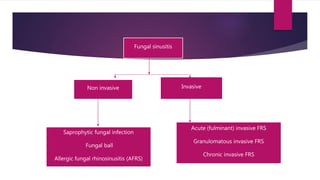
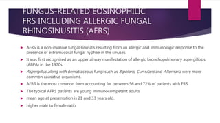
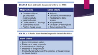
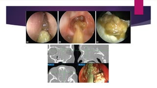
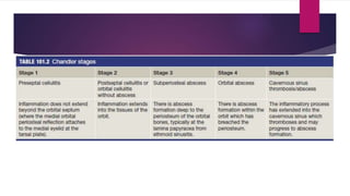
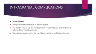
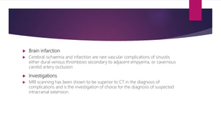
This document discusses various types of fungal sinusitis. It begins by categorizing fungal rhinosinusitis into invasive and non-invasive types based on the presence or absence of fungal invasion of tissue. It then describes the different subtypes of non-invasive and invasive fungal sinusitis in detail, including their pathogenesis, clinical presentation, diagnosis and management. It also discusses potential complications of fungal sinusitis such as orbital or intracranial infections that can arise from local or distant spread beyond the paranasal sinuses.
Deviated nasal septum and other septal conditionskrishnakoirala4
Slides prepared by highly experienced ENT teacher, Dr. Krishna Koirala from Nepal , for teaching undergraduate and postgraduate MBBS students in the field of otorhinolaryngology. A clear and concise explanation of the basic concepts in the subject matter concerned.
Steroids and their use in ENT
Get in touch with us at:
www.facebook.com/drraiammar
www.twitter.com/drraiammar
www.instagram.com/drraiammar
www.linkedin.com/in/drraiammar
www.themedicall.com/blog/auther/drraiammar/
For Any Book or Notes Visit Our Website:
www.allmedicaldata.wordpress.com
www.drraiammar.blogspot.com
YouTube Channel :
https://www.youtube.com/channel/UCu-oR9V3OdFNTJW5yqXWXxA
BY:
DR RAI M. AMMAR MADNI
Medically Important Aspergillus species.pptxNawangSherpa6
The Presentation here is about Medically important Aspergillus species. How does it infect the Human host? What are it's clinical manifestations and How can we diagnose for their infection and potential application for other studies.
ASPERGILLOSIS, MUCORMYCOSIS AND HISTOPLASMOSIS.pptxMkindi Mkindi
There are nearly 100 viruses of the herpes group that infect many different animal species.
Official name of herpesviruses that commonly infect human is Humans herpesvirus (HHV)
herpes simplex virus types 1 (HHV 1)
Herpes simplex virus type 2 (HHV 2)
Varicella-zoster virus (HHV 3)
Epstein-Barr virus, (HHV 4)
Cytomegalovirus (HHV 5)
Human herpesvirus 6 (HHV 6)
Human herpesvirus 7 (HHV 7)
Human herpesvirus 8 (HHV 8) (Kaposi's sarcoma-associated herpesvirus).
Herpes B virus of monkeys can also infect humans
hELMINTHS#corona virus#Aspergillosis#BUGANDO#CUHAS#CUHAS#CUHAS
New Drug Discovery and Development .....NEHA GUPTA
The "New Drug Discovery and Development" process involves the identification, design, testing, and manufacturing of novel pharmaceutical compounds with the aim of introducing new and improved treatments for various medical conditions. This comprehensive endeavor encompasses various stages, including target identification, preclinical studies, clinical trials, regulatory approval, and post-market surveillance. It involves multidisciplinary collaboration among scientists, researchers, clinicians, regulatory experts, and pharmaceutical companies to bring innovative therapies to market and address unmet medical needs.
Recomendações da OMS sobre cuidados maternos e neonatais para uma experiência pós-natal positiva.
Em consonância com os ODS – Objetivos do Desenvolvimento Sustentável e a Estratégia Global para a Saúde das Mulheres, Crianças e Adolescentes, e aplicando uma abordagem baseada nos direitos humanos, os esforços de cuidados pós-natais devem expandir-se para além da cobertura e da simples sobrevivência, de modo a incluir cuidados de qualidade.
Estas diretrizes visam melhorar a qualidade dos cuidados pós-natais essenciais e de rotina prestados às mulheres e aos recém-nascidos, com o objetivo final de melhorar a saúde e o bem-estar materno e neonatal.
Uma “experiência pós-natal positiva” é um resultado importante para todas as mulheres que dão à luz e para os seus recém-nascidos, estabelecendo as bases para a melhoria da saúde e do bem-estar a curto e longo prazo. Uma experiência pós-natal positiva é definida como aquela em que as mulheres, pessoas que gestam, os recém-nascidos, os casais, os pais, os cuidadores e as famílias recebem informação consistente, garantia e apoio de profissionais de saúde motivados; e onde um sistema de saúde flexível e com recursos reconheça as necessidades das mulheres e dos bebês e respeite o seu contexto cultural.
Estas diretrizes consolidadas apresentam algumas recomendações novas e já bem fundamentadas sobre cuidados pós-natais de rotina para mulheres e neonatos que recebem cuidados no pós-parto em unidades de saúde ou na comunidade, independentemente dos recursos disponíveis.
É fornecido um conjunto abrangente de recomendações para cuidados durante o período puerperal, com ênfase nos cuidados essenciais que todas as mulheres e recém-nascidos devem receber, e com a devida atenção à qualidade dos cuidados; isto é, a entrega e a experiência do cuidado recebido. Estas diretrizes atualizam e ampliam as recomendações da OMS de 2014 sobre cuidados pós-natais da mãe e do recém-nascido e complementam as atuais diretrizes da OMS sobre a gestão de complicações pós-natais.
O estabelecimento da amamentação e o manejo das principais intercorrências é contemplada.
Recomendamos muito.
Vamos discutir essas recomendações no nosso curso de pós-graduação em Aleitamento no Instituto Ciclos.
Esta publicação só está disponível em inglês até o momento.
Prof. Marcus Renato de Carvalho
www.agostodourado.com
Basavarajeeyam is an important text for ayurvedic physician belonging to andhra pradehs. It is a popular compendium in various parts of our country as well as in andhra pradesh. The content of the text was presented in sanskrit and telugu language (Bilingual). One of the most famous book in ayurvedic pharmaceutics and therapeutics. This book contains 25 chapters called as prakaranas. Many rasaoushadis were explained, pioneer of dhatu druti, nadi pareeksha, mutra pareeksha etc. Belongs to the period of 15-16 century. New diseases like upadamsha, phiranga rogas are explained.
MANAGEMENT OF ATRIOVENTRICULAR CONDUCTION BLOCK.pdfJim Jacob Roy
Cardiac conduction defects can occur due to various causes.
Atrioventricular conduction blocks ( AV blocks ) are classified into 3 types.
This document describes the acute management of AV block.
HOT NEW PRODUCT! BIG SALES FAST SHIPPING NOW FROM CHINA!! EU KU DB BK substit...GL Anaacs
Contact us if you are interested:
Email / Skype : kefaya1771@gmail.com
Threema: PXHY5PDH
New BATCH Ku !!! MUCH IN DEMAND FAST SALE EVERY BATCH HAPPY GOOD EFFECT BIG BATCH !
Contact me on Threema or skype to start big business!!
Hot-sale products:
NEW HOT EUTYLONE WHITE CRYSTAL!!
5cl-adba precursor (semi finished )
5cl-adba raw materials
ADBB precursor (semi finished )
ADBB raw materials
APVP powder
5fadb/4f-adb
Jwh018 / Jwh210
Eutylone crystal
Protonitazene (hydrochloride) CAS: 119276-01-6
Flubrotizolam CAS: 57801-95-3
Metonitazene CAS: 14680-51-4
Payment terms: Western Union,MoneyGram,Bitcoin or USDT.
Deliver Time: Usually 7-15days
Shipping method: FedEx, TNT, DHL,UPS etc.Our deliveries are 100% safe, fast, reliable and discreet.
Samples will be sent for your evaluation!If you are interested in, please contact me, let's talk details.
We specializes in exporting high quality Research chemical, medical intermediate, Pharmaceutical chemicals and so on. Products are exported to USA, Canada, France, Korea, Japan,Russia, Southeast Asia and other countries.
Flu Vaccine Alert in Bangalore Karnatakaaddon Scans
As flu season approaches, health officials in Bangalore, Karnataka, are urging residents to get their flu vaccinations. The seasonal flu, while common, can lead to severe health complications, particularly for vulnerable populations such as young children, the elderly, and those with underlying health conditions.
Dr. Vidisha Kumari, a leading epidemiologist in Bangalore, emphasizes the importance of getting vaccinated. "The flu vaccine is our best defense against the influenza virus. It not only protects individuals but also helps prevent the spread of the virus in our communities," he says.
This year, the flu season is expected to coincide with a potential increase in other respiratory illnesses. The Karnataka Health Department has launched an awareness campaign highlighting the significance of flu vaccinations. They have set up multiple vaccination centers across Bangalore, making it convenient for residents to receive their shots.
To encourage widespread vaccination, the government is also collaborating with local schools, workplaces, and community centers to facilitate vaccination drives. Special attention is being given to ensuring that the vaccine is accessible to all, including marginalized communities who may have limited access to healthcare.
Residents are reminded that the flu vaccine is safe and effective. Common side effects are mild and may include soreness at the injection site, mild fever, or muscle aches. These side effects are generally short-lived and far less severe than the flu itself.
Healthcare providers are also stressing the importance of continuing COVID-19 precautions. Wearing masks, practicing good hand hygiene, and maintaining social distancing are still crucial, especially in crowded places.
Protect yourself and your loved ones by getting vaccinated. Together, we can help keep Bangalore healthy and safe this flu season. For more information on vaccination centers and schedules, residents can visit the Karnataka Health Department’s official website or follow their social media pages.
Stay informed, stay safe, and get your flu shot today!
Title: Sense of Smell
Presenter: Dr. Faiza, Assistant Professor of Physiology
Qualifications:
MBBS (Best Graduate, AIMC Lahore)
FCPS Physiology
ICMT, CHPE, DHPE (STMU)
MPH (GC University, Faisalabad)
MBA (Virtual University of Pakistan)
Learning Objectives:
Describe the primary categories of smells and the concept of odor blindness.
Explain the structure and location of the olfactory membrane and mucosa, including the types and roles of cells involved in olfaction.
Describe the pathway and mechanisms of olfactory signal transmission from the olfactory receptors to the brain.
Illustrate the biochemical cascade triggered by odorant binding to olfactory receptors, including the role of G-proteins and second messengers in generating an action potential.
Identify different types of olfactory disorders such as anosmia, hyposmia, hyperosmia, and dysosmia, including their potential causes.
Key Topics:
Olfactory Genes:
3% of the human genome accounts for olfactory genes.
400 genes for odorant receptors.
Olfactory Membrane:
Located in the superior part of the nasal cavity.
Medially: Folds downward along the superior septum.
Laterally: Folds over the superior turbinate and upper surface of the middle turbinate.
Total surface area: 5-10 square centimeters.
Olfactory Mucosa:
Olfactory Cells: Bipolar nerve cells derived from the CNS (100 million), with 4-25 olfactory cilia per cell.
Sustentacular Cells: Produce mucus and maintain ionic and molecular environment.
Basal Cells: Replace worn-out olfactory cells with an average lifespan of 1-2 months.
Bowman’s Gland: Secretes mucus.
Stimulation of Olfactory Cells:
Odorant dissolves in mucus and attaches to receptors on olfactory cilia.
Involves a cascade effect through G-proteins and second messengers, leading to depolarization and action potential generation in the olfactory nerve.
Quality of a Good Odorant:
Small (3-20 Carbon atoms), volatile, water-soluble, and lipid-soluble.
Facilitated by odorant-binding proteins in mucus.
Membrane Potential and Action Potential:
Resting membrane potential: -55mV.
Action potential frequency in the olfactory nerve increases with odorant strength.
Adaptation Towards the Sense of Smell:
Rapid adaptation within the first second, with further slow adaptation.
Psychological adaptation greater than receptor adaptation, involving feedback inhibition from the central nervous system.
Primary Sensations of Smell:
Camphoraceous, Musky, Floral, Pepperminty, Ethereal, Pungent, Putrid.
Odor Detection Threshold:
Examples: Hydrogen sulfide (0.0005 ppm), Methyl-mercaptan (0.002 ppm).
Some toxic substances are odorless at lethal concentrations.
Characteristics of Smell:
Odor blindness for single substances due to lack of appropriate receptor protein.
Behavioral and emotional influences of smell.
Transmission of Olfactory Signals:
From olfactory cells to glomeruli in the olfactory bulb, involving lateral inhibition.
Primitive, less old, and new olfactory systems with different path
TEST BANK for Operations Management, 14th Edition by William J. Stevenson, Ve...kevinkariuki227
TEST BANK for Operations Management, 14th Edition by William J. Stevenson, Verified Chapters 1 - 19, Complete Newest Version.pdf
Title: Sense of Taste
Presenter: Dr. Faiza, Assistant Professor of Physiology
Qualifications:
MBBS (Best Graduate, AIMC Lahore)
FCPS Physiology
ICMT, CHPE, DHPE (STMU)
MPH (GC University, Faisalabad)
MBA (Virtual University of Pakistan)
Learning Objectives:
Describe the structure and function of taste buds.
Describe the relationship between the taste threshold and taste index of common substances.
Explain the chemical basis and signal transduction of taste perception for each type of primary taste sensation.
Recognize different abnormalities of taste perception and their causes.
Key Topics:
Significance of Taste Sensation:
Differentiation between pleasant and harmful food
Influence on behavior
Selection of food based on metabolic needs
Receptors of Taste:
Taste buds on the tongue
Influence of sense of smell, texture of food, and pain stimulation (e.g., by pepper)
Primary and Secondary Taste Sensations:
Primary taste sensations: Sweet, Sour, Salty, Bitter, Umami
Chemical basis and signal transduction mechanisms for each taste
Taste Threshold and Index:
Taste threshold values for Sweet (sucrose), Salty (NaCl), Sour (HCl), and Bitter (Quinine)
Taste index relationship: Inversely proportional to taste threshold
Taste Blindness:
Inability to taste certain substances, particularly thiourea compounds
Example: Phenylthiocarbamide
Structure and Function of Taste Buds:
Composition: Epithelial cells, Sustentacular/Supporting cells, Taste cells, Basal cells
Features: Taste pores, Taste hairs/microvilli, and Taste nerve fibers
Location of Taste Buds:
Found in papillae of the tongue (Fungiform, Circumvallate, Foliate)
Also present on the palate, tonsillar pillars, epiglottis, and proximal esophagus
Mechanism of Taste Stimulation:
Interaction of taste substances with receptors on microvilli
Signal transduction pathways for Umami, Sweet, Bitter, Sour, and Salty tastes
Taste Sensitivity and Adaptation:
Decrease in sensitivity with age
Rapid adaptation of taste sensation
Role of Saliva in Taste:
Dissolution of tastants to reach receptors
Washing away the stimulus
Taste Preferences and Aversions:
Mechanisms behind taste preference and aversion
Influence of receptors and neural pathways
Impact of Sensory Nerve Damage:
Degeneration of taste buds if the sensory nerve fiber is cut
Abnormalities of Taste Detection:
Conditions: Ageusia, Hypogeusia, Dysgeusia (parageusia)
Causes: Nerve damage, neurological disorders, infections, poor oral hygiene, adverse drug effects, deficiencies, aging, tobacco use, altered neurotransmitter levels
Neurotransmitters and Taste Threshold:
Effects of serotonin (5-HT) and norepinephrine (NE) on taste sensitivity
Supertasters:
25% of the population with heightened sensitivity to taste, especially bitterness
Increased number of fungiform papillae
Ethanol (CH3CH2OH), or beverage alcohol, is a two-carbon alcohol
that is rapidly distributed in the body and brain. Ethanol alters many
neurochemical systems and has rewarding and addictive properties. It
is the oldest recreational drug and likely contributes to more morbidity,
mortality, and public health costs than all illicit drugs combined. The
5th edition of the Diagnostic and Statistical Manual of Mental Disorders
(DSM-5) integrates alcohol abuse and alcohol dependence into a single
disorder called alcohol use disorder (AUD), with mild, moderate,
and severe subclassifications (American Psychiatric Association, 2013).
In the DSM-5, all types of substance abuse and dependence have been
combined into a single substance use disorder (SUD) on a continuum
from mild to severe. A diagnosis of AUD requires that at least two of
the 11 DSM-5 behaviors be present within a 12-month period (mild
AUD: 2–3 criteria; moderate AUD: 4–5 criteria; severe AUD: 6–11 criteria).
The four main behavioral effects of AUD are impaired control over
drinking, negative social consequences, risky use, and altered physiological
effects (tolerance, withdrawal). This chapter presents an overview
of the prevalence and harmful consequences of AUD in the U.S.,
the systemic nature of the disease, neurocircuitry and stages of AUD,
comorbidities, fetal alcohol spectrum disorders, genetic risk factors, and
pharmacotherapies for AUD.
2. Introduction
Fungal rhinosinusitis (FRS) can be categorized into two broad groups
Invasive
non-invasive
This is based on the presence or absence of fungus in the tissue (mucosa, blood vessel or
bone) respectively.
4. SAPROPHYTIC FUNGAL INFECTION
visible fungal colonization of mucus crusts seen within the nose and paranasal sinuses on
nasoendoscopy.
mechanism
dysfunction in mucociliary transportation from surgery
crust formation
platform for growth of fungal spores
precursors to fungal balls if left untreated.
patients are usually asymptomatic or may present with a foul smelling odour
Endoscopic cleaning of the infected crust with or without continued self-irrigation with saline is
usually the only treatment required
5.
6. Fungal ball
dense accumulation of extramucosal fungal hyphae, usually within one sinus, most
commonly the maxillary sinus.
most common organism Aspergillus
Fungal balls are seen more commonly in immunocompetent, middle-aged and
elderly females,
often with a history of previous dental procedure, especially dental fillings.
7. diagnosis
radiological findings
sinus opacification often with areas of
hyperattenuation
cheesy or clay-like debris within the sinus
accumulation of fungal hyphae without
evidence of tissue fungal invasion seen
microscopically
non-specific chronic inflammation of the
sinus and the absence of eosinophil
predominance, granuloma or allergic
mucin
8. Management wide opening of the involved sinus and complete removal of the
fungal debris.
Examination of the involved sinuses with angled scopes is crucial to ensure
complete surgical extirpation.
Subsequent regular surveillance in the clinic is necessary.
Oral or topical antifungals are not necessary
9. FUNGUS-RELATED EOSINOPHILIC
FRS INCLUDING ALLERGIC FUNGAL
RHINOSINUSITIS (AFRS)
AFRS is a non-invasive fungal sinusitis resulting from an allergic and immunologic response to the
presence of extramucosal fungal hyphae in the sinuses.
It was first recognized as an upper airway manifestation of allergic bronchopulmonary aspergillosis
(ABPA) in the 1970s.
Aspergillus along with dematiaceous fungi such as Bipolaris, Curvularis and Alternaria were more
common causative organisms.
AFRS is the most common form accounting for between 56 and 72% of patients with FRS.
The typical AFRS patients are young immunocompetent adults
mean age at presentation is 21 and 33 years old.
higher male to female ratio
10. Pathophysiology
underlying pathophysiology remains unknown and controversial.
number of popular theories have evolved.
Manning et al. proposed a mechanism derived from the ABPA model
Demonstrating raised fungal-specific IgE antibodies and IgG antibodies in AFRS patients, it is believed that
an atopic host exposed to fungi resulted in antigenic stimulation by a combination of Gell and Coombs
type I and type III hypersensitivity.
alternative theory was later proposed by Panikou et al stating that eosinophilic chemotaxis in response to
extramucosal fungi was the hallmark of the inflammatory reaction in AFRS.
eosinophilic theory does not explain the triggering factor for eosinophil chemotaxis.
The term eosinophilic mucin rhinosinusitis (EMRS) was used to describe non-allergic fungal sinusitis.
These patients were felt to have histological features similar to AFRS but without the presence of fungus.
The underlying mechanism was believed to be a systemic dysregulation of immunologic controls resulting
in upper and lower airway eosinophilia.
11.
12. Investigation
IMMUNOLOGIC TEST
Patients with AFRS have an elevated IgE level 50 and >1000 IU/ml.
The average total IgE level was about 550 IU/mL.
total serum IgE level may be useful in monitoring clinical activity in
the management of AFRS.
An in-vivo (skin prick) or invitro (RAST) test can be utilized to demonstrate fungal
specific IgE as a diagnostic criteria for AFRS
13.
14.
15. Histology
hallmark of AFRS is the presence of
allergic mucin.
Grossly, it is thick, tenacious and highly
viscous in consistency.
Hence, the terms ‘peanut butter’ and
‘axle-grease are often used to
describe the characteristic appearance
of the mucus.
Histologically, allergic mucin consists
of an eosinophilic mucin with necrotic
eosinophils, inflammatory cells,
Charcot-Leyden crystals (the by-
product of eosinophil) and fungal
hyphae.
16. FUNGAL CULTURE
A positive fungal culture provides supporting evidence in the diagnosis of AFRS. However, its
absence does not exclude the diagnosis of AFRS.
The presence of a positive fungal culture in AFRS patients ranged from 49 to 100%
17. Management
SURGICAL TREATMENT
Unlike the management of classical CRS, surgery is usually the first line treatment in the management
of AFRS.
Meticulous and complete endoscopic sinus surgery is the gold standard for the surgical extirpation of
polypoid disease and allergic mucin in an attempt to restore ventilation and drainage of the sinuses
Removal of allergic mucin and fungal debris eliminates the antigenic factor that incites the disease in
an atopic host.
Surgery also provides wide access for surveillance, clinical debridement and application of topical
medication
18.
19. Medical treatment
SYSTEMIC MEDICATIONS
I. Corticosteroids
Oral steroids are useful in the perioperative period ofpatients with AFRS. In the pre-operative period, a short
course of coritcosteroids reduce intra-operative bleeding and size of the polyps
II. Antifungals
Oral antifungals are considered a viable treatment option for patients with recalcitrant AFRS. They are also
used as a steroid-sparing medication, allowing some patients to be weaned off from long-term oral
corticosteroid therapy
oral itraconazole (200–400 mg daily) has potential benefits as a steroid sparing alternative and in prolonging
time to disease recurrence.
Oral itraconazole is associated with risk of elevated liver enzymes, congestive heart failure, nausea, rash,
headache, malaise, fatigue and oedema
Cessation of treatment is usually sufficient for the elevated liver enzymes to revert back to normal.
20. TOPICAL MEDICATION
I. Corticosteroid
Topical corticosteroids are used as standard treatment for patients with AFRS.
They are most effective in the postoperative period when open sinus cavities and
middle meati allow access to the paranasal sinuses.
The benefit of topical over systemic steroid lies in the ability of topical steroid to
achieve the highest drug concentration in the target tissue (sinonasal mucosa) without
the undesirable systemic side effects
These include metered-dose nasal sprays such as mometasone furoate, fluticasone
propionate, fluticasone furoate, budesonide, beclomethasone dipropionate
monohydrate, ciclesonide, flumisolide and triamcinolone acetonide.
21. II. Antifungals
As AFRS is a different disease entity resulting from an immunologic hyperreaction to extramucosal
fungus, topical antifungals should hypothetically reduce the immunologic reaction of an atopic host
decreasing the antigenic load.
fluconazole nasal spray, combined oral itraconazole and fluconazole nasal irrigation
larger RCTs will be required to establish the role of topical antifungals in the management of AFRS.
Non-standard topical nasal steroid (e.g. budesonide), oral antifungals and immunotherapy are
in cases of refractory AFRS.
Future treatment strategy
ANTI-IMMUNOGLOBULIN E (IGE) THERAPY
Omalizumab (Xolair) is a humanized monoclonal anti- IgE antibody that has been used as an
adjuvant treatment in severe atopic asthma, allergic rhinitis and CRS with nasal polyposis
22. ACUTE (FULMINANT) INVASIVE FRS
Acute or fulminant invasive FRS is a life-threatening disease present usually in
immunocompromised patients with impaired neutrophilic response.
These patients include those with uncontrolled diabetes mellitus, acquired immunodeficiency
syndrome (AIDS), iatrogenic immunosuppression, organ transplantation and haematological
malignancies.
This condition is characterized by the presence of hyphal invasion of sinus tissue and a time course
of less than 4 weeks
Histological features include mycotic infiltration of blood vessels, vasculitis with thrombosis, tissue
infarction, haemorrhage and acute neutrophilic infiltrate.
Aspergillus species and the fungi in the order of mucorale (e.g. Rhizopus, Rhizomucor and Mucor)
are the most commonly implicated species
23. clinical symptoms include fever, cough, crusting of the nasal mucosa, epistaxis and headaches,a
high index of suspicion of this disease entity should be present in any immunosuppressed
patients with localizing sinonasal symptoms.
In the early stages, nasoendoscopic findings may be as subtle as the presenting symptoms.
Alteration in mucosal appearance such as a discoloration, granulation and ulceration are the
most consistent physical findings.
CT scan is the initial radiologic investigation of choice
Compared to AFRS, invasive FRS tends to have more focal bony erosions, lacks expansion of
the sinuses, has more limited sinus disease and has more disease outside of the sinuses than
within when there is intra-orbital or intra-cranial extension.
24. Treatment
Surgery is necessary to halt or slow progression of the disease (allowing time for bone marrow
recovery), to reduce fungal load and to provide a tissue culture.
Prior to definitive identification of the causative fungi, empirical treatment with intravenous
amphotericin B, a broad-spectrum antifungal agent, has been recommended.
Once a causative fungal species has been identified, the use of the triazoles (fluconazole,
itraconazole and variconazole) can be considered
The triazoles are effective in the treatment of invasive FRS without the associated nephrotoxicity
seen in standard amphotericin B
25. GRANULOMATOUS INVASIVE FRS
This disease entity is defined by invasive fungal infection lasting more than 12 weeks.
The causative agent is almost exclusively Aspergillus flavus.
Patients are typically immunocompetent and the predominant clinical features include proptosis
with an enlarging mass in the cheek, nose, paranasal sinus and orbit
CT findings are not different to that of chronic invasive FRS although they have a tendency for
multiple sinus involvement
The distinguishing feature from chronic invasive FRS is histological findings of fungal tissue
invasion and a granulomatous reaction with considerable fibrosis.
This is evident from the presence of non-caseating granulomas with foreign body or Langerhans-
type giant cells, occasional vasculitis and sparse hyphae
Treatment includes complete surgical removal and antifungal agents.
26. CHRONIC INVASIVE FRS
Chronic invasive FRS is a slowly destructive disease with a time-course of more than 12 weeks
duration.
Patients are usually immunocompetent or have subtle abnormalities in the immune system from
diabetes mellitus, chronic low dose corticosteroid use and AIDS.
The most common fungi implicated is Aspergillus fumigatus
The clinical picture of chronic invasive FRS is similar to that of granulomatous invasive FRS.
The ethmoid and sphenoid sinuses are most commonly involved.
On histology, chronic invasive FRS demonstrates invasion of fungi into the sinonasal mucosa with a
dense accumulation of fungal hyphae, occasional vascular invasion, and chronic or sparse
inflammatory reaction.
There is no difference in the prognosis or the management of both chronic invasive and
granulomatous invasive FRS.
28. Complications of rhinosinusitis result from progression of acute or chronic
infection beyond the paranasal sinuses, potentially causing significant morbidity
from either local or distant spread.
Complications of rhinosinusitis are more accentuated in children and adolescents
because of their thinner, more porous bony septa and sinus walls, open suture
lines and larger vascular foramina.
29. CLASSIFICATION
Orbital
intra-cranial
bony or chronic.
Complications may be caused by either local progression or distant spread via the
bloodstream.
Local progression is typically through areas where the surrounding bone is thin such as
the porous lamina papyracea, where there is a direct anatomical connection or through
osteitic bone.
30.
31. Direct routes of spread occur through neurovascular foramina such as the infraorbital canal, or via
the valveless diploeic veins of Breschet of the frontal, ethmoid and sphenoid bones.
The venous drainage of the sinus mucosa is via these diploeic veins,which communicate with the
dural venous plexus, the absence of valves facilitates retrograde spread of infection.
32. Frontal
Anterior spread of acute frontal sinusitis through the outer table of the skull may
cause a boggy subperiosteal abscess and osteomyelitis. (Pott’s puffy tumour)
Posterior spread of infection can cause acute intracranial complications such as
subdural empyema, meningitis, cerebritis and intracranial abscess.
Subdural empyema is the most common intracranial complication of sinusitis, and the
most common cause of subdural empyema is frontal sinusitis.
Spread of infection inferiorly can lead to orbital cellulitis.
33. Ethmoid
Orbital cellulitis is by far the most frequent acute complication of ethmoid
sinusitis.
It can vary in degree and severity and is typically graded with the Chandler
classification
34. Maxillary
Isolated maxillary rhinosinusitis rarely gives rise to acute complications.
Acute cheek swelling usually results from complications of dental disease
Sphenoid
Isolated sphenoid sinusitis is rare, but complications can result in meningitis or cavernous sinus
thrombosis by direct spread.
In cavernous sinus thrombosis infection may spread through veins from the paranasal sinuses
and orbit to the cavernous sinuses as thrombophlebitis or by septic emboli.
Bacteria themselves are pro-thrombotic, the thrombus provides good conditions for growth and
bacteria within the thrombus are shielded by the outer layers from antibiotics which they can
later re-infect.
35. Orbital complications
The onset is noted by swelling around the eye. Oedema results from congestion of veins draining the eyelid and
can be present when the infection is still confined to the sinus.
Cellulitis from untreated sinusitis represents stage 1 of the disease with local spread of inflammatory elements to
the lid.
Postseptal cellulitis (stage 2) is confined to the orbit but has extended through the orbital septum, potentially with
intraconal involvement.
Patients will have eyelid swelling, chemosis and proptosis, potentially with impaired extraocular muscle function
and diplopia
Postseptal cellulitis typically results from transmitted pressure from the sinus to orbital veins with leakage of
inflammatory elements into the orbit.
It is difficult to clinically differentiate postseptal cellulitis from subperiosteal abscess (stage 3)
CT scanning is mandatory here and reflects the fact that the classification system pre-dates the ready availability of
scanning.
36.
37.
38.
39. EXAMINATION
Clinical endoscopic examination of the nose should be performed to help determine the site and extent
of disease.
In the case of orbital cellulitis, a formal assessment of the
degree of chemosis
range of eye movements
degree of proptosis
relative afferent pupillary defect
visual acuity (using a Snellen chart)
colour vision (using Ishihara plates)
inspection of the optic disc should be made.
For intracranial complications a full neurological examination should be completed.
40. INVESTIGATIONS
Radiological
The aim of these investigations is to:
• confirm the diagnosis of the complication
• define the extent and site of the complication
• help plan treatment including surgical approach
• confirm that there is no other covert complication
present
• Monitor the response to treatment.
41. Contrast enhanced computed tomography (CT) is advised as first-line imaging
because of its superiority in demonstrating bony anatomy and pathology of the
orbit and sinuses with its speed and ease of examination.
MRI provides outstanding soft tissue detail without radiation
42. Below are some of the common signs to look out for each stage of the Chandler classification.
1. Preseptal cellulitis – eyelid swelling and thickening of the preseptal tissues with possible
posterolateral extension to the temporal fossa.
Importantly, CT will not distinguish between oedema, preseptal cellulitis and allergy.
2. Postseptal cellulitis – the findings as above often with induration of the extraconal, intraconal
and retrobulbar fat without abscess.
3. Subperiosteal abscess – CT scans show a typical lenticular rim-enhancing collection adjacent
to the lamina papyracea with a fat plane between it and the displaced medial rectus muscle
Lateral deviation and proptosis of the globe may be visible on axial images.
Sight threatening complications of optic neuritis or optic nerve ischemia may also be present
without radiological signs. Subperiosteal abscess secondary to frontal sinusitis typically forms an
abscess superiorly with anteroinferior globe displacement
43.
44. 4. Orbital abscess – Scans may show multiple rimenhancing orbital abscesses
with surrounding cellulitis and a resultant proptosis.
5. Cavernous sinus thrombosis – It is important to note that in the early stages
CT scans appear normal.
CT and MR venography are complementary in the diagnosis of cavernous sinus
thrombosis.
There may also be thickening and increased signal intensity in the extraocular
muscles in T2-weighted MRI
45. Treatment
Medical management
Unless an abscess is demonstrated by radiological or other investigation, non-surgical
management of rhinosinusitis complications is normally first choice.
The exception is when vision is affected by pressure on the optic nerve from surrounding
inflammation without abscess formation.
The aim of medical management is to control and eliminate both the disease process of the
complication and of the primary rhinosinusitis.
Antibiotics form the mainstay of medical treatment
Broad-spectrum antibiotics are advised for severe complications of sinusitis to cover the likely
organisms including S. pneumonia, S. anginosus and other Streptococcus species, H.
influenzae, S. aureus, Moraxella catarrhalis and anaerobic bacteria (Prevotella,
Porphyromonas, Fusobacterium and Peptostreptococcus species
46. Surgical management
If there is no significant clinical improvement in the first 24 hours of medical
treatment, or there is any clinical deterioration surgical intervention should be
considered.
The surgical treatment of patients with complications of rhinosinusitis can be
divided into procedures necessary to manage the complication and surgery for
the rhinosinusitis.
47.
48. INTRACRANIAL COMPLICATIONS
Brain abscess
complication of either local or distant spread.
The frontal sinuses are the most common source followed by the ethmoid,
sphenoid and maxillary sinuses.
Haematogenous spread is the most likely mechanism of distant spread.
49. Subdural empyema
one of the commonest intracranial complications of rhinosinusitis, typically from haematogenous
spread
Subdural empyemas present with meningeal irritation and neurological signs such as seizures or
focal deficits.
Serious neurological injury can occur if not treated rapidly and aggressively with combined
treatment and neurosurgical drainage to decompress the brain and evacuate the empyema.
Extradural empyema
Extradural empyemas tend to be less symptomatic as the brain is protected by the dura mater.
signs are less marked and specific and are often only present when the collection reaches a size to
cause mass effect.
50. Brain infarction
Cerebral ischaemia and infarction are rare vascular complications of sinusitis
either dural venous thrombosis secondary to adjacent empyema, or cavernous
carotid artery occlusion
Investigations
MRI scanning has been shown to be superior to CT in the diagnosis of
complications and is the investigation of choice for the diagnosis of suspected
intracranial extension.
51.
52. BONY COMPLICATIONS
Pott’s puffy tumour
Subperiosteal cellulitis or abscess of the frontal bone associated with frontal
osteomyelitis and presenting with headache, swelling and, on occasion, a
discharging frontal fistula.
The infection can spread posteriorly giving rise to intracranial sepsis either by
erosion of the posterior table or more likely by septic thrombophlebitis via the
diploeic veins
Management requires drainage of pus from the frontal sinuses, achieved either
endoscopically or conventionally through a frontal sinus trephination via an
incision in the superomedial aspect of the orbit.
53.
54. CHRONIC COMPLICATIONS
Mucoceles are chronic, slowly expanding lesions in any of the sinuses that may
result in bony erosion and can extend beyond the sinus
55. Rhino orbital mucormycosis
Rhino-orbital mucormycosis is a rare but life threatening infection that generally occurs in
patients with diabetes mellitus and other immune deficiency conditions.
Rhino-orbital and Rhino-cerebral are two form of the disease.
As such the condition is a medical emergency.
Early recognition and treatment are essential because it may lead to death in few days.
Fungal infection of nasal cavity is uncommon but is being seen with increasing frequency
in patients with immune deficiency.
56. Etiology
Mucormycosis are a group of invasive infections which are caused by filamentous fungi of the order,
Mucorales of the Mucoraceae Family.
Rhino-orbital mucormycosis is an aggressive, angioinvasive fungal infection which is seen
inimmunocompromised hosts.
The risk factors are poorly controlled Diabetes mellitus, haematological malignancies and a prolonged
corticosteroid treatment.
The infections which are caused by members of the order mucorales are primarily opportunistic
infections and they represent the third leading cause of invasive fungal infections
following Aspergillus and Candida species.
57. It is an acute opportunistic infection which is caused by a broad, non septate,
saprophytic fungus which is found in soil, air, bread mould, rotten fruit and
vegetables.
It can be cultured from the mouth, nasal tract, throat and the faeces of healthy
persons.
The fungus belongs to the Phycomycetes class, whose most common genera
are Mucor, Rhizopus, Absidia and Basidiobolus.
Contact with the micro organism occurs through spore inhalation.
58. Pathophysiology
The reduced ability of serum to bind iron at a low pH may be the basic defect in the body’s defense
systems.
The high iron, glucose-rich acid medium facilitates fungal growth
The human resistance to fungal infections rests on the body’s ability to restrict the availability of iron
to the invading fungus, by binding it to proteins such as apotransferrin.
The fungal hyphae produce a substance called rhizoferrin, which binds iron avidly.
This iron-rhizoferrin complex is then taken up by the fungus and it becomes available for the vital
intracellular processes.
Diabetic patients are predisposed to mucormycosis because of the decreased ability of their
neutrophils to phagocytose and adhere to the endothelial walls.
Furthermore, the acidosis and hyperglycaemia provide an excellent environment for the fungus to
grow
59. The infection spreads along the vascular and neuronal structures and it infiltrates
the walls of the blood vessels.
It causes erosion of the bony walls of the ethmoid sinuses and it may spread into
the orbit and the retro-orbital area and in the brain (cerebro-rhino-orbital
mucormycosis).
Death may occur due to the cerebral abscesses.
60. Clinical features
Fever > 48 hrs despite adequate medical therapy
Blood stained nasal discharge
Facial pain, facial swelling
Orbital swelling opthalmoplegia, decreased vision
Proptosis CN VI palsy
Cavernous sinus thrombosis is a late sign of intracranial extension
Pulmonary mucormycosis – cough hemoptysis,SOB
Gastrointestinal mucormycosis – abdominal pain, hematemesis
Renal mucormycosis – fever, flank pain
Cutaneous mucormycosis – painful blackish hardened areas
61. Anterior rhinoscopy
Alteration of nasal mucosa appearance
White discoloration of pale mucosa indicates tissue ischemia secondary to
angiocentric invasion
Black discoloration indicates tissue necrosis
Decreased mucosal bleeding
Anaesthetic regions
62. Investigations
Biopsy of polyp or unhealthy mucosa to make definitive diagnosis
Ct and MRI scans with contrast of the sinuses inclusive of orbit and brain.
Opthal opinion for visual acquity and fundus examination
Neurosurgeon to rule out intracranial and cavernous sinus involvement
63. Treatment
Emergency surgical debridement is the treatment of choice
In some cases surgery may be disfiguring because it may involve removal of
palate, nasal cavity or eye structures
Medial maxillectomy may be necessary
Anti fungal agents
Amphotericin B
Derived from streptomyces nodosus
It is poorly absorbed orally hence given intravenously
64. Mechanism of action
It gets bound to ergosterol in the cell membrane of fungus and increases the
permeability of cell membrane andcell contents leak out leading to death of cell
Dose: 1-3 mg/kg/bodyweight given as slow IV
Test dose of 1mg in 50 ml of 5% dextrose given IV over 20-30 min
30 mins later 20 mg IV is given
Next day 40 mg IV is administered.