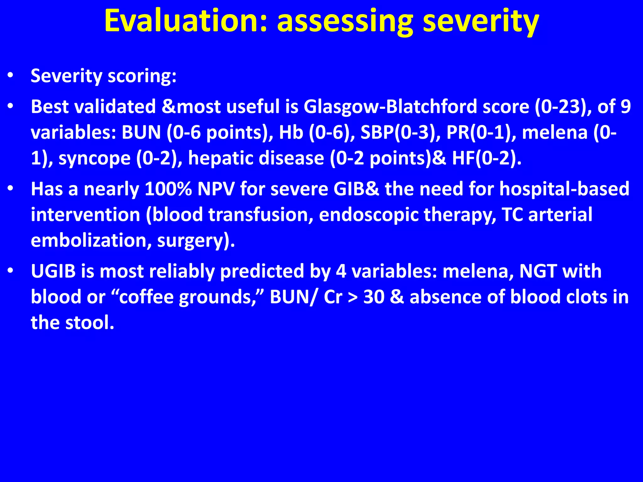
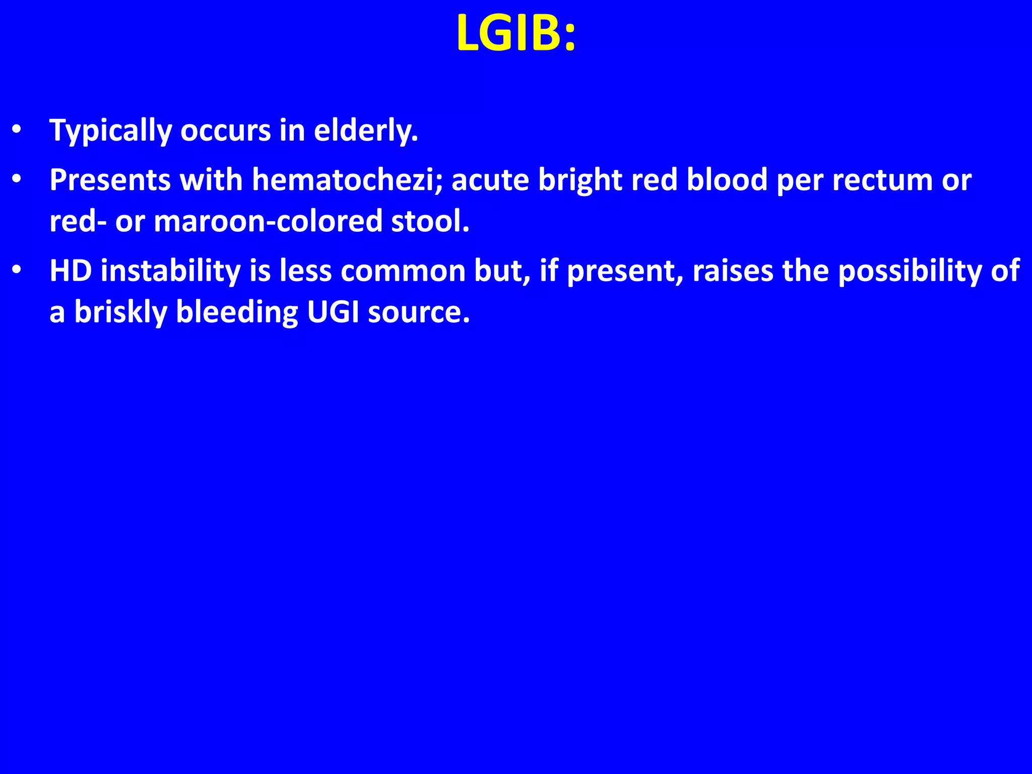
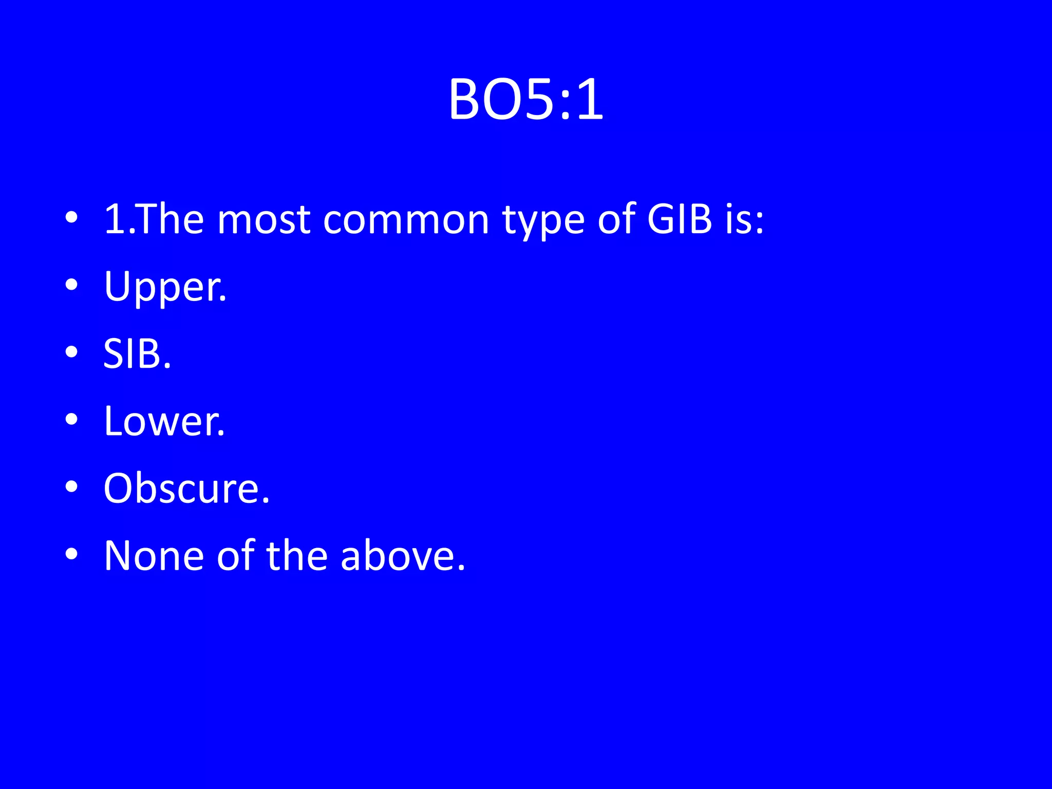
Dr. Mohamed Alshekhani
Professor in Medicine
MBChB-CABM-FRCP-EBGH
2016
1
GIB
Introduction:
• Upper :80%
• Lower :15%
• SIB:5%
• Proximal or distal to the ligament of Treitz.
Upper Gastrointestinal Bleeding
• Presents with:
• Hematemesis (bright-red or “coffee-ground” emesis)
• Melena (black, tarry-appearing stool)
• Or very rarely hematochezia or bright red blood per rectum due to
briskly UGIB, which is associated with increased mortality.
UGIB: Causes
• 80% is due to 4 causes:
• PUD
• EV
• Esophagitis
• Mallory-Weiss tear.
• Bleeding in 80% stops spontaneously
• 20% have persistent or recurrent bleeding, increasing mortality.
UGIB: Causes
• Slow &/or chronic bleeding:
• Suggested by history of IDA.
• Typical of erosive disease: tumor, esophageal ulcer, portal
hypertensive gastropathy, Cameron lesion (5%, eroded large hiatal
hernias)& angiodysplasia.
UGIB:Causes
• Causes of brisk&/or severe upper GIB that increase mortality.
• Peptic ulcer
• Esophagogastric varices
• Dieulafoy lesion
• Aortoenteric fistula
• Hemobilia: usually from liver or biliary procedural complication or
gallstone complications, tumors &angiodysplasia.
• Hemosuccus pancreaticus: (pseudoaneurysm/aneurysm)
• Neoplasm
• Esophageal lesions
• Gastric GIST
UGIB : evaluation by History
• H/O chronic alcohol abuse: a clue to the possibility of VH.
• Chronic dyspepsia: PUD.
• NSAIDs use: PUD.
• H/O Aortic aneurysm repair: aortoenteric fistula.
UGIB : evaluation by History
• Predictors of severe GIB are:
• Hematemesis
• Comorbidities (such as cirrhosis or malignancy)
• HD instability
• Hb <8 g/dL (80 g/L).
• bleeding source.
UGIB : evaluation aims
• Assessing severity.
• Differentiating upper from lower GIB sources.
• Determining the need for interventions.
Evaluation: assessing severity
• Outpatient management is usually appropriate when the following
criteria are met:
• BUN<18.2 mg/dL (6.5 mmol/L)
• Normal Hb
• Systolic BP>109 mm Hg
• PR< or equal to 100/min
• Absence of: melena, Syncope,Liver disease,Cardiac failure.
Evaluation: assessing severity
• Risk-stratification tools guides decisions regarding:
• Hospital admission.
• Discharge home from ER.
• Urgent endoscopy (within 12 hours)
• Non-urgent endoscopy (within 24 hours)
Evaluation: assessing severity
• Severity scoring:
• Best validated &most useful is Glasgow-Blatchford score (0-23), of 9
variables: BUN (0-6 points), Hb (0-6), SBP(0-3), PR(0-1), melena (0-
1), syncope (0-2), hepatic disease (0-2 points)& HF(0-2).
• Has a nearly 100% NPV for severe GIB& the need for hospital-based
intervention (blood transfusion, endoscopic therapy, TC arterial
embolization, surgery).
• UGIB is most reliably predicted by 4 variables: melena, NGT with
blood or “coffee grounds,” BUN/ Cr > 30 & absence of blood clots in
the stool.
UGIB Management:
• 1. Pre-endoscopic care (resuscitation, hemodynamic monitoring, PPI
therapy, attention to coagulopathy)
• 2. Early endoscopic evaluation (with excellent endoscopic vision) &
treatment.
• 3. Postendoscopic care & risk reduction.
UGIB Management:
• Pre-endoscopic Care:
• 1.Resuscitated with crystalloids to reach physiologic endpoints (PR
<100/min, SBP>100 mm Hg&resolution of orthostasis).
• 2.Blood transfusion indicated:
• A. HD instability &ongoing bleeding or susceptibility to
complications from hypoxia (for example IHD).
• B. Target Hb < 7 g/dL, if HD stable with no active or massive
bleeding.
• 3.Early (pre-endoscopic) PPI does not improve clinical outcomes
(bleeding, surgery, mortality) but is safe & reduces the likelihood of
detecting ulcers with high-risk stigmata & need for endoscopic trt.
• 4.Coagulopathy (INR >1.5) corrected with FRP not vit K (delayed full
therapeutic effect) in actively bleeding receiving anticoags.
• 5.Octreotide & antibiotics should be given before endoscopy for
suspected variceal bleeding.
UGIB Management:
• NGT is not required for diagnosis, prognosis, visualization, or
therapeutic effect.
• Routine use of prokinetics is not recommended except when
patients are suspected of having large amounts of blood in the
UGIT; in such cases, erythromycin can be given prior to upper
endoscopy.
UGIB Management:
• Endoscopic treatment:
• Upper endoscopy within 24 hours of presentation in patients with
features of UGIB.
• Endoscopy within 12 hours is generally recommended only for
patients with suspected variceal bleeding.
• Low-risk ulcers not requiring endoscopic intervention are clean-
based or have a non-protuberant pigmented spot.
• Intermediate-risk ulcers have adherent clots & should be vigorously
irrigated to dislodge the clot & reclassified based on appearance.
• High-risk ulcers that require endoscopic treatment: active arterial
spurting or a non-bleeding visible vessel &.
• Routine second-look endoscopy is not required after UGIB unless
rebleeding occurs or the initial examination was incomplete.
UGIB Management:
• Post-endoscopic Care & Risk Reduction:
• Post endoscopic PPI improves outcome after endoscopic
interventions.
• PUD tested for H pylori &If positive, eradication done &confirmed.
• If negative, retesting done with an alternative method BZ of false-
negative results with bleeding, PPI, or concomitant antibiotics.
• Aspirin should be resumed within 3 - 5 days for patients with
established CVD.
• Long-term PPI may not be necessary for aspirin users who undergo
H. pylori testing &eradication.
• Long-term, daily PPI should be offered to aspirin users who are H.
pylori negative or those who use concomitant NSAIDs,
anticoagulants, glucocorticoids, or other antiplatelets.
Variceal bleeding Management:
• Octreotide / telipresin infusion & antibiotics are given even if this is
suspected.
• FLuid resuscitation is preferred with crystaloids.
• Endoscopic intervention can be done safely even with INR up to 2.5
& above that, correction done with FFP.
• Endoscopic band ligation is preferred over sclerotherpay for acute
esophageal variceal bleeding.
• For bleeding gastric varices cyanoacrylate sclerotherpay is preferred
over band ligation.
• When the above measures fail, temponade with esophgeal balloons
as Baltimore-Sengestaken tube is used as bridge to more definitive
therapies as TIPS or surgery.
• NS Beta-blockers are used after the control of the bleeding.
LGIB:
• Typically occurs in elderly.
• Presents with hematochezi; acute bright red blood per rectum or
red- or maroon-colored stool.
• HD instability is less common but, if present, raises the possibility of
a briskly bleeding UGI source.
LGIB:
• Causes:
• Diverticulosis 30%
• Colitis 24%
• Ischemic 12%
• IBD 9%
• Radiation 3%
• Hemorrhoids 14%
• Postpolypectomy 8%
• Colon polyps or cancer 6%
• Rectal ulcer 6%
• Angiodysplasia 3%
• Other 6%.
LGIB:
• Causes of severe LGIB:
• Diverticulosis
• Aortoenteric fistula
• Colonic or rectal varices
• Dieulafoy lesions
• Neoplasm
• Colitis
• Ischemic
• IBD
• Infectious
• Intussusception
• Meckel diverticulum
• Angiodysplasia
SIB or Obscure GIB
• Relatively uncommon ; 5–10% of GIB.
• with advances in SI imaging(VCE, deep enteroscopy& radioimaging)
the cause of bleeding in SI identified in most patients.
• OGIB should be reserved for patients in whom a source of bleeding
cannot be identified anywhere in the GI tract.
• SIB should be considered in patients with GI bleeding after
performance of a normal upper & lower endoscopic exams.
• Second-look exams using upper endoscopy, push enteroscopy&/or
colonoscopy can be performed if indicated before SB evaluation.
• VCE should be considered a first-line procedure for SIB& should be
performed before deep enteroscopy if there is no contraindication.
• Any method of deep enteroscopy can be used when endoscopic
evaluation& therapy are required.
SIB or Obscure GIB
• CTE should be performed in patients with suspected obstruction
before VCE or after negative VCE exams.
• When there is acute overt hemorrhage in the unstable patient,
angiography should be performed emergently.
• In patients with occult hemorrhage or stable patients with active
overt bleeding, multiphasic computed tomography should be
performed after VCE or CTE to identify the source of bleeding &
guide further management.
• If a source of bleeding is identified in the small bowel that is
associated with significant ongoing anemia and/or active bleeding,
the patient should be managed with endoscopic therapy.
• Conservative management is recommended for patients without a
source found after SB investigation, whereas repeat diagnostic
investigations are recommended for patients with initial negative
SB evaluations & ongoing overt or occult bleeding.
BO5:1
• 1.The most common type of GIB is:
• Upper.
• SIB.
• Lower.
• Obscure.
• None of the above.
BO5:2
• 2.The least common type of GIB is:
• Upper.
• SIB.
• Lower.
• Overt.
• None of the above.
BO5:3
• 3.The most common presentation of UGIB is:
• Fresh hematemesis.
• Melena.
• Hematochesia.
• Cofee-ground vomiting.
• None of the above.
BO5:4
• 4.The most common presentation of LGIB is:
• Fresh hematemesis.
• Melena.
• Hematochesia.
• Cofee-ground vomiting.
• None of the above.
BO5:5
• 5.The following can be a presentation of UGIB
except:
• Fresh hematemesis.
• Melena.
• Hematochesia.
• Cofee-ground vomiting.
• None of the above.
BO5:6
• 6.The following are essential component of
significant UGIB except:
• IV access.
• PPI.
• Upper endoscopy.
• NG Tube.
• Blood grouping and cross match.
BO5:7
• 7.The prokinetic of choice in severe upper GIB
is:
• IV metochlorpromide.
• IV erythromycin.
• Oral erythromycin.
• Oral domperidone.
• None of the above.
BO5:8
• 8.Interventions that improve survival in severe
upper GIB include all except:
• Endoscopic hemostasis if indicated.
• Pre-endoscopy PPI.
• Post-endoscopic PPI.
• Radiological intervention.
• Surgery.
BO5:9
• 9.The need for endoscopic intervention is
decided by:
• Forrest classification.
• Blackford scoring.
• Glasgo scoring.
• Apgar scoring.
• Non of the above.
BO5:10
• 10.The following endoscopic lesions warrants
endoscopic intervention during UGIB except:
• Spurting vessel.
• Oozing lesion.
• Visible vessel.
• Adherent clot.
• Ulcer with pigmentation.
BO5:11
• 11.The endoscopic intervention of choice for
severe UGIB is:
• Single.
• Dual.
• Triple.
• All.
• None.
BO5:12
• 12.H Pylori testing during UGIB is frequently:
• False positive.
• False negative.
• Accurate.
• Need need not to be repeated.
• All of the above.
BO5:13
• 13.The first step in managing severe LGIB is:
• Colonoscopy.
• Exclude upper GI source.
• Surgery.
• IV PPI.
• All of the above.
BO5:14
• 14.The best approach in managing severe LGIB
which can not be hemoynamicly be stabilized
is:
• Angiographic intervention.
• Colonoscopy.
• Surgery.
• IV PPI.
• All of the above.
BO5:15
• 15.Endoscopy in non-variceal UGIB is
indicated within:
• 12 hours.
• 24 hours.
• 36 hours.
• 48 hours.
• 72 hours.
BO5:16
• 16.Endoscopy in variceal UGIB is indicated
within:
• 12 hours.
• 24 hours.
• 36 hours.
• 48 hours.
• 72 hours.
BO5:17
• 17.The following should be given in suspected
variceal UGIB except:
• Somatostatin.
• Telepressin.
• PPI.
• Antibiotics.
• Crystaloides.

GIT 4th GIB16.

  • 1.
    Dr. Mohamed Alshekhani Professorin Medicine MBChB-CABM-FRCP-EBGH 2016 1 GIB
  • 2.
    Introduction: • Upper :80% •Lower :15% • SIB:5% • Proximal or distal to the ligament of Treitz.
  • 3.
    Upper Gastrointestinal Bleeding •Presents with: • Hematemesis (bright-red or “coffee-ground” emesis) • Melena (black, tarry-appearing stool) • Or very rarely hematochezia or bright red blood per rectum due to briskly UGIB, which is associated with increased mortality.
  • 4.
    UGIB: Causes • 80%is due to 4 causes: • PUD • EV • Esophagitis • Mallory-Weiss tear. • Bleeding in 80% stops spontaneously • 20% have persistent or recurrent bleeding, increasing mortality.
  • 5.
    UGIB: Causes • Slow&/or chronic bleeding: • Suggested by history of IDA. • Typical of erosive disease: tumor, esophageal ulcer, portal hypertensive gastropathy, Cameron lesion (5%, eroded large hiatal hernias)& angiodysplasia.
  • 6.
    UGIB:Causes • Causes ofbrisk&/or severe upper GIB that increase mortality. • Peptic ulcer • Esophagogastric varices • Dieulafoy lesion • Aortoenteric fistula • Hemobilia: usually from liver or biliary procedural complication or gallstone complications, tumors &angiodysplasia. • Hemosuccus pancreaticus: (pseudoaneurysm/aneurysm) • Neoplasm • Esophageal lesions • Gastric GIST
  • 7.
    UGIB : evaluationby History • H/O chronic alcohol abuse: a clue to the possibility of VH. • Chronic dyspepsia: PUD. • NSAIDs use: PUD. • H/O Aortic aneurysm repair: aortoenteric fistula.
  • 8.
    UGIB : evaluationby History • Predictors of severe GIB are: • Hematemesis • Comorbidities (such as cirrhosis or malignancy) • HD instability • Hb <8 g/dL (80 g/L). • bleeding source.
  • 9.
    UGIB : evaluationaims • Assessing severity. • Differentiating upper from lower GIB sources. • Determining the need for interventions.
  • 10.
    Evaluation: assessing severity •Outpatient management is usually appropriate when the following criteria are met: • BUN<18.2 mg/dL (6.5 mmol/L) • Normal Hb • Systolic BP>109 mm Hg • PR< or equal to 100/min • Absence of: melena, Syncope,Liver disease,Cardiac failure.
  • 11.
    Evaluation: assessing severity •Risk-stratification tools guides decisions regarding: • Hospital admission. • Discharge home from ER. • Urgent endoscopy (within 12 hours) • Non-urgent endoscopy (within 24 hours)
  • 12.
    Evaluation: assessing severity •Severity scoring: • Best validated &most useful is Glasgow-Blatchford score (0-23), of 9 variables: BUN (0-6 points), Hb (0-6), SBP(0-3), PR(0-1), melena (0- 1), syncope (0-2), hepatic disease (0-2 points)& HF(0-2). • Has a nearly 100% NPV for severe GIB& the need for hospital-based intervention (blood transfusion, endoscopic therapy, TC arterial embolization, surgery). • UGIB is most reliably predicted by 4 variables: melena, NGT with blood or “coffee grounds,” BUN/ Cr > 30 & absence of blood clots in the stool.
  • 13.
    UGIB Management: • 1.Pre-endoscopic care (resuscitation, hemodynamic monitoring, PPI therapy, attention to coagulopathy) • 2. Early endoscopic evaluation (with excellent endoscopic vision) & treatment. • 3. Postendoscopic care & risk reduction.
  • 14.
    UGIB Management: • Pre-endoscopicCare: • 1.Resuscitated with crystalloids to reach physiologic endpoints (PR <100/min, SBP>100 mm Hg&resolution of orthostasis). • 2.Blood transfusion indicated: • A. HD instability &ongoing bleeding or susceptibility to complications from hypoxia (for example IHD). • B. Target Hb < 7 g/dL, if HD stable with no active or massive bleeding. • 3.Early (pre-endoscopic) PPI does not improve clinical outcomes (bleeding, surgery, mortality) but is safe & reduces the likelihood of detecting ulcers with high-risk stigmata & need for endoscopic trt. • 4.Coagulopathy (INR >1.5) corrected with FRP not vit K (delayed full therapeutic effect) in actively bleeding receiving anticoags. • 5.Octreotide & antibiotics should be given before endoscopy for suspected variceal bleeding.
  • 15.
    UGIB Management: • NGTis not required for diagnosis, prognosis, visualization, or therapeutic effect. • Routine use of prokinetics is not recommended except when patients are suspected of having large amounts of blood in the UGIT; in such cases, erythromycin can be given prior to upper endoscopy.
  • 16.
    UGIB Management: • Endoscopictreatment: • Upper endoscopy within 24 hours of presentation in patients with features of UGIB. • Endoscopy within 12 hours is generally recommended only for patients with suspected variceal bleeding. • Low-risk ulcers not requiring endoscopic intervention are clean- based or have a non-protuberant pigmented spot. • Intermediate-risk ulcers have adherent clots & should be vigorously irrigated to dislodge the clot & reclassified based on appearance. • High-risk ulcers that require endoscopic treatment: active arterial spurting or a non-bleeding visible vessel &. • Routine second-look endoscopy is not required after UGIB unless rebleeding occurs or the initial examination was incomplete.
  • 18.
    UGIB Management: • Post-endoscopicCare & Risk Reduction: • Post endoscopic PPI improves outcome after endoscopic interventions. • PUD tested for H pylori &If positive, eradication done &confirmed. • If negative, retesting done with an alternative method BZ of false- negative results with bleeding, PPI, or concomitant antibiotics. • Aspirin should be resumed within 3 - 5 days for patients with established CVD. • Long-term PPI may not be necessary for aspirin users who undergo H. pylori testing &eradication. • Long-term, daily PPI should be offered to aspirin users who are H. pylori negative or those who use concomitant NSAIDs, anticoagulants, glucocorticoids, or other antiplatelets.
  • 19.
    Variceal bleeding Management: •Octreotide / telipresin infusion & antibiotics are given even if this is suspected. • FLuid resuscitation is preferred with crystaloids. • Endoscopic intervention can be done safely even with INR up to 2.5 & above that, correction done with FFP. • Endoscopic band ligation is preferred over sclerotherpay for acute esophageal variceal bleeding. • For bleeding gastric varices cyanoacrylate sclerotherpay is preferred over band ligation. • When the above measures fail, temponade with esophgeal balloons as Baltimore-Sengestaken tube is used as bridge to more definitive therapies as TIPS or surgery. • NS Beta-blockers are used after the control of the bleeding.
  • 23.
    LGIB: • Typically occursin elderly. • Presents with hematochezi; acute bright red blood per rectum or red- or maroon-colored stool. • HD instability is less common but, if present, raises the possibility of a briskly bleeding UGI source.
  • 24.
    LGIB: • Causes: • Diverticulosis30% • Colitis 24% • Ischemic 12% • IBD 9% • Radiation 3% • Hemorrhoids 14% • Postpolypectomy 8% • Colon polyps or cancer 6% • Rectal ulcer 6% • Angiodysplasia 3% • Other 6%.
  • 25.
    LGIB: • Causes ofsevere LGIB: • Diverticulosis • Aortoenteric fistula • Colonic or rectal varices • Dieulafoy lesions • Neoplasm • Colitis • Ischemic • IBD • Infectious • Intussusception • Meckel diverticulum • Angiodysplasia
  • 28.
    SIB or ObscureGIB • Relatively uncommon ; 5–10% of GIB. • with advances in SI imaging(VCE, deep enteroscopy& radioimaging) the cause of bleeding in SI identified in most patients. • OGIB should be reserved for patients in whom a source of bleeding cannot be identified anywhere in the GI tract. • SIB should be considered in patients with GI bleeding after performance of a normal upper & lower endoscopic exams. • Second-look exams using upper endoscopy, push enteroscopy&/or colonoscopy can be performed if indicated before SB evaluation. • VCE should be considered a first-line procedure for SIB& should be performed before deep enteroscopy if there is no contraindication. • Any method of deep enteroscopy can be used when endoscopic evaluation& therapy are required.
  • 29.
    SIB or ObscureGIB • CTE should be performed in patients with suspected obstruction before VCE or after negative VCE exams. • When there is acute overt hemorrhage in the unstable patient, angiography should be performed emergently. • In patients with occult hemorrhage or stable patients with active overt bleeding, multiphasic computed tomography should be performed after VCE or CTE to identify the source of bleeding & guide further management. • If a source of bleeding is identified in the small bowel that is associated with significant ongoing anemia and/or active bleeding, the patient should be managed with endoscopic therapy. • Conservative management is recommended for patients without a source found after SB investigation, whereas repeat diagnostic investigations are recommended for patients with initial negative SB evaluations & ongoing overt or occult bleeding.
  • 34.
    BO5:1 • 1.The mostcommon type of GIB is: • Upper. • SIB. • Lower. • Obscure. • None of the above.
  • 35.
    BO5:2 • 2.The leastcommon type of GIB is: • Upper. • SIB. • Lower. • Overt. • None of the above.
  • 36.
    BO5:3 • 3.The mostcommon presentation of UGIB is: • Fresh hematemesis. • Melena. • Hematochesia. • Cofee-ground vomiting. • None of the above.
  • 37.
    BO5:4 • 4.The mostcommon presentation of LGIB is: • Fresh hematemesis. • Melena. • Hematochesia. • Cofee-ground vomiting. • None of the above.
  • 38.
    BO5:5 • 5.The followingcan be a presentation of UGIB except: • Fresh hematemesis. • Melena. • Hematochesia. • Cofee-ground vomiting. • None of the above.
  • 39.
    BO5:6 • 6.The followingare essential component of significant UGIB except: • IV access. • PPI. • Upper endoscopy. • NG Tube. • Blood grouping and cross match.
  • 40.
    BO5:7 • 7.The prokineticof choice in severe upper GIB is: • IV metochlorpromide. • IV erythromycin. • Oral erythromycin. • Oral domperidone. • None of the above.
  • 41.
    BO5:8 • 8.Interventions thatimprove survival in severe upper GIB include all except: • Endoscopic hemostasis if indicated. • Pre-endoscopy PPI. • Post-endoscopic PPI. • Radiological intervention. • Surgery.
  • 42.
    BO5:9 • 9.The needfor endoscopic intervention is decided by: • Forrest classification. • Blackford scoring. • Glasgo scoring. • Apgar scoring. • Non of the above.
  • 43.
    BO5:10 • 10.The followingendoscopic lesions warrants endoscopic intervention during UGIB except: • Spurting vessel. • Oozing lesion. • Visible vessel. • Adherent clot. • Ulcer with pigmentation.
  • 44.
    BO5:11 • 11.The endoscopicintervention of choice for severe UGIB is: • Single. • Dual. • Triple. • All. • None.
  • 45.
    BO5:12 • 12.H Pyloritesting during UGIB is frequently: • False positive. • False negative. • Accurate. • Need need not to be repeated. • All of the above.
  • 46.
    BO5:13 • 13.The firststep in managing severe LGIB is: • Colonoscopy. • Exclude upper GI source. • Surgery. • IV PPI. • All of the above.
  • 47.
    BO5:14 • 14.The bestapproach in managing severe LGIB which can not be hemoynamicly be stabilized is: • Angiographic intervention. • Colonoscopy. • Surgery. • IV PPI. • All of the above.
  • 48.
    BO5:15 • 15.Endoscopy innon-variceal UGIB is indicated within: • 12 hours. • 24 hours. • 36 hours. • 48 hours. • 72 hours.
  • 49.
    BO5:16 • 16.Endoscopy invariceal UGIB is indicated within: • 12 hours. • 24 hours. • 36 hours. • 48 hours. • 72 hours.
  • 50.
    BO5:17 • 17.The followingshould be given in suspected variceal UGIB except: • Somatostatin. • Telepressin. • PPI. • Antibiotics. • Crystaloides.