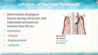
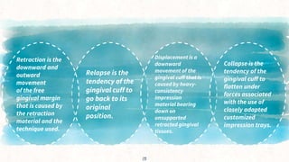
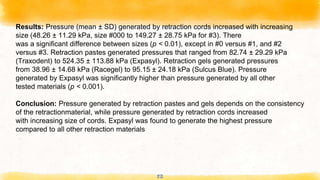
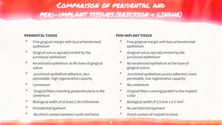
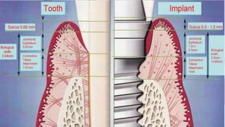
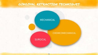
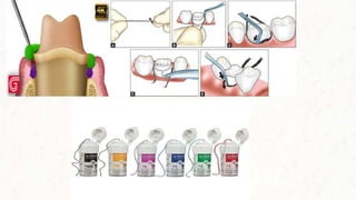
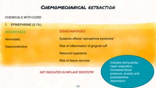
SlideShare a Scribd company logo
Gingival retraction
in implants
presented by
dr. bhavesh kumar jha
pg ii YEAR
contents
INTRODUCTION
COMPARISON OF PERIDENTAL AND PERI
IMPLANT TISSUES
REQIREMENT OF IMPRESSION
GINGIVAL RETRACTION TECHNIQUES
ADVANCEMENT IN GINGIVAL RETRACTION
TECHNIQUES
DISCUSSION
CONCLUSION
REFERENCES
2
INTRODUCTION
▸ Implant dentistry has seen rapid progress in recent years. Its
increased use in the treatment of partially edentulous patients
has led to two restorative techniques: screw retained implant
restorations and cement-retained restorations.
3
introduction
Cement-retained prostheses are the restoration of
choice for many patients who receive implants for
several reasons, including
esthetics,
occlusal stability,
overcoming angulation problems and
fabrication of a passively fitting restoration.
4
introduction
Several impression techniques are used in
implant dentistry, and some require gingival displacement
while making impressions.
5
Baharav H, Laufer BZ, Langer Y, Cardash HS. The effect of displacement
time on gingival crevice width. Int J Prosthodont 1997;10(3):
248-253.
To ensure accuracy with polyvinyl siloxane impression materials,
clinicians must maintain a minimum bulk of 0.2-millimeter
thickness in the sulcus area, which they can achieve by
retracting the gingiva for at least four minutes before making the
impression.
6
Donovan and Chee described a variety of gingival
displacement techniques, but there is no
articles that specifically reviewed gingival retraction
techniques in implant dentistry.
Since the architecture of the gingival crevice surrounding
natural teeth is different biologically from that around
implants, we wanted to know if conventional retraction
techniques could be applied safely to peri-implant tissue.
Gingival retraction techniques for implants versus teeth :Current status
Vincent Bennani, Donald Schwass, Nicholas Chandler
7
PERIDENTAL TISSUE
▸ Free gingival margin with buccal keratinized
epithelium
▸ Gingival sulcus apically limited by the
junctional epithelium
▸ Keratinized epithelium at the base of gingival
sulcus
▸ Junctional epithelium adherent, less
permeable, high regenerative capacity
▸ Cementum
▸ Gingival fibers inserting perpendicularly in the
cementum
▸ Biological width of at least 2.04 millimetres
▸ Periodontal ligament
▸ No direct contact between tooth and bone
Comparison of peridental and
peri-implant tissues.(ERICSSON & LINDHE)
PERI-IMPLANT TISSUE
▸ Free gingival margin with buccal keratinized
epithelium
▸ Gingival sulcus apically limited by the
junctional epithelium
▸ No keratinized epithelium at the base of
gingival sulcus
▸ Junctional epithelium poorly adherent, more
permeable, low regenerative capacity
▸ No cementum
▸ Gingival fibers running parallel to the implant
collar
▸ Biological width of 2.5 mm ± 0.5 mm*
▸ No periodontal ligament
▸ Direct contact of implant to bone
8
9
Requirements of impression
EXPOSE ACCESS ISOLATE
10
“
Gingival retraction or displacement is the deflection
of the marginal gingiva away from the tooth. ‘tissue
dilation’-GPT 9
11
GINGIVAL RETRACTION TECHNIQUES
▸ Deformation of gingival
tissues during retraction and
impression procedures
involves four forces:
▸ retraction
▸ relapse
▸ displacement
▸ collapse
12
Relapse is the
tendency of the
gingival cuff to
go back to its
original
position.
Retraction is the
downward and
outward
movement
of the free
gingival margin
that is caused by
the retraction
material and the
technique used.
Displacement is a
downward
movement of the
gingival cuff that is
caused by heavy-
consistency
Impression
material bearing
down on
unsupported
retracted gingival
tissues.
13
Collapse is the
tendency of the
gingival cuff to
flatten under
forces associated
with the use of
closely adapted
customized
impression trays.
GINGIVAL RETRACTION TECHNIQUES
▸ Peri-implant fiber structure does not provide the
same level of support for gingival tissues when the
retraction agents are removed .
▸ Thus, more collapsing forces occur on retracted
tissues of implants as compared to peridental
retracted tissues.
▸ Particularly true in situations in which depth of
sulcus is greater than average, such as when implant
is placed deeply.
14
The aim of gingival
retraction is to atraumatically allow access for the
impression material beyond the abutment margins
and to create space so that the impression
material is sufficiently thick so as to be tear-resistant
15
GINGIVAL RETRACTION TECHNIQUES
16
CHEMICOMECHANICAL
MECHANICAL
SURGICAL
MECHANICAL RETRACTION
17
CORD
(may be twisted, knitted or braided)
Single-cord technique VS Dual-cord technique
Retraction cords were developed for use with natural teeth. They provide
more effective control of gingival hemorrhage and exudate when used in
conjunction with medicaments than when used with no medicaments.
The dual-cord technique in which the first cord remains in the sulcus
reduces the tendency for the gingival cuff to recoil and partially displace
the setting impression material.
Maps
Placement of retraction cords can cause injury to the sulcular
epithelium and underlying connective tissues, as shown by the results
of experiments involving dogs’ teeth. The filaments or fibers of
conventional cords also may cause residual contamination of sulcus
wounds, creating foreign body reactions and exacerbating
inflammation.
18
Healing of the sulcus can take seven to 10 days. Use of
minimal force is necessary when packing cords to protect
Sharpey fibers and application of excessive force is
inappropriate because it may cause crevicular bleeding,
gingival inflammation and shrinkage of marginal tissues.
▸ use of cords around implants is questionable since the junctional
epithelium is not as adherent, is more permeable and has a lower
regenerative capacity than the junctional epithelium around teeth.
19
20
ADVANTAGES
1. Inexpensive
2. Achieves varying
degrees of
retraction
3. Can be used with
chemical adjuncts
DISADVANTAGES
1. Painful
2. Rapid collapse of sulcus
after removal
3. Risk of traumatizing
epithelial attachment
4. No hemostasis without
chemical agent
5. Placement is time-
consuming
6. Risk of sulcus contamination
21
Chemomechanical retraction
22
CHEMICALS WITH CORD
1. EPINEPHRINE (0.1%)
ADVANTAGES
Hemostatic
Vasoconstrictive
DISADVANTAGES
Systemic effects “epinephrine syndrome”
Risk of inflammation of gingival cuff
Rebound hyperemia
Risk of tissue necrosis
NOT INDICATED IN IMPLANT DENTISTRY
includes tachycardia,
rapid respiration,
increased blood
pressure, anxiety and
postoperative
depression.
Dose-related effects of epinephrine on human gingival blood flow and crevicular fluid production used as
a soaking solution for chemo-mechanical tissue retraction
Maria Csillag 1, Gabriella Nyiri, Janos Vag, Arpad Fazekas
23
Purpose: The aim of this study was to identify the effective concentration of epinephrine
that may prevent the hyperemic response and consequently keep the crevicular fluid
production low after cord removal without local or systemic side effects.
Material and methods: Seventeen healthy human subjects had their crevicular fluid
volume and gingival blood flow measured by Periotron and laser Doppler flowmetry,
respectively, before and after cord removal at the left maxillary central incisor. The right
maxillary incisor served as the control. Retraction cords were presoaked in physiological
saline or various concentrations (0.001%, 0.01%, and 0.1% w/v) of epinephrine solution.
Double repeated-measures analysis of variance with the Fisher Least Significant
Difference post hoc test was used to statistically evaluate the blood flow values (mean +/-
SE, alpha=.05), and the Wilcoxon matched pair test was used for crevicular fluid values,
given as median (25-75 percentile, alpha=.01).
24
Results: In the saline group, cord removal resulted in elevated blood flow (140% +/- 11%,
P<.001) and crevicular fluid production (300% (130%-470%), P<.05). After cord removal
in the 0.01% and 0.1% epinephrine groups, blood flow remained low for the measured
period (43%-70%, P<.05). The crevicular fluid production transiently increased in the
0.01% epinephrine group (170% (140%-380%), P<.001), but then returned to baseline
level and remained low as for the 0.1% group. No systemic vascular effect was detected
in any groups.
Conclusion: The prolonged increase in crevicular fluid production and hyperemic
response after cord removal can be prevented by application of 0.01% epinephrine
solution without systematic changes.
Chemomechanical retraction
25
2.Synthetic sympathomimetic agents
ADVANTAGES
Hemostatic
Vasoconstrictive
More effective than epinephrine with
the absence of systemic effects
DISADVANTAGES
Rebound hyperemia
Risk of inflammation of gingival cuff
Risk of tissue necrosis
NOT INDICATED IN IMPLANT DENTISTRY
Chemomechanical retraction
26
3. Aluminum sulphate and aluminium potassium sulphate
ADVANTAGES
Hemostasis
Least inflammation of all agents used
with cords
Little sulcus collapse after cord removal
DISADVANTAGES
Offensive taste
Risk of sulcus contamination
Risk of necrosis if in high concentration
METHOD COULD BE USED BUT NOT RECOMMENDED IN IMPLANT DENTISTRY
Chemomechanical retraction
27
4. Aluminum chloride
ADVANTAGES
No systemic effects
Least irritating of all chemicals
Hemostasis
Little sulcus collapse after cord removal
DISADVANTAGES
Less vasoconstriction than epinephrine
Risk of sulcus contamination
Modifies surface detail reproduction
Inhibits set of polyvinyl siloxane and
polyether impressions
METHOD COULD BE USED BUT NOT RECOMMENDED IN IMPLANT DENTISTRY
Chemomechanical retraction
28
5. Ferric sulphate
ADVANTAGES
Hemostasis
DISADVANTAGES
Tissue discoloration
Acidic taste
Risk of sulcus contamination
Inhibits set of polyvinyl siloxane and
polyether impressions
METHOD COULD BE USED BUT NOT RECOMMENDED IN IMPLANT DENTISTRY
DRAWBACK
▸ The two main drawbacks of using chemicals with retraction
cords are:
▸ occurrence of rebound hyperemia that often occurs after cord
removal, which affects how effectively clinicians can make
impressions .
▸ inflammatory reactions induced by these chemicals, which can
affect the subepithelial connective tissue.
29
Chemomechanical retraction
30
Chemicals in an injectable matrix
1. Aluminum chloride with kaolin
ADVANTAGES
Reduced risk of inflammation (injectable form)
Nontraumatizing to junctional epithelium
Hydrophilic
Ease of placement
Painless
No adverse effects
DISADVANTAGES
Inhibits set of polyvinyl siloxane and polyether
impressions
More expensive
Less effective with very subgingival margins
METHOD RECOMMENDED IN IMPLANT DENTISTRY
Chemomechanical retraction
31
Inert matrix
Polyvinyl siloxane
ADVANTAGES
No risk of inflammation or irritation
Nontraumatizing
Ease of placement
Painless
No adverse effects
DISADVANTAGES
Limited capacity for hemostasis (no active
chemistry)
Less effective with subgingival margins
METHOD RECOMMENDED IN IMPLANT DENTISTRY
SURGICAL RETRACTION
32
Laser
ADVANTAGES
Excellent hemostasis–carbon dioxide (CO2) laser safe
for implants as reflected by metal
Reduced tissue shrinkage
Relatively painless
Sterilizes sulcus
DISAVANTAGES
Neodymium:yttrium-aluminum-garnet
laser contraindicated with implants
Erbium:yttrium-aluminum-garnet laser
reflected by metal but not as good at
hemostasis as CO2 laser
CO2 laser provides no tactile feedback,
leading to risk of damage to junctional
epithelium
METHOD COULD BE USED BUT NOT RECOMMENDED IN IMPLANT DENTISTRY
SURGICAL RETRACTION
33
Electrosurgery
ADVANTAGES
Efficient precise hemostasis
DISADVANTAGES
Contraindicated with implant (risk of arcing)
Gingival sulcus too small for two electrodes,
impractical in implant dentistry
NOT INDICATED IN IMPLANT DENTISTRY
Rotary curettage
34
ADVANTAGES
Fast
Ability to reduce excessive tissue
Ability to recontour gingival outline
DISADVANTAGES
Causes considerable haemorrhage
Contraindicated with implants
High risk of the bur damaging the implant surface
Risk of tissue retraction exposing implant threads
High risk of traumatizing the epithelial attachment
NOT INDICATED IN IMPLANT DENTISTRY
Advancements in Gingival Retraction Techniques
35
1. Expasyl (SDS/KERR)
Expasyl is considered a viable alternative to a conventional retraction cord. It is
a viscous paste used for all techniques which necessitates gingival retraction .
Expasyl is a biocompatible material which presents with advantages of having
excellent retraction with longer shelf life.
Minimal pressure required to displace the tissues.
It produces hemostasis and controls crevicular seepage
Advancements in Gingival Retraction Techniques
36
Expasyl (Kerr) is an aluminum chloride (AlCl3) based paste-like
material syringed into the sulcus with autoclavable stainless steel
dispenser, acting both as a chemical hemostatic agent and mechanical
retraction material (chemomechanical method).
Advancements in Gingival Retraction Techniques
37
2. Magic Foamcord (Coltene/Whale dent)
Magic Foamcord presents with efficient hemostasis and minimal damage to
tissues while retraction
Magic foam cord is a polymeric material which is introduced into the gingival
sulcus and allowed to set.
Circular foams are supplied along with the material which is contoured to the
shape of gingival sulcus which is available in three sizes to accommodate
different teeth.
Advancements in Gingival Retraction Techniques
38
The patient is advised to bite on a cap (Comprecap) while maintaining the
pressure on for 3 minutes.
The material slightly expands during setting and produces exceptional lateral
and vertical displacement. The cap and foam are removed after 5 minutes and
the tooth is set for the final impression
Advancements in Gingival Retraction Techniques
▸ ADVANTAGES
▸ Magic foam cord is less
traumatic to tissues than
conventional retraction cord.
▸ The Color of foam aids in
visualization. The material is
easy to separate from the
sulcus. It has adequate working
time
▸ DISADVANTAGES
▸ no hemostasis provided, expensive
No improvement is observed in
working time or quality of retraction
compared with conventional cord.
▸ It is considered to less effective on
subgingival margins. Intraoral tips
provided may be too bulky to
adequately inject material into
gingival sulcus.
39
Advancements in Gingival Retraction Techniques
40
3. Gingitrac (Centrix)
It is an effective gingival retraction system based on vinyl polysiloxane material
with aluminium sulfate astringent.
It truly harnesses the power of pressure, astringency and time unlike traumatic
cord techniques or messy paste alternatives.
Unlike with retraction cord, the coagulum will not stick to the silicone GingiTrac,
so there is no bleeding when it is removed.
A GingiCap is used for single preparation retraction which works in less than 5
minutes, without hands in mouth and blanches the gingiva till the vestibule.
Advancements in Gingival Retraction Techniques
41
Advancements in Gingival Retraction Techniques
42
4. Gel-Cord
Gelcord comprises of- 25% Aluminum Sulfate Gel. Unlike liquid astringents it
stays put when placed for maximum hemostasis.
No reports of tissue necrosis have been reported.
The gel is rubbed mildly into the hemorrhaging area. Gelcord is flavored well for
greater patient acceptance and brightly colored for better visualization.
It provides enough lubrication for the initial cord to slide easily into the sulcus.
Advancements in Gingival Retraction Techniques
43
Advancements in Gingival Retraction Techniques
44
5. Tissue Goo
Tissue Goo is a gel that contains active ingredient is 25% aluminum sulfate.
Aluminum sulfate does not cauterize, but rather acts similar to a coagulant to
arrest the bleeding.
It also acts as a lubricant while placement of the cord.
Tissue Goo will not impede with the set impression material.
Advancements in Gingival Retraction Techniques
45
6. G Cuff
A Canadian company, named Stomatotech, launched a disposable plastic collar for
gingival retraction which is inserted on the apical end of the abutment before the
abutment is engaged to the implant.
Once the impression is retrieved from the mouth, the plastic collar is drawn out and
removed permanently.
The plastic creates a valve preventing the liquids from contaminating the area of the
finish line of the abutment.
G-Cuffs major intention is to maintain soft tissue surrounding the implant abutment
permitting the impression (conventional or digital) to have an access to the surface of the
abutment required for the optimal restoration.
Advancements in Gingival Retraction Techniques
46
Advancements in Gingival Retraction Techniques
47
7. Retraction Capsule
The recently introduced 3M™ ESPE™ Retraction Capsule is 15% aluminum
chloride retraction paste. It is packaged in unit-dose capsules with an extra-fine
tip that fits directly into the sulcus.
When compared with retraction cords, the retraction procedure with this
material can be up to 50% faster.
The significantly fine tip of the capsule offersimproved access into the sulcus
and interproximal areas.
Advancements in Gingival Retraction Techniques
48
Clinical evaluation of the effect of two gingival retraction systems, gingival cuff and gingival retraction
paste, on peri-implant soft tissue
Sugandha Gupta, Pankaj Dhawan, Pankaj Madhukar, Piyush Tandan, Aman Sachdeva
49
Aims: The aim of this study was to clinically evaluate the host tissue response around
oral implants using two gingival retraction systems, namely, G-Cuff™ and Traxodent®
Materials and Methods: Twenty cases were selected and divided into two groups:
Group A – ten patients in whom gingival retraction was done using G-Cuff™ and Group B
– ten patients in whom gingival retraction was done using Traxodent®. Patients with
immobile, stable, and planned cement-retained implant prosthesis were enrolled in the
study. Both Group A and B patients, peri-implant soft tissues were analyzed three times:
preretraction, postretraction, and after 7 days for the various parameters.
50
Statistical Analysis Used: The data obtained were statistically analyzed using Kruskal–
Wallis test, Pearson's Chi-square test and Mann–Whitney test.
Results: The use of G-Cuff™ resulted in decrease in the mean of the probing depth
values after 7 days from 1.30 to 1.13 mm. The values of the probing depth for the
Traxodent® group showed a slight increase from 1.30 mm to 1.60 and 1.57 mm at
immediately and 7 days after retraction. The mucosal index increased for G-Cuff™ and
Traxodent® group. Bleeding on probing significantly decreased in Traxodent® group.
Conclusion: There was difference in the host tissue response by the two types of
retraction agents in relation to some parameters, and also, the level of pain and
discomfort by the use of G-Cuff™ was found in few cases. Based on the results of short-
term evaluations, chemical cordless retraction system functioned statistically well in terms
of hemostasis.
Comparison of Gingival Retraction Materials Using a New Gingival Sulcus Model
Marco Dederichs, Mina D. Fahmy, Harald Kuepper,& Arndt Guentsch,
51
Purpose: To investigate the pressure generated by different retraction materials using
a novel gingival sulcus model.
Materials and Methods: A gingival sulcus model was made using a polymer frame
filledwith silicon.Apressure sensor and a sulcus-fluid simulationwere embedded into
the silicon chamber to evaluate the pressure generated by different retractionmaterials.
Six sizes of Ultrapak retraction cords (Ultradent, sizes #000 - 3), 4 retraction pastes
(Expazen, Expasyl, Acteon, Access Edge, Traxodent) and 2 retraction gels (Sulcus
Blue, Racegel) were analyzed. The mean andmedian pressure, interquartile range, and
standard deviation (SD) of n = 10 repeated measurements were calculated. Statistical
analysis was conducted by Kruskal-Wallis test for differences between the main
groups of retraction materials, and Mann-Whitney U-test was performed to analyze
differences between the single retraction materials.
52
Results: Pressure (mean ± SD) generated by retraction cords increased with increasing
size (48.26 ± 11.29 kPa, size #000 to 149.27 ± 28.75 kPa for #3). There
was a significant difference between sizes (p < 0.01), except in #0 versus #1, and #2
versus #3. Retraction pastes generated pressures that ranged from 82.74 ± 29.29 kPa
(Traxodent) to 524.35 ± 113.88 kPa (Expasyl). Retraction gels generated pressures
from 38.96 ± 14.68 kPa (Racegel) to 95.15 ± 24.18 kPa (Sulcus Blue). Pressure
generated by Expasyl was significantly higher than pressure generated by all other
tested materials (p < 0.001).
Conclusion: Pressure generated by retraction pastes and gels depends on the consistency
of the retractionmaterial, while pressure generated by retraction cords increased
with increasing size of cords. Expasyl was found to generate the highest pressure
compared to all other retraction materials
53
DISCUSSION
The mechanical retraction of gingival tissues by using cords around implant
restorations can lead to ulceration of the junctional epithelium.
The forces used in cord placement are likely to exceed peri-implant tissues’
capacity to resist them.
Once patients’ gingival epithelial structure is damaged, there is significant risk of
permanent recession and loss of attachment developing.
Thus, the use of mechanical retraction with cords may be contraindicated around
implants, except in situations in which patients’ sulcus depths are shallow, their
mucosal health is impeccable and a robust, thick periodontal biotype is present.
54
The addition of chemical adjuncts to retraction cords further complicates the
situation and may lead to increased inflammation of the subsulcular tissues.
The lacerated sulcus provides reduced protection against the penetration of
chemicals into deeper subepithelial cell layers and against systemic
dissemination when the vascular bed is exposed.
Surgical retraction procedures, however, are destructive and involve excision
of tissue.
Peri-implant mucosa does not have the same capacity for regeneration as
peridental mucosa.
55
The correct use of lasers with appropriate wavelengths may be applicable in
some, but not all, implant situations during retraction and when making
impressions.
the aluminum chloride in the injectable matrix offers the best outcome of the
chemical choices to date.
Although injectable matrices are promising as a gingival rétraction technique
for implant situations, further development is needed. Compared with research
on implant fixtures, there is relatively little research to guide clinicians
regarding how to restore implants and about which gingival retraction
techniques to use around implant abutments.
56
CONCLUSION
The literature concerning gingival retraction for impressions in fixed prosthodontics is
extensive.
By contrast, little has been published about the challenges presented by the unique anatomy
surrounding
implants.
As implants become mainstream treatments for tooth loss, this topic will warrant further
research.
57
REFERENCES
1. Rieder CE. Copings on tooth and implant abutments for superstructure prostheses. Int J Periodontics Restorative Dent
1990;
10(6):436-453.
2. Misch CE. Screw-retained versus cement-retained implant-supported prostheses. Pract Periodontics Aesthet Dent
1995;7(9):15-18.
3. Bidez MW, Misch CE. Force transfer in implant dentistry: basic concepts and principles. J Oral Implantol 1992;18(3):264-
274.
4. Guichet DL. Load transfer in screw- and cement-retained implant fixed partial denture design: proceedings of the fourth
International Symposium on Implant Dentistry—focus on esthetics, San Diego, Calif., January 27 through 29, 1994. J
Prosthet Dent 1994;72(6):631.
5. Singer A, Serfaty V. Cement-retained implant-supported fixed partial dentures: a 6-month to 3-year follow-up. Int J Oral
Maxillofac
Implants 1996;11(5):645-649.
7. Laufer BZ, Baharav H, Cardash HS. The linear accuracy of impressions and stone dies as affected by the thickness of
the impression margin. Int J Prosthodont 1994;7(3):247-252.
8. Baharav H, Kupershmidt I, Laufer BZ, Cardash HS. The effect of sulcular width on the linear accuracy of impression
materials in the
presence of an undercut. Int J Prosthodont 2004;17(5):585-589.
9. Baharav H, Laufer BZ, Langer Y, Cardash HS. The effect of displacement time on gingival crevice width. Int J
Prosthodont 1997;10(3): 248-253.
10. Laufer BZ, Baharav H, Langer Y, Cardash HS. The closure of the gingival crevice following gingival retraction for
impression making. J Oral Rehabil 1997;24(9):629-635.
11. Ruel J, Schuessler PJ, Malament K, Mori D. Effect of retraction procedures on the periodontium in humans. J Prosthet
Dent 1980; 44(5):508-515.
THANK YOU
58

More Related Content

What's hot

Implant quality scale ; osseointegration, success criteria and basic guides
Implant quality scale ; osseointegration, success criteria and basic guidesImplant quality scale ; osseointegration, success criteria and basic guides
Implant quality scale ; osseointegration, success criteria and basic guides
Vinay Kadavakolanu
 
Gingival retraction.pptx
Gingival retraction.pptxGingival retraction.pptx
Gingival retraction.pptx
Dr.shiva sai vemula
 
horizontal jaw relation in complete denture
 horizontal jaw relation in complete denture horizontal jaw relation in complete denture
horizontal jaw relation in complete denture
dipalmawani91
 
Guided bone regeneration
Guided bone regenerationGuided bone regeneration
Guided bone regeneration
Bhaumik Thakkar
 
7. RETENTION IN COMPLETE DENTURE.pptx
7. RETENTION IN COMPLETE DENTURE.pptx7. RETENTION IN COMPLETE DENTURE.pptx
7. RETENTION IN COMPLETE DENTURE.pptx
SusovanGiri6
 
Attachments In Prosthodontics
Attachments In ProsthodonticsAttachments In Prosthodontics
Attachments In Prosthodontics
Self employed
 
PLATFORM SWITCHING
PLATFORM SWITCHINGPLATFORM SWITCHING
PLATFORM SWITCHING
shari kurup
 
Balanced occlusion aditi ghai
Balanced occlusion aditi ghaiBalanced occlusion aditi ghai
Balanced occlusion aditi ghai
Aditi Ghai
 
Gingival Retraction
Gingival Retraction Gingival Retraction
Gingival Retraction
Harshil Modi
 
Implant abutment and implant abutment connections
Implant abutment and implant abutment connectionsImplant abutment and implant abutment connections
Implant abutment and implant abutment connections
DR.BHAVESH JHA
 
Journal club presentation on tooth supported overdentures
Journal club presentation on tooth supported overdentures Journal club presentation on tooth supported overdentures
Journal club presentation on tooth supported overdentures
NAMITHA ANAND
 
dental Monoblock obturation technique or concept in endodontics
dental Monoblock obturation technique or concept in endodonticsdental Monoblock obturation technique or concept in endodontics
dental Monoblock obturation technique or concept in endodontics
Ahmed Ali
 
Loading protocols in implant
Loading protocols in implantLoading protocols in implant
Loading protocols in implant
PiyaliBhattacharya10
 
Obturation
ObturationObturation
Obturation
Dr. Abhisek Guria
 
Resin bonded FPDs
Resin bonded FPDsResin bonded FPDs
Resin bonded FPDs
benju sharma
 
Complete overdenture(3)
Complete overdenture(3)Complete overdenture(3)
Complete overdenture(3)
Ahmed Samir
 
Root canal obturation timing materials and techniques
Root canal obturation timing materials and techniquesRoot canal obturation timing materials and techniques
Root canal obturation timing materials and techniques
Silas Toka
 
implant supported fixed restorations
implant supported fixed restorationsimplant supported fixed restorations
implant supported fixed restorations
Taban Ameen
 
Gingival tissue management
Gingival tissue management Gingival tissue management
Gingival tissue management
Abhijeet Pallewar
 
Bone Graft in Periodontal Treatment
Bone Graft in Periodontal TreatmentBone Graft in Periodontal Treatment
Bone Graft in Periodontal Treatment
Cing Sian Dal
 

What's hot (20)

Implant quality scale ; osseointegration, success criteria and basic guides
Implant quality scale ; osseointegration, success criteria and basic guidesImplant quality scale ; osseointegration, success criteria and basic guides
Implant quality scale ; osseointegration, success criteria and basic guides
 
Gingival retraction.pptx
Gingival retraction.pptxGingival retraction.pptx
Gingival retraction.pptx
 
horizontal jaw relation in complete denture
 horizontal jaw relation in complete denture horizontal jaw relation in complete denture
horizontal jaw relation in complete denture
 
Guided bone regeneration
Guided bone regenerationGuided bone regeneration
Guided bone regeneration
 
7. RETENTION IN COMPLETE DENTURE.pptx
7. RETENTION IN COMPLETE DENTURE.pptx7. RETENTION IN COMPLETE DENTURE.pptx
7. RETENTION IN COMPLETE DENTURE.pptx
 
Attachments In Prosthodontics
Attachments In ProsthodonticsAttachments In Prosthodontics
Attachments In Prosthodontics
 
PLATFORM SWITCHING
PLATFORM SWITCHINGPLATFORM SWITCHING
PLATFORM SWITCHING
 
Balanced occlusion aditi ghai
Balanced occlusion aditi ghaiBalanced occlusion aditi ghai
Balanced occlusion aditi ghai
 
Gingival Retraction
Gingival Retraction Gingival Retraction
Gingival Retraction
 
Implant abutment and implant abutment connections
Implant abutment and implant abutment connectionsImplant abutment and implant abutment connections
Implant abutment and implant abutment connections
 
Journal club presentation on tooth supported overdentures
Journal club presentation on tooth supported overdentures Journal club presentation on tooth supported overdentures
Journal club presentation on tooth supported overdentures
 
dental Monoblock obturation technique or concept in endodontics
dental Monoblock obturation technique or concept in endodonticsdental Monoblock obturation technique or concept in endodontics
dental Monoblock obturation technique or concept in endodontics
 
Loading protocols in implant
Loading protocols in implantLoading protocols in implant
Loading protocols in implant
 
Obturation
ObturationObturation
Obturation
 
Resin bonded FPDs
Resin bonded FPDsResin bonded FPDs
Resin bonded FPDs
 
Complete overdenture(3)
Complete overdenture(3)Complete overdenture(3)
Complete overdenture(3)
 
Root canal obturation timing materials and techniques
Root canal obturation timing materials and techniquesRoot canal obturation timing materials and techniques
Root canal obturation timing materials and techniques
 
implant supported fixed restorations
implant supported fixed restorationsimplant supported fixed restorations
implant supported fixed restorations
 
Gingival tissue management
Gingival tissue management Gingival tissue management
Gingival tissue management
 
Bone Graft in Periodontal Treatment
Bone Graft in Periodontal TreatmentBone Graft in Periodontal Treatment
Bone Graft in Periodontal Treatment
 

Similar to Gingival retraction in implant

Gingival tissue management
Gingival tissue managementGingival tissue management
Gingival tissue management
Aarthi Govindaraju
 
omfs journal club ppt on bone ridge augmentation
omfs journal club ppt on bone ridge augmentationomfs journal club ppt on bone ridge augmentation
omfs journal club ppt on bone ridge augmentation
Akhil Sankar
 
Gingival tissue management
Gingival tissue managementGingival tissue management
Gingival tissue management
somendrasaraswat2
 
Periimplantitis
PeriimplantitisPeriimplantitis
Periimplantitis
JahnaviYadav5
 
gingival retraction seminar pptx gingival displacment methods ppt
gingival retraction seminar pptx gingival displacment methods pptgingival retraction seminar pptx gingival displacment methods ppt
gingival retraction seminar pptx gingival displacment methods ppt
KanmaniAthi
 
Complication & failure of dental implants / cosmetic dentistry training
Complication & failure of dental implants / cosmetic dentistry trainingComplication & failure of dental implants / cosmetic dentistry training
Complication & failure of dental implants / cosmetic dentistry training
Indian dental academy
 
Gingival retraction
Gingival retractionGingival retraction
Gingival retraction
Sayli Patil
 
vdocuments.net_surgical-periodontal-therapy-56ebcbc25be52.ppt
vdocuments.net_surgical-periodontal-therapy-56ebcbc25be52.pptvdocuments.net_surgical-periodontal-therapy-56ebcbc25be52.ppt
vdocuments.net_surgical-periodontal-therapy-56ebcbc25be52.ppt
Rutu Dabhi
 
Endodontic surgery
Endodontic surgeryEndodontic surgery
Endodontic surgery
Dr Bhavik Miyani
 
per flap with vest deep.pptx
per flap with vest deep.pptxper flap with vest deep.pptx
per flap with vest deep.pptx
mayankgupta672202
 
LaserFistulaEndodoncia2018.pdf
LaserFistulaEndodoncia2018.pdfLaserFistulaEndodoncia2018.pdf
LaserFistulaEndodoncia2018.pdf
andreiberdichewsky
 
Peri implantitis treatment with regenerative approach
Peri implantitis treatment with regenerative approachPeri implantitis treatment with regenerative approach
Peri implantitis treatment with regenerative approach
ajayashreep
 
Surgical re treatment ( an overview)
Surgical re treatment ( an overview)Surgical re treatment ( an overview)
Surgical re treatment ( an overview)
Hamza Tahir
 
Gingival recession
Gingival recessionGingival recession
Gingival recession
Imen Kassoma
 
Treatment of gingival recession using coronally advanced flap
Treatment of gingival recession using coronally advanced flapTreatment of gingival recession using coronally advanced flap
Treatment of gingival recession using coronally advanced flap
Shruti Maroo
 
Implant surgeries to overcome anatomic difficulties ii / dental implant cour...
Implant surgeries to overcome anatomic difficulties  ii / dental implant cour...Implant surgeries to overcome anatomic difficulties  ii / dental implant cour...
Implant surgeries to overcome anatomic difficulties ii / dental implant cour...
Indian dental academy
 
143rd publication sjodr- 3rd name
143rd publication  sjodr- 3rd name143rd publication  sjodr- 3rd name
143rd publication sjodr- 3rd name
CLOVE Dental OMNI Hospitals Andhra Hospital
 
140th publication sjodr- 3rd name
140th publication  sjodr- 3rd name140th publication  sjodr- 3rd name
140th publication sjodr- 3rd name
CLOVE Dental OMNI Hospitals Andhra Hospital
 
3RD PUBLICATION - JCDR - Dr. RAHUL VC TIWARI, SIBAR INSTITUTE OF DENTAL SCIE...
3RD  PUBLICATION - JCDR - Dr. RAHUL VC TIWARI, SIBAR INSTITUTE OF DENTAL SCIE...3RD  PUBLICATION - JCDR - Dr. RAHUL VC TIWARI, SIBAR INSTITUTE OF DENTAL SCIE...
3RD PUBLICATION - JCDR - Dr. RAHUL VC TIWARI, SIBAR INSTITUTE OF DENTAL SCIE...
CLOVE Dental OMNI Hospitals Andhra Hospital
 
Surgical Endodontics
Surgical Endodontics Surgical Endodontics
Surgical Endodontics
DrOsamaMushtaq
 

Similar to Gingival retraction in implant (20)

Gingival tissue management
Gingival tissue managementGingival tissue management
Gingival tissue management
 
omfs journal club ppt on bone ridge augmentation
omfs journal club ppt on bone ridge augmentationomfs journal club ppt on bone ridge augmentation
omfs journal club ppt on bone ridge augmentation
 
Gingival tissue management
Gingival tissue managementGingival tissue management
Gingival tissue management
 
Periimplantitis
PeriimplantitisPeriimplantitis
Periimplantitis
 
gingival retraction seminar pptx gingival displacment methods ppt
gingival retraction seminar pptx gingival displacment methods pptgingival retraction seminar pptx gingival displacment methods ppt
gingival retraction seminar pptx gingival displacment methods ppt
 
Complication & failure of dental implants / cosmetic dentistry training
Complication & failure of dental implants / cosmetic dentistry trainingComplication & failure of dental implants / cosmetic dentistry training
Complication & failure of dental implants / cosmetic dentistry training
 
Gingival retraction
Gingival retractionGingival retraction
Gingival retraction
 
vdocuments.net_surgical-periodontal-therapy-56ebcbc25be52.ppt
vdocuments.net_surgical-periodontal-therapy-56ebcbc25be52.pptvdocuments.net_surgical-periodontal-therapy-56ebcbc25be52.ppt
vdocuments.net_surgical-periodontal-therapy-56ebcbc25be52.ppt
 
Endodontic surgery
Endodontic surgeryEndodontic surgery
Endodontic surgery
 
per flap with vest deep.pptx
per flap with vest deep.pptxper flap with vest deep.pptx
per flap with vest deep.pptx
 
LaserFistulaEndodoncia2018.pdf
LaserFistulaEndodoncia2018.pdfLaserFistulaEndodoncia2018.pdf
LaserFistulaEndodoncia2018.pdf
 
Peri implantitis treatment with regenerative approach
Peri implantitis treatment with regenerative approachPeri implantitis treatment with regenerative approach
Peri implantitis treatment with regenerative approach
 
Surgical re treatment ( an overview)
Surgical re treatment ( an overview)Surgical re treatment ( an overview)
Surgical re treatment ( an overview)
 
Gingival recession
Gingival recessionGingival recession
Gingival recession
 
Treatment of gingival recession using coronally advanced flap
Treatment of gingival recession using coronally advanced flapTreatment of gingival recession using coronally advanced flap
Treatment of gingival recession using coronally advanced flap
 
Implant surgeries to overcome anatomic difficulties ii / dental implant cour...
Implant surgeries to overcome anatomic difficulties  ii / dental implant cour...Implant surgeries to overcome anatomic difficulties  ii / dental implant cour...
Implant surgeries to overcome anatomic difficulties ii / dental implant cour...
 
143rd publication sjodr- 3rd name
143rd publication  sjodr- 3rd name143rd publication  sjodr- 3rd name
143rd publication sjodr- 3rd name
 
140th publication sjodr- 3rd name
140th publication  sjodr- 3rd name140th publication  sjodr- 3rd name
140th publication sjodr- 3rd name
 
3RD PUBLICATION - JCDR - Dr. RAHUL VC TIWARI, SIBAR INSTITUTE OF DENTAL SCIE...
3RD  PUBLICATION - JCDR - Dr. RAHUL VC TIWARI, SIBAR INSTITUTE OF DENTAL SCIE...3RD  PUBLICATION - JCDR - Dr. RAHUL VC TIWARI, SIBAR INSTITUTE OF DENTAL SCIE...
3RD PUBLICATION - JCDR - Dr. RAHUL VC TIWARI, SIBAR INSTITUTE OF DENTAL SCIE...
 
Surgical Endodontics
Surgical Endodontics Surgical Endodontics
Surgical Endodontics
 

Recently uploaded

Top Effective Soaps for Fungal Skin Infections in India
Top Effective Soaps for Fungal Skin Infections in IndiaTop Effective Soaps for Fungal Skin Infections in India
Top Effective Soaps for Fungal Skin Infections in India
SwisschemDerma
 
Adhd Medication Shortage Uk - trinexpharmacy.com
Adhd Medication Shortage Uk - trinexpharmacy.comAdhd Medication Shortage Uk - trinexpharmacy.com
Adhd Medication Shortage Uk - trinexpharmacy.com
reignlana06
 
CHEMOTHERAPY_RDP_CHAPTER 1_ANTI TB DRUGS.pdf
CHEMOTHERAPY_RDP_CHAPTER 1_ANTI TB DRUGS.pdfCHEMOTHERAPY_RDP_CHAPTER 1_ANTI TB DRUGS.pdf
CHEMOTHERAPY_RDP_CHAPTER 1_ANTI TB DRUGS.pdf
rishi2789
 
Integrating Ayurveda into Parkinson’s Management: A Holistic Approach
Integrating Ayurveda into Parkinson’s Management: A Holistic ApproachIntegrating Ayurveda into Parkinson’s Management: A Holistic Approach
Integrating Ayurveda into Parkinson’s Management: A Holistic Approach
Ayurveda ForAll
 
Tests for analysis of different pharmaceutical.pptx
Tests for analysis of different pharmaceutical.pptxTests for analysis of different pharmaceutical.pptx
Tests for analysis of different pharmaceutical.pptx
taiba qazi
 
CHEMOTHERAPY_RDP_CHAPTER 4_ANTI VIRAL DRUGS.pdf
CHEMOTHERAPY_RDP_CHAPTER 4_ANTI VIRAL DRUGS.pdfCHEMOTHERAPY_RDP_CHAPTER 4_ANTI VIRAL DRUGS.pdf
CHEMOTHERAPY_RDP_CHAPTER 4_ANTI VIRAL DRUGS.pdf
rishi2789
 
TEST BANK For An Introduction to Brain and Behavior, 7th Edition by Bryan Kol...
TEST BANK For An Introduction to Brain and Behavior, 7th Edition by Bryan Kol...TEST BANK For An Introduction to Brain and Behavior, 7th Edition by Bryan Kol...
TEST BANK For An Introduction to Brain and Behavior, 7th Edition by Bryan Kol...
rightmanforbloodline
 
Histololgy of Female Reproductive System.pptx
Histololgy of Female Reproductive System.pptxHistololgy of Female Reproductive System.pptx
Histololgy of Female Reproductive System.pptx
AyeshaZaid1
 
Top-Vitamin-Supplement-Brands-in-India List
Top-Vitamin-Supplement-Brands-in-India ListTop-Vitamin-Supplement-Brands-in-India List
Top-Vitamin-Supplement-Brands-in-India List
SwisschemDerma
 
A Classical Text Review on Basavarajeeyam
A Classical Text Review on BasavarajeeyamA Classical Text Review on Basavarajeeyam
A Classical Text Review on Basavarajeeyam
Dr. Jyothirmai Paindla
 
Part II - Body Grief: Losing parts of ourselves and our identity before, duri...
Part II - Body Grief: Losing parts of ourselves and our identity before, duri...Part II - Body Grief: Losing parts of ourselves and our identity before, duri...
Part II - Body Grief: Losing parts of ourselves and our identity before, duri...
bkling
 
Abortion PG Seminar Power point presentation
Abortion PG Seminar Power point presentationAbortion PG Seminar Power point presentation
Abortion PG Seminar Power point presentation
AksshayaRajanbabu
 
Local Advanced Lung Cancer: Artificial Intelligence, Synergetics, Complex Sys...
Local Advanced Lung Cancer: Artificial Intelligence, Synergetics, Complex Sys...Local Advanced Lung Cancer: Artificial Intelligence, Synergetics, Complex Sys...
Local Advanced Lung Cancer: Artificial Intelligence, Synergetics, Complex Sys...
Oleg Kshivets
 
Hemodialysis: Chapter 4, Dialysate Circuit - Dr.Gawad
Hemodialysis: Chapter 4, Dialysate Circuit - Dr.GawadHemodialysis: Chapter 4, Dialysate Circuit - Dr.Gawad
Hemodialysis: Chapter 4, Dialysate Circuit - Dr.Gawad
NephroTube - Dr.Gawad
 
8 Surprising Reasons To Meditate 40 Minutes A Day That Can Change Your Life.pptx
8 Surprising Reasons To Meditate 40 Minutes A Day That Can Change Your Life.pptx8 Surprising Reasons To Meditate 40 Minutes A Day That Can Change Your Life.pptx
8 Surprising Reasons To Meditate 40 Minutes A Day That Can Change Your Life.pptx
Holistified Wellness
 
Top 10 Best Ayurvedic Kidney Stone Syrups in India
Top 10 Best Ayurvedic Kidney Stone Syrups in IndiaTop 10 Best Ayurvedic Kidney Stone Syrups in India
Top 10 Best Ayurvedic Kidney Stone Syrups in India
Swastik Ayurveda
 
Diabetic nephropathy diagnosis treatment
Diabetic nephropathy diagnosis treatmentDiabetic nephropathy diagnosis treatment
Diabetic nephropathy diagnosis treatment
arahmanzai5
 
share - Lions, tigers, AI and health misinformation, oh my!.pptx
share - Lions, tigers, AI and health misinformation, oh my!.pptxshare - Lions, tigers, AI and health misinformation, oh my!.pptx
share - Lions, tigers, AI and health misinformation, oh my!.pptx
Tina Purnat
 
Chapter 11 Nutrition and Chronic Diseases.pptx
Chapter 11 Nutrition and Chronic Diseases.pptxChapter 11 Nutrition and Chronic Diseases.pptx
Chapter 11 Nutrition and Chronic Diseases.pptx
Earlene McNair
 
The Best Ayurvedic Antacid Tablets in India
The Best Ayurvedic Antacid Tablets in IndiaThe Best Ayurvedic Antacid Tablets in India
The Best Ayurvedic Antacid Tablets in India
Swastik Ayurveda
 

Recently uploaded (20)

Top Effective Soaps for Fungal Skin Infections in India
Top Effective Soaps for Fungal Skin Infections in IndiaTop Effective Soaps for Fungal Skin Infections in India
Top Effective Soaps for Fungal Skin Infections in India
 
Adhd Medication Shortage Uk - trinexpharmacy.com
Adhd Medication Shortage Uk - trinexpharmacy.comAdhd Medication Shortage Uk - trinexpharmacy.com
Adhd Medication Shortage Uk - trinexpharmacy.com
 
CHEMOTHERAPY_RDP_CHAPTER 1_ANTI TB DRUGS.pdf
CHEMOTHERAPY_RDP_CHAPTER 1_ANTI TB DRUGS.pdfCHEMOTHERAPY_RDP_CHAPTER 1_ANTI TB DRUGS.pdf
CHEMOTHERAPY_RDP_CHAPTER 1_ANTI TB DRUGS.pdf
 
Integrating Ayurveda into Parkinson’s Management: A Holistic Approach
Integrating Ayurveda into Parkinson’s Management: A Holistic ApproachIntegrating Ayurveda into Parkinson’s Management: A Holistic Approach
Integrating Ayurveda into Parkinson’s Management: A Holistic Approach
 
Tests for analysis of different pharmaceutical.pptx
Tests for analysis of different pharmaceutical.pptxTests for analysis of different pharmaceutical.pptx
Tests for analysis of different pharmaceutical.pptx
 
CHEMOTHERAPY_RDP_CHAPTER 4_ANTI VIRAL DRUGS.pdf
CHEMOTHERAPY_RDP_CHAPTER 4_ANTI VIRAL DRUGS.pdfCHEMOTHERAPY_RDP_CHAPTER 4_ANTI VIRAL DRUGS.pdf
CHEMOTHERAPY_RDP_CHAPTER 4_ANTI VIRAL DRUGS.pdf
 
TEST BANK For An Introduction to Brain and Behavior, 7th Edition by Bryan Kol...
TEST BANK For An Introduction to Brain and Behavior, 7th Edition by Bryan Kol...TEST BANK For An Introduction to Brain and Behavior, 7th Edition by Bryan Kol...
TEST BANK For An Introduction to Brain and Behavior, 7th Edition by Bryan Kol...
 
Histololgy of Female Reproductive System.pptx
Histololgy of Female Reproductive System.pptxHistololgy of Female Reproductive System.pptx
Histololgy of Female Reproductive System.pptx
 
Top-Vitamin-Supplement-Brands-in-India List
Top-Vitamin-Supplement-Brands-in-India ListTop-Vitamin-Supplement-Brands-in-India List
Top-Vitamin-Supplement-Brands-in-India List
 
A Classical Text Review on Basavarajeeyam
A Classical Text Review on BasavarajeeyamA Classical Text Review on Basavarajeeyam
A Classical Text Review on Basavarajeeyam
 
Part II - Body Grief: Losing parts of ourselves and our identity before, duri...
Part II - Body Grief: Losing parts of ourselves and our identity before, duri...Part II - Body Grief: Losing parts of ourselves and our identity before, duri...
Part II - Body Grief: Losing parts of ourselves and our identity before, duri...
 
Abortion PG Seminar Power point presentation
Abortion PG Seminar Power point presentationAbortion PG Seminar Power point presentation
Abortion PG Seminar Power point presentation
 
Local Advanced Lung Cancer: Artificial Intelligence, Synergetics, Complex Sys...
Local Advanced Lung Cancer: Artificial Intelligence, Synergetics, Complex Sys...Local Advanced Lung Cancer: Artificial Intelligence, Synergetics, Complex Sys...
Local Advanced Lung Cancer: Artificial Intelligence, Synergetics, Complex Sys...
 
Hemodialysis: Chapter 4, Dialysate Circuit - Dr.Gawad
Hemodialysis: Chapter 4, Dialysate Circuit - Dr.GawadHemodialysis: Chapter 4, Dialysate Circuit - Dr.Gawad
Hemodialysis: Chapter 4, Dialysate Circuit - Dr.Gawad
 
8 Surprising Reasons To Meditate 40 Minutes A Day That Can Change Your Life.pptx
8 Surprising Reasons To Meditate 40 Minutes A Day That Can Change Your Life.pptx8 Surprising Reasons To Meditate 40 Minutes A Day That Can Change Your Life.pptx
8 Surprising Reasons To Meditate 40 Minutes A Day That Can Change Your Life.pptx
 
Top 10 Best Ayurvedic Kidney Stone Syrups in India
Top 10 Best Ayurvedic Kidney Stone Syrups in IndiaTop 10 Best Ayurvedic Kidney Stone Syrups in India
Top 10 Best Ayurvedic Kidney Stone Syrups in India
 
Diabetic nephropathy diagnosis treatment
Diabetic nephropathy diagnosis treatmentDiabetic nephropathy diagnosis treatment
Diabetic nephropathy diagnosis treatment
 
share - Lions, tigers, AI and health misinformation, oh my!.pptx
share - Lions, tigers, AI and health misinformation, oh my!.pptxshare - Lions, tigers, AI and health misinformation, oh my!.pptx
share - Lions, tigers, AI and health misinformation, oh my!.pptx
 
Chapter 11 Nutrition and Chronic Diseases.pptx
Chapter 11 Nutrition and Chronic Diseases.pptxChapter 11 Nutrition and Chronic Diseases.pptx
Chapter 11 Nutrition and Chronic Diseases.pptx
 
The Best Ayurvedic Antacid Tablets in India
The Best Ayurvedic Antacid Tablets in IndiaThe Best Ayurvedic Antacid Tablets in India
The Best Ayurvedic Antacid Tablets in India
 

Gingival retraction in implant

  • 1. Gingival retraction in implants presented by dr. bhavesh kumar jha pg ii YEAR
  • 2. contents INTRODUCTION COMPARISON OF PERIDENTAL AND PERI IMPLANT TISSUES REQIREMENT OF IMPRESSION GINGIVAL RETRACTION TECHNIQUES ADVANCEMENT IN GINGIVAL RETRACTION TECHNIQUES DISCUSSION CONCLUSION REFERENCES 2
  • 3. INTRODUCTION ▸ Implant dentistry has seen rapid progress in recent years. Its increased use in the treatment of partially edentulous patients has led to two restorative techniques: screw retained implant restorations and cement-retained restorations. 3
  • 4. introduction Cement-retained prostheses are the restoration of choice for many patients who receive implants for several reasons, including esthetics, occlusal stability, overcoming angulation problems and fabrication of a passively fitting restoration. 4
  • 5. introduction Several impression techniques are used in implant dentistry, and some require gingival displacement while making impressions. 5
  • 6. Baharav H, Laufer BZ, Langer Y, Cardash HS. The effect of displacement time on gingival crevice width. Int J Prosthodont 1997;10(3): 248-253. To ensure accuracy with polyvinyl siloxane impression materials, clinicians must maintain a minimum bulk of 0.2-millimeter thickness in the sulcus area, which they can achieve by retracting the gingiva for at least four minutes before making the impression. 6
  • 7. Donovan and Chee described a variety of gingival displacement techniques, but there is no articles that specifically reviewed gingival retraction techniques in implant dentistry. Since the architecture of the gingival crevice surrounding natural teeth is different biologically from that around implants, we wanted to know if conventional retraction techniques could be applied safely to peri-implant tissue. Gingival retraction techniques for implants versus teeth :Current status Vincent Bennani, Donald Schwass, Nicholas Chandler 7
  • 8. PERIDENTAL TISSUE ▸ Free gingival margin with buccal keratinized epithelium ▸ Gingival sulcus apically limited by the junctional epithelium ▸ Keratinized epithelium at the base of gingival sulcus ▸ Junctional epithelium adherent, less permeable, high regenerative capacity ▸ Cementum ▸ Gingival fibers inserting perpendicularly in the cementum ▸ Biological width of at least 2.04 millimetres ▸ Periodontal ligament ▸ No direct contact between tooth and bone Comparison of peridental and peri-implant tissues.(ERICSSON & LINDHE) PERI-IMPLANT TISSUE ▸ Free gingival margin with buccal keratinized epithelium ▸ Gingival sulcus apically limited by the junctional epithelium ▸ No keratinized epithelium at the base of gingival sulcus ▸ Junctional epithelium poorly adherent, more permeable, low regenerative capacity ▸ No cementum ▸ Gingival fibers running parallel to the implant collar ▸ Biological width of 2.5 mm ± 0.5 mm* ▸ No periodontal ligament ▸ Direct contact of implant to bone 8
  • 9. 9
  • 11. “ Gingival retraction or displacement is the deflection of the marginal gingiva away from the tooth. ‘tissue dilation’-GPT 9 11
  • 12. GINGIVAL RETRACTION TECHNIQUES ▸ Deformation of gingival tissues during retraction and impression procedures involves four forces: ▸ retraction ▸ relapse ▸ displacement ▸ collapse 12
  • 13. Relapse is the tendency of the gingival cuff to go back to its original position. Retraction is the downward and outward movement of the free gingival margin that is caused by the retraction material and the technique used. Displacement is a downward movement of the gingival cuff that is caused by heavy- consistency Impression material bearing down on unsupported retracted gingival tissues. 13 Collapse is the tendency of the gingival cuff to flatten under forces associated with the use of closely adapted customized impression trays.
  • 14. GINGIVAL RETRACTION TECHNIQUES ▸ Peri-implant fiber structure does not provide the same level of support for gingival tissues when the retraction agents are removed . ▸ Thus, more collapsing forces occur on retracted tissues of implants as compared to peridental retracted tissues. ▸ Particularly true in situations in which depth of sulcus is greater than average, such as when implant is placed deeply. 14
  • 15. The aim of gingival retraction is to atraumatically allow access for the impression material beyond the abutment margins and to create space so that the impression material is sufficiently thick so as to be tear-resistant 15
  • 17. MECHANICAL RETRACTION 17 CORD (may be twisted, knitted or braided) Single-cord technique VS Dual-cord technique Retraction cords were developed for use with natural teeth. They provide more effective control of gingival hemorrhage and exudate when used in conjunction with medicaments than when used with no medicaments. The dual-cord technique in which the first cord remains in the sulcus reduces the tendency for the gingival cuff to recoil and partially displace the setting impression material.
  • 18. Maps Placement of retraction cords can cause injury to the sulcular epithelium and underlying connective tissues, as shown by the results of experiments involving dogs’ teeth. The filaments or fibers of conventional cords also may cause residual contamination of sulcus wounds, creating foreign body reactions and exacerbating inflammation. 18
  • 19. Healing of the sulcus can take seven to 10 days. Use of minimal force is necessary when packing cords to protect Sharpey fibers and application of excessive force is inappropriate because it may cause crevicular bleeding, gingival inflammation and shrinkage of marginal tissues. ▸ use of cords around implants is questionable since the junctional epithelium is not as adherent, is more permeable and has a lower regenerative capacity than the junctional epithelium around teeth. 19
  • 20. 20 ADVANTAGES 1. Inexpensive 2. Achieves varying degrees of retraction 3. Can be used with chemical adjuncts DISADVANTAGES 1. Painful 2. Rapid collapse of sulcus after removal 3. Risk of traumatizing epithelial attachment 4. No hemostasis without chemical agent 5. Placement is time- consuming 6. Risk of sulcus contamination
  • 21. 21
  • 22. Chemomechanical retraction 22 CHEMICALS WITH CORD 1. EPINEPHRINE (0.1%) ADVANTAGES Hemostatic Vasoconstrictive DISADVANTAGES Systemic effects “epinephrine syndrome” Risk of inflammation of gingival cuff Rebound hyperemia Risk of tissue necrosis NOT INDICATED IN IMPLANT DENTISTRY includes tachycardia, rapid respiration, increased blood pressure, anxiety and postoperative depression.
  • 23. Dose-related effects of epinephrine on human gingival blood flow and crevicular fluid production used as a soaking solution for chemo-mechanical tissue retraction Maria Csillag 1, Gabriella Nyiri, Janos Vag, Arpad Fazekas 23 Purpose: The aim of this study was to identify the effective concentration of epinephrine that may prevent the hyperemic response and consequently keep the crevicular fluid production low after cord removal without local or systemic side effects. Material and methods: Seventeen healthy human subjects had their crevicular fluid volume and gingival blood flow measured by Periotron and laser Doppler flowmetry, respectively, before and after cord removal at the left maxillary central incisor. The right maxillary incisor served as the control. Retraction cords were presoaked in physiological saline or various concentrations (0.001%, 0.01%, and 0.1% w/v) of epinephrine solution. Double repeated-measures analysis of variance with the Fisher Least Significant Difference post hoc test was used to statistically evaluate the blood flow values (mean +/- SE, alpha=.05), and the Wilcoxon matched pair test was used for crevicular fluid values, given as median (25-75 percentile, alpha=.01).
  • 24. 24 Results: In the saline group, cord removal resulted in elevated blood flow (140% +/- 11%, P<.001) and crevicular fluid production (300% (130%-470%), P<.05). After cord removal in the 0.01% and 0.1% epinephrine groups, blood flow remained low for the measured period (43%-70%, P<.05). The crevicular fluid production transiently increased in the 0.01% epinephrine group (170% (140%-380%), P<.001), but then returned to baseline level and remained low as for the 0.1% group. No systemic vascular effect was detected in any groups. Conclusion: The prolonged increase in crevicular fluid production and hyperemic response after cord removal can be prevented by application of 0.01% epinephrine solution without systematic changes.
  • 25. Chemomechanical retraction 25 2.Synthetic sympathomimetic agents ADVANTAGES Hemostatic Vasoconstrictive More effective than epinephrine with the absence of systemic effects DISADVANTAGES Rebound hyperemia Risk of inflammation of gingival cuff Risk of tissue necrosis NOT INDICATED IN IMPLANT DENTISTRY
  • 26. Chemomechanical retraction 26 3. Aluminum sulphate and aluminium potassium sulphate ADVANTAGES Hemostasis Least inflammation of all agents used with cords Little sulcus collapse after cord removal DISADVANTAGES Offensive taste Risk of sulcus contamination Risk of necrosis if in high concentration METHOD COULD BE USED BUT NOT RECOMMENDED IN IMPLANT DENTISTRY
  • 27. Chemomechanical retraction 27 4. Aluminum chloride ADVANTAGES No systemic effects Least irritating of all chemicals Hemostasis Little sulcus collapse after cord removal DISADVANTAGES Less vasoconstriction than epinephrine Risk of sulcus contamination Modifies surface detail reproduction Inhibits set of polyvinyl siloxane and polyether impressions METHOD COULD BE USED BUT NOT RECOMMENDED IN IMPLANT DENTISTRY
  • 28. Chemomechanical retraction 28 5. Ferric sulphate ADVANTAGES Hemostasis DISADVANTAGES Tissue discoloration Acidic taste Risk of sulcus contamination Inhibits set of polyvinyl siloxane and polyether impressions METHOD COULD BE USED BUT NOT RECOMMENDED IN IMPLANT DENTISTRY
  • 29. DRAWBACK ▸ The two main drawbacks of using chemicals with retraction cords are: ▸ occurrence of rebound hyperemia that often occurs after cord removal, which affects how effectively clinicians can make impressions . ▸ inflammatory reactions induced by these chemicals, which can affect the subepithelial connective tissue. 29
  • 30. Chemomechanical retraction 30 Chemicals in an injectable matrix 1. Aluminum chloride with kaolin ADVANTAGES Reduced risk of inflammation (injectable form) Nontraumatizing to junctional epithelium Hydrophilic Ease of placement Painless No adverse effects DISADVANTAGES Inhibits set of polyvinyl siloxane and polyether impressions More expensive Less effective with very subgingival margins METHOD RECOMMENDED IN IMPLANT DENTISTRY
  • 31. Chemomechanical retraction 31 Inert matrix Polyvinyl siloxane ADVANTAGES No risk of inflammation or irritation Nontraumatizing Ease of placement Painless No adverse effects DISADVANTAGES Limited capacity for hemostasis (no active chemistry) Less effective with subgingival margins METHOD RECOMMENDED IN IMPLANT DENTISTRY
  • 32. SURGICAL RETRACTION 32 Laser ADVANTAGES Excellent hemostasis–carbon dioxide (CO2) laser safe for implants as reflected by metal Reduced tissue shrinkage Relatively painless Sterilizes sulcus DISAVANTAGES Neodymium:yttrium-aluminum-garnet laser contraindicated with implants Erbium:yttrium-aluminum-garnet laser reflected by metal but not as good at hemostasis as CO2 laser CO2 laser provides no tactile feedback, leading to risk of damage to junctional epithelium METHOD COULD BE USED BUT NOT RECOMMENDED IN IMPLANT DENTISTRY
  • 33. SURGICAL RETRACTION 33 Electrosurgery ADVANTAGES Efficient precise hemostasis DISADVANTAGES Contraindicated with implant (risk of arcing) Gingival sulcus too small for two electrodes, impractical in implant dentistry NOT INDICATED IN IMPLANT DENTISTRY
  • 34. Rotary curettage 34 ADVANTAGES Fast Ability to reduce excessive tissue Ability to recontour gingival outline DISADVANTAGES Causes considerable haemorrhage Contraindicated with implants High risk of the bur damaging the implant surface Risk of tissue retraction exposing implant threads High risk of traumatizing the epithelial attachment NOT INDICATED IN IMPLANT DENTISTRY
  • 35. Advancements in Gingival Retraction Techniques 35 1. Expasyl (SDS/KERR) Expasyl is considered a viable alternative to a conventional retraction cord. It is a viscous paste used for all techniques which necessitates gingival retraction . Expasyl is a biocompatible material which presents with advantages of having excellent retraction with longer shelf life. Minimal pressure required to displace the tissues. It produces hemostasis and controls crevicular seepage
  • 36. Advancements in Gingival Retraction Techniques 36 Expasyl (Kerr) is an aluminum chloride (AlCl3) based paste-like material syringed into the sulcus with autoclavable stainless steel dispenser, acting both as a chemical hemostatic agent and mechanical retraction material (chemomechanical method).
  • 37. Advancements in Gingival Retraction Techniques 37 2. Magic Foamcord (Coltene/Whale dent) Magic Foamcord presents with efficient hemostasis and minimal damage to tissues while retraction Magic foam cord is a polymeric material which is introduced into the gingival sulcus and allowed to set. Circular foams are supplied along with the material which is contoured to the shape of gingival sulcus which is available in three sizes to accommodate different teeth.
  • 38. Advancements in Gingival Retraction Techniques 38 The patient is advised to bite on a cap (Comprecap) while maintaining the pressure on for 3 minutes. The material slightly expands during setting and produces exceptional lateral and vertical displacement. The cap and foam are removed after 5 minutes and the tooth is set for the final impression
  • 39. Advancements in Gingival Retraction Techniques ▸ ADVANTAGES ▸ Magic foam cord is less traumatic to tissues than conventional retraction cord. ▸ The Color of foam aids in visualization. The material is easy to separate from the sulcus. It has adequate working time ▸ DISADVANTAGES ▸ no hemostasis provided, expensive No improvement is observed in working time or quality of retraction compared with conventional cord. ▸ It is considered to less effective on subgingival margins. Intraoral tips provided may be too bulky to adequately inject material into gingival sulcus. 39
  • 40. Advancements in Gingival Retraction Techniques 40 3. Gingitrac (Centrix) It is an effective gingival retraction system based on vinyl polysiloxane material with aluminium sulfate astringent. It truly harnesses the power of pressure, astringency and time unlike traumatic cord techniques or messy paste alternatives. Unlike with retraction cord, the coagulum will not stick to the silicone GingiTrac, so there is no bleeding when it is removed. A GingiCap is used for single preparation retraction which works in less than 5 minutes, without hands in mouth and blanches the gingiva till the vestibule.
  • 41. Advancements in Gingival Retraction Techniques 41
  • 42. Advancements in Gingival Retraction Techniques 42 4. Gel-Cord Gelcord comprises of- 25% Aluminum Sulfate Gel. Unlike liquid astringents it stays put when placed for maximum hemostasis. No reports of tissue necrosis have been reported. The gel is rubbed mildly into the hemorrhaging area. Gelcord is flavored well for greater patient acceptance and brightly colored for better visualization. It provides enough lubrication for the initial cord to slide easily into the sulcus.
  • 43. Advancements in Gingival Retraction Techniques 43
  • 44. Advancements in Gingival Retraction Techniques 44 5. Tissue Goo Tissue Goo is a gel that contains active ingredient is 25% aluminum sulfate. Aluminum sulfate does not cauterize, but rather acts similar to a coagulant to arrest the bleeding. It also acts as a lubricant while placement of the cord. Tissue Goo will not impede with the set impression material.
  • 45. Advancements in Gingival Retraction Techniques 45 6. G Cuff A Canadian company, named Stomatotech, launched a disposable plastic collar for gingival retraction which is inserted on the apical end of the abutment before the abutment is engaged to the implant. Once the impression is retrieved from the mouth, the plastic collar is drawn out and removed permanently. The plastic creates a valve preventing the liquids from contaminating the area of the finish line of the abutment. G-Cuffs major intention is to maintain soft tissue surrounding the implant abutment permitting the impression (conventional or digital) to have an access to the surface of the abutment required for the optimal restoration.
  • 46. Advancements in Gingival Retraction Techniques 46
  • 47. Advancements in Gingival Retraction Techniques 47 7. Retraction Capsule The recently introduced 3M™ ESPE™ Retraction Capsule is 15% aluminum chloride retraction paste. It is packaged in unit-dose capsules with an extra-fine tip that fits directly into the sulcus. When compared with retraction cords, the retraction procedure with this material can be up to 50% faster. The significantly fine tip of the capsule offersimproved access into the sulcus and interproximal areas.
  • 48. Advancements in Gingival Retraction Techniques 48
  • 49. Clinical evaluation of the effect of two gingival retraction systems, gingival cuff and gingival retraction paste, on peri-implant soft tissue Sugandha Gupta, Pankaj Dhawan, Pankaj Madhukar, Piyush Tandan, Aman Sachdeva 49 Aims: The aim of this study was to clinically evaluate the host tissue response around oral implants using two gingival retraction systems, namely, G-Cuff™ and Traxodent® Materials and Methods: Twenty cases were selected and divided into two groups: Group A – ten patients in whom gingival retraction was done using G-Cuff™ and Group B – ten patients in whom gingival retraction was done using Traxodent®. Patients with immobile, stable, and planned cement-retained implant prosthesis were enrolled in the study. Both Group A and B patients, peri-implant soft tissues were analyzed three times: preretraction, postretraction, and after 7 days for the various parameters.
  • 50. 50 Statistical Analysis Used: The data obtained were statistically analyzed using Kruskal– Wallis test, Pearson's Chi-square test and Mann–Whitney test. Results: The use of G-Cuff™ resulted in decrease in the mean of the probing depth values after 7 days from 1.30 to 1.13 mm. The values of the probing depth for the Traxodent® group showed a slight increase from 1.30 mm to 1.60 and 1.57 mm at immediately and 7 days after retraction. The mucosal index increased for G-Cuff™ and Traxodent® group. Bleeding on probing significantly decreased in Traxodent® group. Conclusion: There was difference in the host tissue response by the two types of retraction agents in relation to some parameters, and also, the level of pain and discomfort by the use of G-Cuff™ was found in few cases. Based on the results of short- term evaluations, chemical cordless retraction system functioned statistically well in terms of hemostasis.
  • 51. Comparison of Gingival Retraction Materials Using a New Gingival Sulcus Model Marco Dederichs, Mina D. Fahmy, Harald Kuepper,& Arndt Guentsch, 51 Purpose: To investigate the pressure generated by different retraction materials using a novel gingival sulcus model. Materials and Methods: A gingival sulcus model was made using a polymer frame filledwith silicon.Apressure sensor and a sulcus-fluid simulationwere embedded into the silicon chamber to evaluate the pressure generated by different retractionmaterials. Six sizes of Ultrapak retraction cords (Ultradent, sizes #000 - 3), 4 retraction pastes (Expazen, Expasyl, Acteon, Access Edge, Traxodent) and 2 retraction gels (Sulcus Blue, Racegel) were analyzed. The mean andmedian pressure, interquartile range, and standard deviation (SD) of n = 10 repeated measurements were calculated. Statistical analysis was conducted by Kruskal-Wallis test for differences between the main groups of retraction materials, and Mann-Whitney U-test was performed to analyze differences between the single retraction materials.
  • 52. 52 Results: Pressure (mean ± SD) generated by retraction cords increased with increasing size (48.26 ± 11.29 kPa, size #000 to 149.27 ± 28.75 kPa for #3). There was a significant difference between sizes (p < 0.01), except in #0 versus #1, and #2 versus #3. Retraction pastes generated pressures that ranged from 82.74 ± 29.29 kPa (Traxodent) to 524.35 ± 113.88 kPa (Expasyl). Retraction gels generated pressures from 38.96 ± 14.68 kPa (Racegel) to 95.15 ± 24.18 kPa (Sulcus Blue). Pressure generated by Expasyl was significantly higher than pressure generated by all other tested materials (p < 0.001). Conclusion: Pressure generated by retraction pastes and gels depends on the consistency of the retractionmaterial, while pressure generated by retraction cords increased with increasing size of cords. Expasyl was found to generate the highest pressure compared to all other retraction materials
  • 53. 53 DISCUSSION The mechanical retraction of gingival tissues by using cords around implant restorations can lead to ulceration of the junctional epithelium. The forces used in cord placement are likely to exceed peri-implant tissues’ capacity to resist them. Once patients’ gingival epithelial structure is damaged, there is significant risk of permanent recession and loss of attachment developing. Thus, the use of mechanical retraction with cords may be contraindicated around implants, except in situations in which patients’ sulcus depths are shallow, their mucosal health is impeccable and a robust, thick periodontal biotype is present.
  • 54. 54 The addition of chemical adjuncts to retraction cords further complicates the situation and may lead to increased inflammation of the subsulcular tissues. The lacerated sulcus provides reduced protection against the penetration of chemicals into deeper subepithelial cell layers and against systemic dissemination when the vascular bed is exposed. Surgical retraction procedures, however, are destructive and involve excision of tissue. Peri-implant mucosa does not have the same capacity for regeneration as peridental mucosa.
  • 55. 55 The correct use of lasers with appropriate wavelengths may be applicable in some, but not all, implant situations during retraction and when making impressions. the aluminum chloride in the injectable matrix offers the best outcome of the chemical choices to date. Although injectable matrices are promising as a gingival rétraction technique for implant situations, further development is needed. Compared with research on implant fixtures, there is relatively little research to guide clinicians regarding how to restore implants and about which gingival retraction techniques to use around implant abutments.
  • 56. 56 CONCLUSION The literature concerning gingival retraction for impressions in fixed prosthodontics is extensive. By contrast, little has been published about the challenges presented by the unique anatomy surrounding implants. As implants become mainstream treatments for tooth loss, this topic will warrant further research.
  • 57. 57 REFERENCES 1. Rieder CE. Copings on tooth and implant abutments for superstructure prostheses. Int J Periodontics Restorative Dent 1990; 10(6):436-453. 2. Misch CE. Screw-retained versus cement-retained implant-supported prostheses. Pract Periodontics Aesthet Dent 1995;7(9):15-18. 3. Bidez MW, Misch CE. Force transfer in implant dentistry: basic concepts and principles. J Oral Implantol 1992;18(3):264- 274. 4. Guichet DL. Load transfer in screw- and cement-retained implant fixed partial denture design: proceedings of the fourth International Symposium on Implant Dentistry—focus on esthetics, San Diego, Calif., January 27 through 29, 1994. J Prosthet Dent 1994;72(6):631. 5. Singer A, Serfaty V. Cement-retained implant-supported fixed partial dentures: a 6-month to 3-year follow-up. Int J Oral Maxillofac Implants 1996;11(5):645-649. 7. Laufer BZ, Baharav H, Cardash HS. The linear accuracy of impressions and stone dies as affected by the thickness of the impression margin. Int J Prosthodont 1994;7(3):247-252. 8. Baharav H, Kupershmidt I, Laufer BZ, Cardash HS. The effect of sulcular width on the linear accuracy of impression materials in the presence of an undercut. Int J Prosthodont 2004;17(5):585-589. 9. Baharav H, Laufer BZ, Langer Y, Cardash HS. The effect of displacement time on gingival crevice width. Int J Prosthodont 1997;10(3): 248-253. 10. Laufer BZ, Baharav H, Langer Y, Cardash HS. The closure of the gingival crevice following gingival retraction for impression making. J Oral Rehabil 1997;24(9):629-635. 11. Ruel J, Schuessler PJ, Malament K, Mori D. Effect of retraction procedures on the periodontium in humans. J Prosthet Dent 1980; 44(5):508-515.