CALL ON ➥9907093804 🔝 Call Girls Hadapsar ( Pune) Girls Service
LaserFistulaEndodoncia2018.pdf
1. ORIGINAL RESEARCH PAPER
ENDODONTIC MANAGEMENT OF EXTRAORAL SINUS TRACT WITH DIODE
LASER AND PROPOLIS.
Dr.Pritesh Kisanlal
Agrawal
Senior Lecturer, Department Of Conservative Dentistry and Endodontics, ACPM Dental
College&Hospital,Dhule,Maharashtra,India.424001.
ABSTRACT
Clinically extraoral sinus tract may be confused with many other clinical conditions. Proper diagnosis and management of odontogenic cutaneous
tract is of paramount importance. This case report presents a case of a twenty year old girl reporting to the department with cutaneous sinus tract in
the chin region. Management of odontogenic cutaneous tract was done by endodontic therapy. In this case, laser and propolis were used for
intracanaldisinfectionshowing promisingresults.
KEYWORDS
Odontogenic Cutaneous Sinus Tract, Propolis, Laser
Introduction:
The sinus tract is defined as a channel leading from an enclosed area of
1
inflammation to an epithelial surface. The odontogenic tract may open
intraorally or extraorally. The site of a sinus tract depends on the
location of the perforation in the cortical plate and its relationship to
2
facial-muscle attachments. Extraorally common locations are cheek,
1
chin and angle of the mandible. The most common cause of a
3
cutaneous sinus tract is a chronic periradicular abscess. Odontogenic
cutaneous sinus tract may be confused with traumatic lesions, foreign
body lesions, squamous cell carcinoma, congenital fistula, salivary
2
gland fistula etc. Hence early diagnosis is essential to achieve early
healing and prevent unnecessary treatment. Initially it was thought that
these sinus tracts are lined with epithelium but Bender & Seltzer
(1961) and Grossman (1981) reported that such tracts are generally
4
lined with granulation tissue. If not treated properly this leads to
cutaneous scarring and dimpling adversely affecting the facial
aesthetics. Proper root canal treatment of the responsible teeth leads to
resolution of the sinus tract in most of the cases.This requires thorough
mechanical and chemical disinfection of the root canal with the help of
irrigantsandintracanalmedicaments.
Apart from traditional phenol based disinfectants and calcium
hydroxide, nowadays, laser and natural products like propolis have
also been used for intracanal disinfection with satisfactory results.
Considering the shortcomings of calcium hydroxide in eradication of
E. faecalis newer materials has been under research. Propolis is a
byproduct of honey bee having antibacterial ,antifungal and antiviral
properties, promotes cartilaginous and bone tissue regeneration, and
5
possesses anestheticandimmunomodulatoryproperties.
Lasersystemshavegainedwidespreadacceptanceinendodontictherapy
because of their effectiveness in cleaning and disinfecting the root canal
lumen. In the present case conventional root canal therapy combined
with gallium- aluminum- arsenide diode laser (GaAlAs) disinfection
and propolis as an intracanal medicament lead to complete healing of the
extraoralcutaneoussinustract.
Case Report
A twenty year old female patient reported to the department with a
chief complaint of pus discharge & draining sinus through chin region
extraorally. Patient was having this problem since 6 months. Initially
she had pain in lower anterior tooth region but the pain subsided after
taking medicine. Then she developed sinus tract in chin region
extraorally. She visited local doctor who prescribed antibiotics for the
same but the problem did not resolve. Then she was referred to a
dermatologist who also treated her for one month but the problem
continued.Finallyshe was referredtoourdepartment.
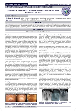
Figure.1: Clinical pictures: (A) Preoperative photograph showing
extraoral sinus tract. (B) photograph at second visit (C) 6 months
followup showing completeresolutionof thelesion.
On clinical examination extraoral sinus tract was seen in the chin
region. On palpation, a thumb-tip-sized nodal swelling around the
fistula was found (Fig.1 A). Intraoral clinical examination showed
discolored 31, 32 & 41. Slight tenderness on percussion was present
with 31 and 32. Radiographic examination showed periapical
radiolucency with 31, 32 & 41 (Fig.2 A). On electric pulp vitality
testingnoresponsewas seenwith31, 32 &41.
Sinus tracing showed association of the sinus tract with 31 & 32. The
diagnosis of chronic periapical abscess with an extraoral sinus tract
was established. The treatment plan decided was root canal treatment
with31, 32&41.
Access opening was done with round bur and endo access bur.
Biomechanical preparation was performed by hybrid instrumentation
technique using hand K files (Mani Inc) and Protaper (Dentsply
Maillefer, Ballaigues, Switzerland) rotary files. Irrigation was done
with 5% sodium hypochlorite followed by 17 % liquid EDTA
(Canalarge, Ammdent; Amrit Chemicals and Minerals Agency,
Mohali, Punjab, India). Agitation of the irrigant was done with
endoactivator. Later on laser was used for intracanal disinfection
following which propolis (Hitech India Naturals Limited) was placed
as an intracanal medicament for 2 weeks. No systemic antibiotics were
giventothepatient.
Intracanal irradiation was performed using 980 nm GaAlAs Diode
Laser (Denlase-980/7, China Daheng Group, Inc.) with a 200
micrometer fiberoptic tip and set at a power of 2.5 W. Using an
oscillatory technique, the diode fiber was introduced 1 mm short of the
apex and recessed in helicoidal movements at a speed of approxi-
mately 1 mm/s, and repeated four times at intervals of 10 s between
6
eachone.
After 2 weeks when the patient came for second visit there was
significant healing seen in that area with no pus discharge (Fig.1 B).
Also the teeth have become asymptomatic now. The intracanal
medicament was removed and obturation was done by warm vertical
compaction technique (E and Q Plus system; Meta Biomed Inc,
Chalfont, PA) (Fig.2 B). The patient was kept under follow up. Six
months follow up radiograph shows good periapical healing (Fig.2 C).
Clinically healed sinus was seen with scar formation (Fig.1 C).
Cosmeticsurgicaltreatmentwas advisedtothepatientforthesame.
Figure 2. Intraoral Radiographs: (A) Preoperative ; (B)
ImmediatePostoperative;(C) 6-Months Recall.
INTERNATIONAL JOURNAL OF SCIENTIFIC RESEARCH
Dental Science
22 International Journal of Scientific Research
Volume-7 | Issue-4 | April-2018 | PRINT ISSN No 2277 - 8179
2. Volume-7 | Issue-4 | April-2018
Discussion:
Diagnosis of odontogenic cutaneous tract by differentiation from other
similar conditions is important. Odontogenic cutaneous tract develops
due to pulp necrosis & periapical abscess. It may be confused with
various other conditions like squamous cell carcinoma, fungal &
bacterial infections, congenital fistulas etc. Sinus tracing helps us to
confirm the diagnosis and the offending tooth. Initially it was said that
removal of the sinus tract is essential for treatment, but proper
endodontic treatment is the only thing needed. Here also the patient
received only non surgical root canal treatment with satisfactory
results.
The success of endodontic therapy depends on complete eradication of
microbes from the root canal system. However only mechanical
instrumentation and conventional irrigants or medicaments may not be
able to reach all the areas of the root canal system. Calcium hydroxide
is unable to eradicate some bacterial species like Enterococcus (E.)
faecalis, the main microorganism found in root canal therapy failures.
Hence newer materials have been tried to overcome the shortcomings.
Of the newly found medications, propolis has attracted attention as a
natural antimicrobial agent. Propolis is a byproduct of honeybees
7
having antibacterial, antiviral, and antifungal properties. Propolis has
7
significant efficacy in killing E. Faecalis. Also propolis is 10 times less
potent as a cytotoxic agent than Calcium hydroxide. It is also much less
5
cytotoxictotheperiapicaltisssues as comparedtocalciumhydroxide.
Lasers have been shown to effectively sterilize the root canal. Laser
treatment may effectively replace conventional techniques because of
its improved disinfection efficacy, better penetrability, more effective
root canal cleaning, and elimination of the need to use toxic sol-
vents GaAlAs -gallium aluminum arsenide allows for greater
8
.
absorption by water in dental tissues and results in greater laser light
penetration through dentin making it possible to act on microorgan-
isms present in the dentinal tubules. Diode laser irradiation has a depth
of penetration up to 1,000 micrometer into dentinal tubules when
compared to the penetration power of chemical disinfectants, which is
6
limitedto100 micrometer.
Systemic antibiotic therapy is not needed for the healing of the tract. It
(1, 9)
results only in the temporary resolution of the symptoms. Six
months later, the cutaneous lesion had completely healed with a linear
scar formation. Unlike intraoral sinus tracts, extraoral tracts will heal
with granulation tissue thus leaving a cutaneous scar. Therefore, the
patient needs to be advised of a possible surgical revision of the scar.
Usually, the surgical revision is uneventful and enhances cosmetic
1
result.
Conclusion:
The present case shows that dental etiology should always be
considered in cases of odontogenic cutaneous tract to prevent
unnecessary treatments. In this case conventional root canal therapy
combined with laser disinfection and propolis as an intracanal
medicament lead to complete healing of the extraoral cutaneous sinus
tract.
References:
1) Cohenca N, Karni S, Rotstein I. Extraoral sinus tract misdiagnosed as an endodontic
lesion.J Endod.2003Dec;29(12):841-3.
2) Agız Dışı Deri Fistüllerinin Cerrahi Olmayan Endodontik Tedavi ile Iyilestirilmesi:Alt
Olgu Bildirisi. Treatment of extraoral cutaneous sinus tracts with non-surgical
endodonticintervention:reportof sixcases.J IstanbulUnivFacDent2015;49(2):35-40.
3) Lee SH, Yun SJ. Odontogenic cutaneous sinus tract presenting as a growing cheek mass
intheemergencydepartment.AmJ EmergMed.2017May;35(5):808.e5-808.e7.
4) Nakamura Y1, Hirayama K, Hossain M, Matsumoto K. A case of an odontogenic
cutaneoussinus tract.IntEndodJ. 1999Aug;32(4):328-31.
5) Ferreira FB, Torres SA, Rosa OP, Ferreira CM, Garcia RB, Marcucci MC, Gomes BP.
Antimicrobial effect of propolis and other substances against selected endodontic
pathogens. Oral Surg Oral Med Oral Pathol Oral Radiol Endod. 2007 Nov;104(5):709-
16.
6) Mithra N. Hegde, Raksha Bhat, Preethesh Shetty. Efficiency of a semiconductor diode
laser in disinfection of the root canal system in endodontics:An in vitro study. Journal of
the International Clinical Dental Research Organization | January-June 2015 | Vol 7 |
Issue1
7) Zare Jahromi M, Toubayani H, Rezaei M. Propolis: a new alternative for root canal
disinfection.IranEndodJ. 2012Summer;7(3):127-33.Epub2012Aug 1.
8) Koustubh M Kulkarni, Lotika Beri, Swapnil Bhosale. Non-surgical management of an
extraoral cutaneous sinus tract of odontogenic origin using Nd:YAG laser: An
endodonticChallenge.JournalofDentalLasers• January-June2012• Issue 1•Vol6
9) TidwellE, Jenkins JD, EllisCD, Hutson B, Cederberg RA. Cutaneous odontogenicsinus
tracttothechin:acasereport.IntEndodJ. 1997Sep;30(5):352-5.
International Journal of Scientific Research 23
PRINT ISSN No 2277 - 8179