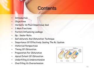
Obturation
Presented by: Dr Abhisek Guria
Dept. of Conservative Dentistry & Endodontics
Contents
• Introduction
• Objectives
• Hermetic Vs Fluid Impervious Seal
• 3 Main Functions
• Factors Influencing Leakage
• Gp : Sealer Ratio
• Def:obturate And Obturation Technique
• Importance Of Effectively Sealing The Rc System
• Historical Perspectives
• Timing Of Obturation
• Preparation For Obturation
• Monoblock Concept
• Apical Extent Of Obturation
• Underfilling & Underextension
• Overfilling Vs Overextension 2
• Evaluation of obturation
• Radiographic evaluation
• Clinical evaluation
• Methods of microleakage evaluation
• Obturating materials
• Ideal requirements
• Earlier root canal filling materials
• Silver cone
• Gutta Percha
• Evolution
• Chemistry
• Dental GP composition
• Phases
• Phase transformation
• Properties
5/11/2020 9:21 AM OBTURATION 3
• Commercial Manufacture
• Coagulation
• Obach’s Technique
• Current Forms
Solid Core Gutta Percha Points:
• Standardized Points:
• Non Standardized Points
Thermoplasticized Gutta Percha:
Chemically Plastisized Gp
• Immuno Cross Reactivity
• Disinfection
• Disinfecting Agents
5/11/2020 9:21 AM OBTURATION 4
Other newer obturating materials
• Resilon
– Components
– Primer:
– Resilon sealer (epiphany)
– Resin matrix
• Endorez
• Activ gp
• Medicated gp
• Smartseal
– Controlled expansion
5/11/2020 9:21 AM OBTURATION 5
5/11/2020 9:21 AM OBTURATION 6
Introduction
• Whether treatment is being undertaken to remove a
vital pulp, manage a necrotic/infected pulp, or revise a
previous root canal treatment, filling the root canal
represents a critical final stage of endodontic
therapy
5/11/2020 9:21 AM OBTURATION 7
Textbook of Endodontology, Lars Bjørndal
Objectives
• The final objective - total obturation of the root canal
space
• It is the sealing off of the complex root canal system
from the PDL & bone which insures the health of the
attachment apparatus
• Key to success: The elimination of microorganisms &
their byproducts Schilder.H. JOE — Volume 32, Number 4, April 2006
5/11/2020 9:21 AM OBTURATION 8
After disinfection, the obturation stage:
– Fill the root canal- hermetic seal from the coronal
orifice of the canal to the apical foramen at the
CDJ
– The responsibility does not end here
– Coronal seal- an integral part of endodontic
treatment & vital role in the treatment’s success
• BRITISH DENTAL JOURNAL Volume 216 No. 6 MAR 21 2014
5/11/2020 9:21 AM OBTURATION 9
Hermetic vs Fluid impervious seal
• According to accepted dictionary definitions, the
word hermetic means sealed against the escape or
entry of air—or made airtight by fusion or sealing.
• Instead, terms such as fluid-tight, fluid-impervious,
or bacteria-tight seals are more contemporary.
5/11/2020 9:21 AM OBTURATION 10
3 main functions
1. Prevent coronal leakage of microorganisms or
potential nutrients to support their growth into the
dead space of the root canal system
2. Prevent periapical or periodontal fluids percolating
into canals and feeding microorganisms
3. Entomb any residual microorganisms that have
survived the debridement & disinfection stages of
treatment
BRITISH DENTAL JOURNAL VOLUME 216 NO. 6 MAR 21 2014
5/11/2020 9:21 AM OBTURATION 11
Core concept :Factors influencing
leakage
• Root canal anatomy and preparation
– Oval, C-shaped and keyhole shaped root canal cross-sectional
profiles
– unsuitable shapes created during instrumentation prevent
the correct application of root canal filling materials.
• Access cavity
– Microorganisms may penetrate an obturated root canal within
a few days/weeks (coronal leakage).
• Smear layer
– Removal using citric acid (10–50%) or EDTA (17%) may
influence leakage
– The effect depends apparently on the sealer used.
12
• Hemostasis/dryness of the root canal.
– The wall must be clean and dry for tight adaptation of the
sealer to the wall.
• Root canal filling material
– Stability, adhesion to dentin and lack of pores.
• Sealer thickness and filling technique.
– Thick layers showed more leakage, because most sealers
contain pores or dissolve more rapidly in thick layers.
• Rinsing regimen
– Removal of the smear layer allows better penetration of the
sealer into dentinal tubules
– Formation of an interdiffusion zone between the sealer and
dentin collagen fibers.
5/11/2020 9:21 AM OBTURATION 13
Biocompatibility
• Root canal filling materials comes in contact with host
tissue- must show acceptable biocompatibility.
• Newer materials actively stimulate tissue
regeneration.
• They are osteoconductive or osteoinductive.
5/11/2020 9:21 AM OBTURATION 14
GP : Sealer
• Maximize volume of core material and minimize sealer.
• No magic filling material that will eliminate the need
for correct diagnosis, careful chemomechanical
preparation, and optimal coronal restoration
5/11/2020 9:21 AM OBTURATION 15
• Obturate—To fill the shaped and debrided canal
space with a temporary or permanent filling material.
• Obturation technique—The method used to fill and
seal a cleaned and shaped root canal using a root canal
sealer and core filling material; sealers are frequently
used as the sole obturating material in deciduous
teeth
• Glossary of Endodontic Terms
5/11/2020 9:21 AM OBTURATION 16
Importance of Effectively Sealing the RC System
• Ingle & colleagues: 58% of treatment failures – incomplete obturation
– Often poorly prepared
– Procedural errors
• Correlation between the quality of obturation & nonhealing: presence of
bacteria Fabricus et al.
• Cleaning & shaping determines: the degree of disinfection & the ability
to obturate the radicular space.
• Reflection of the cleaning and shaping
5/11/2020 9:21 AM OBTURATION 17
Failure of diagnostics
– Not possible to assess
the quality of the seal
with a radiograph
– Porous tubular
structure of dentin
and canal irregularities
– An adequate
radiographic
appearance : may not-
adequate seal
5/11/2020 9:21 AM OBTURATION 18
• Variation in radiographic
interpretation by the
clinician
• Overlying osseous
structures
• Lack of uniformity in the
obturation materials
5/11/2020 9:21 AM OBTURATION 19
Historical Perspectives
• Over 100 years
• Edward Hudson in 1825: Filling with gold foil
• 1847 - Hill Developed “Hill’s-stopping” a restorative material, a
mixture of bleached Guttapercha and carbonate of lime and
quartz.
20
• 1967 -“Hermetic seal” of the canal terminating at the dentinocemental
junction- Grossman’s Principle 9
• “Sealed against the escape or entry of air” or made ‘Airtight by fusion or
sealing’ – inaccurate
• Hermes Trismegistus - seal of wax
• Obturating the canal space: fluid leakage at the apex or the coronal level.
• ‘‘Impermeable seal’’ fluid-tight, fluid-impervious, or bacteria-tight seals
• 1867 - Bowman was the first to use Guttapercha for root canal filling.
5/11/2020 9:21 AM OBTURATION 21
• 1883 - Perry used pointed gold wire wrapped with soft GP, rolled
and packed it into the canal.
• 1887 - S.S White Company was the first to start the commercial
manufacture of GP points.
• 1893 - Rollins used Gutta-percha with pure oxide of mercury
into root canal filling.
• Edmund Kells- first endodontic radiograph in 1899
• 1914 - Callahan introduced softening and dissolution of GP with
the use of rosins in obturation.
5/11/2020 9:21 AM OBTURATION 22
• 1959 – Ingle and Levine were the first persons to propose
standardization of root canal instruments and filling materials and at
their behest, standardized Gutta-percha was introduced to the
profession after 2nd International Conference of Endodontics at
Philadelphia.
• 1976 - A group evolved into the present day International standards
organization (ISO) for approval of specification of root canal
instruments and filling materials.
5/11/2020 9:21 AM OBTURATION 23
Timing of Obturation
• RC system is dry and time permits, obturating at the same visit
is recommended.
• If a dry root canal system not achieved: inter-appointment
dressing.
• Asymptomatic
• Mild/ significant symptoms: asymptomatic upon obturation
5/11/2020 9:21 AM OBTURATION 24
BRITISH DENTAL JOURNAL
VOLUME 21 NO. 6 MAR 21 2014
• Negative bacterial cultures.
• Sjogren et al. 5-year recall
• 94% of cases - negative cultures: successful
• 68% of those filled with positive cultures were successful
• Procedural concerns
• Difficult cases- multiple appointments
• Medical conditions, psychologic state of mind, and fatigue.
5/11/2020 9:21 AM OBTURATION 25
Preparation for Obturation
• Obturation materials designed to bond with dentin,
• Irrigation solutions must be used with consideration of
the condition of the dentin surface that is most suitable
for bonding
• Smear layer: slowly disintegrate & dissolve around leaking
obturation materials
• Sealer penetration into dentinal tubules does not occur
when the smear layer is present
• Colleagues for Excellence. Fall 2009
5/11/2020 9:21 AM OBTURATION 26
Smear layer removal
• 17% disodium EDTA for one minute, followed by a final
rinse of NaOCl
• Sonic and ultrasonic instrumentation
• MTAD
• 50% HNO3
• Tetracycline
• Concern: increased dentin permeability due to
demineralization
5/11/2020 9:21 AM OBTURATION 27
LENGTH / APICAL EXTENT OF
OBTURATION
5/11/2020 9:21 AM OBTURATION 28
• The totality of the 3D filling of the RC is more
important than its vertical extent alone
5/11/2020 9:21 AM OBTURATION 29
Kuttler : Termination should be to the apical constriction, when the
apical constriction exists.
Seltzer et al. The reaction to tissues were milder when instrumenting
short of the apex as compared to instrumenting beyond the apex.
Schilder.H. JOE — Volume 32, Number 4, April 2006
Schaeffer et al. JOE — Volume 31, Number 4, April 2005
• Most North American and European Schools: instrumentation &
obturation should be contained within the root canal
(Cailleteau & Mullaney 1997)
• Weine: A point located 1mm coronal to the apex is close to the area of
the CDJ; 1mm short of the radiographic apex probably acceptable.
• Agreed with Kuttler's study (1955) which identified a smaller diameter
or `apical constriction‘ as the point where the canal preparation should
end and where the deposition of calcified tissue is most desirable.
5/11/2020 9:21 AM OBTURATION 30
Wein’s recommendation
5/11/2020 9:21 AM OBTURATION 31
• Ingle (1973): Obturation at 0.5mm from the radiographic apex;
obturating up to the radiographic terminus of the root actually
results in an overfilling.
• Pecchioni (1983) : “during instrumentation it is better not to go
nearer than 0.5±1mm from the radiographic apex”.
• ‘’While it is very serious and damaging to go beyond this
limit with instrumentation, it is less serious to slightly
overfill the apex, since the common sealers are generally
tolerated and easily resorbable‘
5/11/2020 9:21 AM OBTURATION 32
Nguyen (1985)
indicated the CDJ
as the limit of the
preparation
Guldener (1985):
A working length
which corresponds
to the tooth
length less 0.5mm
for cases with a
necrotic pulp.
In cases of vital
pulp extirpation:
an additional
reduction of 0.5
mm, 1mm short of
the tooth length.
Taylor (1988): a
narrower spot at
the apical level
called `Minor
Diameter' which
he believed to
correspond
histologically to
the CDJ
5/11/2020 9:21 AM OBTURATION 33
Langeland (1957, 1967, 1987, 1995)
• Termination of instrumentation & obturation at the apical
constriction
• “The most frustrating clinical aspect is that no exact distance
from the radiographic apex could be given, because the distance
from the radiographic apex to the apical constriction varies
widely from root to root”
• All endodontic sealers are irritant and resorbable (1974, 1995)
• Ricucci.D. International Endodontic Journal (1998) 31, 384 -393
5/11/2020 9:21 AM OBTURATION 34
Schilder -1967, 1976
• Debridement and obturation to the radiographic apex, which often
results in material being extruded into periradicular tissues
• 3 - 5% NaOCl solution completely removes necrotic organic debris
• Opposed limitation of preparation at the CDJ or at the apical
constriction: too approximate to apply a mathematical or statistical
formula (0.5, 1 or 2 mm).
• Schilder (1987) later requested that canal instrumentation and
obturation should stop at the canal terminus
• Scianamblo (1989): RCs & ramifications can be effectively cleaned
during cleaning and shaping if these systems are properly irrigated with
NaOCl
• Ricucci.D. International Endodontic Journal (1998) 31, 384 -393
5/11/2020 9:21 AM OBTURATION 35
Longitudinal studies
• Strindberg (1956): 775 endodontically treated roots, reviewed up to
10 years after treatment: the highest success rate - obturation
terminated 1mm short of the radiographic apex
• Swartz et al. (1983): 1007 endodontically treated teeth, 1770 canals:
‘ overfilled canals were four times more likely to fail than canals filled
short of the radiographical apex'.
• Friedman et al. (1995): Presence of extruded sealer the success rate
was 56.7% against 81.9% in the absence of extrusion.
5/11/2020 9:21 AM OBTURATION 36
Underfilling & Underextension
• Over and under extension refer solely to the vertical dimension
of the root canal filling, beyond or short of the root apex.
• Underfilling—An incomplete obturation of the root canal space
with resultant voids
• Underfilled tooth : A tooth whose RC system has been
inadequately obturated in any dimension, leaving large reservoirs
for recontamination and infection.
• Schilder.H. JOE — Volume 32, Number 4, April 2006
• Glossary of Endodontic Terms
5/11/2020 9:21 AM OBTURATION 37
Overfilling vs Overextension
• Overfilling—A solid or semi-solid core root canal filling extending
beyond the apical foramen; commonly used to imply that the root canal
space is completely obturated.
• Overextension—A solid or semi-solid core root canal filling extending
beyond the apical foramen, often the result of failure to create an
apical stop during instrumentation; commonly used to imply that the
root canal space is not completely obturated.
• Schilder.H. JOE — Volume 32, Number 4, April 2006
• Glossary of Endodontic Terms
5/11/2020 9:21 AM OBTURATION 38
• No case of endodontic failure due to overfilling, ie.
when the root canal has been obturated in its entirety
& surplus material has been intruded into the apical
periodontium
• Numerous cases of failure of vertical overextensions
of underfilled root canals.
• Schilder.H. JOE — Volume 32, Number 4, April 2006
5/11/2020 9:21 AM OBTURATION 39
Evaluation of obturation
1. Three-dimensionally fills the entire RC system as close to
the CDJ as possible
2. Shape reflecting a continuously tapered funnel: approx.
the same as the external root morphology
3. Radiographically: dense, 3D filling that extends as close
as possible to the CDJ
• Root Canal Obturation: An update
5/11/2020 9:21 AM OBTURATION 40
Clinical evaluation
1. Normal findings to routine tests
2. Concerned about prognosis, the reevaluation visit
should be scheduled in a few weeks
3. Routine reevaluation periods: 6 months and 1 year.
4. If symptoms occur they should call the office for an
appointment
• Colleagues for Excellence. Fall 2009
5/11/2020 9:21 AM OBTURATION 41
Radiographic evaluation
• The length of an ideal fill should be from the minor
constriction to the canal orifice unless a post is
planned.
• The shape of the completed case: obturation
technique being used
• Voids should not be visible on the radiographic image
• Adequate provisional restoration or definitive
• Colleagues for Excellence. Fall 2009
5/11/2020 9:21 AM OBTURATION 42
In vitro studies
• Methods of microleakage evaluation
• Dye penetration
• Radioisotopes
• Electrochemical
• Fluorometrics
• SEM
• Root clarification
• Fluid filtration
• Fluid transport
• Proteins
• Endotoxins
• Glucose penetration
5/11/2020 9:21 AM OBTURATION 43
Tissue toxicity
(1) cytotoxicity evaluation
(2) subcutaneous implantation
(3) intraosseous implantation
(4) in vivo periapical
• Chisholm: ZnO & oil of cloves, 130 years ago, to
dentistry
• Eugenol: quite cytotoxic
• Proven track record
5/11/2020 9:21 AM OBTURATION 44
OBTURATING
MATERIALS
5/11/2020 9:21 AM OBTURATION 45
Ideal requirements
1. It should be easily introduced into the root canal.
2. It should be removed easily from the root canal, if necessary.
3. It should seal the canal laterally as well as apically.
4. It should not shrink after being inserted.
5. It should be impervious to moisture.
6. It should be bacteriostatic or at least not encourage bacterial
growth.
7. It should be radiopaque.
8. It should not stain tooth structure.
9. It should not irritate periradicular tissues.
10. It should be sterile, or easily and quickly sterilized,
immediately before insertion.
5/11/2020 9:21 AM OBTURATION 46
Core concept : Requirements for an ideal root canal filling material
Technical
_ No shrinkage
_ No solubility in tissue fluids, undisturbed setting in the presence of
moisture
_ Good adhesion/adaptation to dentin or combining materials (cones,
sealers)
_ No pores and water sorption
_ No tooth discoloration
Biological
_ No general health problems or allergies for patients and dental
personnel
_ No irritation of local tissues
_ Sterile
_ Antimicrobial – no enhanced bacterial growth
_ Stimulation of the periapical healing process
5/11/2020 9:21 AM OBTURATION 47
Textbook of Endodontology, Lars Bjørndal
• Handling
• Setting in an adequate time, allowing sufficient time for obturation and
radiographic control
• Easy to apply and easy to remove using
solvents, heat, or mechanical instrumentation
• ISO 6876 requires endodontic filling materials to
have a radiopacity >3mm Al equivalent
• According to European regulations (Medical Device Regulations (EU) 2017/745)
root canal filling materials must successfully pass a clinical risk assessment
procedure before they can be marketed.
5/11/2020 9:21 AM OBTURATION 48
Earlier root canal filling materials
• Amalgam
• Asbestos
• Balsam
• Cement
• Copper
• Gold Foil
• Iron
• Lead
• Oxy- Chloride of Zinc
• Paraffin
• Pastes, Plaster of Paris
• Resin, Rubber
• Silverpoints
• Tin foil
5/11/2020 9:21 AM OBTURATION 49
Silver cone
• Jasper introduced - claimed produced the same success rate as
GP and were easier to use.
• The rigidity made them easy to place and permitted more
predictable length control
• Inability to fill the irregularly shaped RC system permitted
leakage .
• When silver points contact tissue fluids or saliva, they corrode.
5/11/2020 9:21 AM OBTURATION 50
• The corrosion products have been found to be
cytotoxic and produced pathosis or impeded periapical
healing.
• Treatment failures were
the result of leakage and
failure to remove the irritants
from the root canal system.
• No longer used in contemporary endodontic practice.
5/11/2020 9:21 AM OBTURATION 51
Gutta Percha
• ‘’GETAH”- meaning gum
• ‘’PERTJA”- name of the tree in Malay language
• The first person to discover this material was
John Tradescant, in 1656
• he named this material as “Mazer wood”.
• But the honour of introduction of this material
goes to Dr. William Montogmerie (1843), who was
a medical officer in Indian service.
5/11/2020 9:21 AM OBTURATION 52
Evolution
• GP was first introduced to dentistry as a temporary
filling material by Edwin Truman.
• ADA specification - No.78.
5/11/2020 9:21 AM OBTURATION 53
Chemistry
• It is a Trans- isomer of poly isoprene.
• Its chemical structure is 1, 4, trans–poly isoprene.
• Its molecular structure is close to that of natural
rubber
5/11/2020 9:21 AM OBTURATION 54
• GUTTA -------- 75-82%
• ALBAN -------- 14-16%
• FLUAVIL --------- 4-6%
• Also contains Tannin, salts and saccharine substance.
• Dental GP composition:
5/11/2020 9:21 AM OBTURATION 55
Phases
• Chemically pure Gutta-percha exists in two distinctly
different crystalline forms (α and β)
• Natural gutta percha from tree- α form
• most commercial available product is in β form
• During the process of manufacture, if the cooling is done
rapidly, ‘β’ form results.
• If it is cooled slowly, less than 0.5°c/hr, ‘α ’ form results.
• Another unstable form (γ) exists, which is amorphous in
nature.
5/11/2020 9:21 AM OBTURATION 56
• α ---- runny, tacky and sticky
(lower viscosity)
• β ---- solid, compactible and
elongatible (higher viscosity)
• γ ----- similar to a (unstable)
5/11/2020 9:21 AM OBTURATION 57
Phase transformation
• When heated from room temperature to 1000 C.
• Beta to alpha: occurs between 42-49 ( this phase
transformation is reversible)
• Alpha to amorphous: occurs between 53-590C
5/11/2020 9:21 AM OBTURATION 58
5/11/2020 9:21 AM OBTURATION 59
Alpha form Beta form
Brittle at room temperature Stable and flexible
Becomes gluey, adhesive and
highly flowable when heated
(low viscosity)
Becomes less adhesive and
flowable when heated
(high viscosity)
Thermoplastisized GP for warm
condensation is in alpha form
In cold condensation it is beta
form
Properties
• Biocompatibility:
• highly biocompatible; does not react with body fluids
• inert irrespective of alkaline or acidic medium
• does not initiate allergic host response.
• Dimensional stability:
• expansion or shrinkage of material occurs only during phase
transformations.
• Ductility and malleability:
• depending on existing phase, material may be ductile or
malleable.
• Melting point: around 600C, starts softening
• Ease of handling: easy to manipulate. When required can be
easily removed from root canal.
• Adaptation to root apex: either in cones or warm,
thermoplasticized form.
5/11/2020 9:21 AM OBTURATION 60
COMMERCIAL MANUFACTURE
• Coagulation
• The sap that oozes out is collected and put
into a pot and boiled with little water.
• kneaded under running water.
• Chemical method of coagulation is by
addition of alcohol and creosote mixture
(20:1), ammonia, limewater or caustic soda
5/11/2020 9:21 AM OBTURATION 61
Obach’s technique
The obtained pulp is mixed with water and heated to 75°c to release the GP
threads and then cooled to 45°c.
The flocculated GP called “yellow Gutta” contains 60% poly isoprene and
40%contaminants .
Yellow Gutta is mixed with cold industrial gasoline at below 0°c
temperature.
This treatment not only flocculates the GP but also dissolves resins and
denatures any residual proteins.
Then dissolved in warm water at 75°c and dirt particulate is allowed to
precipitate.
Residual greenish yellow solution is bleached with activated clay, filtered
to remove any particulate and then steam distilled to remove the gasoline.
“Final ultra pure” GP has gasoline scent, before it is modified with fillers into
its final commercial product formulation.
5/11/2020 9:21 AM OBTURATION 62
CURRENT FORMS
1. Solid core gutta percha points:
• Standardized points:
• manufactured in coherence to instrument taper and apical gauge.
• Stiff due to high ZnO
• Non standardized points:
• manufactured in variable taper
• The tip of point is adjusted with an apical gauge to best fit and
seal the apex.
• Flexibility is enhanced due to low content of zinc oxide.
5/11/2020 9:21 AM OBTURATION 63
2. Thermoplasticized gutta percha:
• Usually available in injectable form (alpha phase).
• These systems provide special heaters which heats till the
temperature when it flows.
• Apical seal is achieved with the plugging of master cone and then
the gutta percha is backfilled.
3. Chemically plastisized Gp
• Gutta percha may be dissolved in various solvents, such as
chloroform, oil of eucalyptus or xylol.
5/11/2020 9:21 AM OBTURATION 64
5/11/2020 9:21 AM OBTURATION 65
Scheme for the dimensions of a standardized gutta-percha cone according to ISO 6877; d1 × 100 = size designation
of gutta-percha cone (ISO 10–ISO 140).
IMMUNO CROSS REACTIVITY
• GUTA PERCHA and NATURAL RUBBER LATEX (NRL), are from
different trees of the same botanical family
• Raw GP contains the rubber molecule trans-polyisoprene
• NRL contains cis-polyisoprene intermingled with a variety of
latex-allergen proteins
• Dental latex materials can cause immediate hypersensitivity
(type I allergy) in dental professionals and patients.
• include ocular, nasal, pulmonary, gastrointestinal, cardiovascular
and genitourinary symptoms
• be localized and systemic
5/11/2020 9:21 AM OBTURATION 66
• Gazelius et al. and Boxer et al. reported that - a potential cross-reactivity
between NRL and gutta percha allergens in latex allergy patients exists.
• Costa et al. investigated the cross-reactivity between gutta percha, gutta
balata, and NRL using a competitive radio-allegro-sorbent test (RAST) inhibition
assay.
• These results demonstrated that the raw or manufactured GP points did not
show detectible cross reactivity.
• However, raw gutta balata extracts cross-reacted with NRL-specific IgE
antibody.
• Using the RAST inhibition and the immunoblot inhibition assays, Hamann et al. -
there is no cross-reactivity between GP and NRL, but raw GP cross-reacted with
serum from patients with type 1 NRL allergy.
67
• Kang PB et al. studied the postulated immuno cross-
reactivity between proteins derived from raw GP (RGP), GP
point (GPP) and natural rubber latex (NRL).
• Determined by ELISA inhibition assay.
• Except for NRL glove extracts, neither extracts from RGP
or GPP were reactive in ELISA inhibition assay.
• The immuno cross-reactivity of gutta percha points Peter B. Kang∗, Kristy Vogt, Stephen E. Gruninger,
Milton Marshall, Chakwan Siew, Daniel M. Meyer Dental materials 23 ( 2007 ) 380–384
5/11/2020 9:21 AM OBTURATION 68
Disinfection
• GP can be contaminated by handling, aerosols and physical
sources during the storage process.
• Why disinfection of GP?
• free from pathogenic microorganisms
• Penetration of bacteria into the blood stream should be avoided
• cannot be sterilized by moist or dry heat - cause alteration of
structure.
• Therefore, a rapid chairside chemical disinfection is needed.
5/11/2020 9:21 AM OBTURATION 69
Why do we need a quick decontamination method?
• Difficult to know beforehand how many
accessory cones will be used.
• 5.25% NaOCl is an effective agent
• CHX was not effective in eliminating
Bacillus subtilis spores after 72 h of
contact
5/11/2020 9:21 AM OBTURATION 70
Disinfection of gutta-percha cones with chlorhexidine and sodium hypochlorite Brenda Paula Figueiredo de
Almeida Gomes et al. Oral SurgOral Med Oral PatholOral Radiol Endod 2005;100:512-7
Disinfecting agent
• Polyvinyl pyrolidone-iodine
• Ethyl alcohol
• Sodium hypochlorite
• Hydrogen peroxide
• Quaternary of ammonium
• Glutaraldehyde
• Chlorhexidine liquid.
5/11/2020 9:21 AM OBTURATION 71
Presented by: Dr Abhisek Guria
Dept. of Conservative Dentistry & Endodontics
SHDCH , Hassan
Sem - 10
Date of Presentation : 08/03/2019
Contents
• Introduction
• Objectives
• Hermetic Vs Fluid Impervious Seal
• 3 Main Functions
• Factors Influencing Leakage
• Gp : Sealer Ratio
• Def:obturate And Obturation Technique
• Importance Of Effectively Sealing The Rc System
• Historical Perspectives
• Timing Of Obturation
• Preparation For Obturation
• Apical Extent Of Obturation
• Underfilling & Underextension
• Overfilling Vs Overextension
73
• Evaluation of obturation
• Radiographic evaluation
• Clinical evaluation
• Methods of microleakage evaluation
• Obturating materials
• Ideal requirements
• Earlier root canal filling materials
• Silver cone
• Gutta Percha
• Evolution
• Chemistry
• Dental GP composition
• Phases
• Phase transformation
• Properties
5/11/2020 9:21 AM OBTURATION 74
• Commercial Manufacture
• Coagulation
• Obach’s Technique
• Current Forms
Solid Core Gutta Percha Points:
• Standardized Points:
• Non Standardized Points
Thermoplasticized Gutta Percha:
Chemically Plastisized Gp
• Immuno Cross Reactivity
• Disinfection
• Disinfecting Agents
5/11/2020 9:21 AM OBTURATION 75
Other newer obturating materials
• Resilon
– Components
– Monoblock Concept
– Primer:
– Resilon sealer (epiphany)
– Resin matrix
• Endorez
• Activ gp
• Medicated gp
• Smartseal
– Controlled expansion
5/11/2020 9:21 AM OBTURATION 76
Obturation techniques
• Lateral compaction
– Procedure
– Advantages
– Limitations
– Variants on cold lateral compaction
– Warm Lateral Compaction Technique
• Warm vertical condensation technique:
– Procedure
– Advantages
– Disadvantages
• Continuous Wave Compaction Technique
– System B™ Obturation
• McSpadden Thermomechanical Compaction
OBTURATION 77
– Procedure
– Advantages
– Limitations
• Thermoplastic injection techniques
– Obtura iii
– Calamus
– Elements
– Hotshot
– Ultrafil 3d
– Obtura ii
• Carrier-based gutta-percha techniques
– Generations
– Thermafil
– Simplifil
– Successfil
– Trifecta technique
– Js quick-fill
5/11/2020 9:21 AM OBTURATION 78
• Chemically Plasticized Gutta Percha
– Chloropercha
– Callahan–Johnson diffusion technique
– Nygaard–Ostby technique
• Custom Cone Technique
• Immediate Obturation
• Single- cone obturations
• Removal of obturation material
– Guttapercha
– Solid core obturators.
– Paste
– Resilon
– Silver points.
5/11/2020 9:21 AM OBTURATION 79
• Removal of single cone GP
• Removal of Condensed GP
– manually
– using rotary system
– using LASER
• Remove solid core obturators
– Metal core obturators
– Removal of plastic core obturators
– Removal of Modified GP core carrier
• Conclusion
• Referrences
5/11/2020 9:21 AM OBTURATION 80
OTHER NEWER
OBTURATING MATERIALS
5/11/2020 9:21 AM OBTURATION 81
Resilon
• It offers solutions to the
problems associated with GP:
– Shrinkage of GP after
application of heat.
– Gap formation between
the sealer and the GP.
• This resilon core material
bonded to the sealer by
polymerization.
5/11/2020 9:21 AM OBTURATION 82
Monoblock concept
5/11/2020 9:21 AM OBTURATION 83
Components
• 1. Primer:
• It is a self etch primer
• contains a sulfonic acid terminated functional
monomer, HEMA
• 2. Resilon sealer (Epiphany)
• a dual-cured, resin-based composite sealer.
• bonds to both the root dentin and Resilon cones.
• The resin matrix is comprised of Bis-GMA,
ethoxylated Bis-GMA, UDMA and hydrophilic
difunctional methacrylates.
5/11/2020 9:21 AM OBTURATION 84
• 3. Resilon core material:
• It is a thermoplastic synthetic polymer based
(polyester) core material
• contains bioactive glass, bismuth oxychloride and
barium sulphate.
• filler content - 65 % by weight.
• Noncytotoxic and nonmutagenic.
5/11/2020 9:21 AM OBTURATION 85
EndoREZ
• It is a hydrophilic, two-component (base and
catalysts), dual curing self priming sealer.
• It has hydrophilic characteristics providing
penetration into dentinal tubules.
• The radiopacity is equivalent to gutta-percha.
• does not compromise the function of dentin bonding
agents or luting resin polymerization.
5/11/2020 9:21 AM OBTURATION 86
• Designed to be used with EndoREZ Points and/or gutta percha.
• EndoREZ Points are standard ISO-sized gutta percha points
overlayed with a thin resin coating, which bonds chemically with
EndoREZ sealer.
OBTURATION 87
Activ GP
• a core material containing GP which is impregnated
and coated with glass ionomer.
• sealer is a traditional glass ionomer sealer which can
adhere chemically and micromechanically to the Activ
GP cones and bond to the dentin.
• Coating helps bonding between gutta-percha and glass
ionomer sealer.
5/11/2020 9:21 AM OBTURATION 88
• Activ GP Sealer - 12 minute
working time
• no need for a bonding agent
(primer)
5/11/2020 9:21 AM OBTURATION 89
Medicated GP
1. Calcium hydroxide containing GP
2. Chlorhexidine diacetate containing GP
3. Iodoform containing GP
5/11/2020 9:21 AM OBTURATION 90
Smartseal
• Based on polymer technology.
• Principle - the hydrophilic nature of the obturating
points which can absorb surrounding moisture and
expand resulting in filling of voids and spaces.
5/11/2020 9:21 AM OBTURATION 91
constructed in two parts
• Central Core
• point with the flexibility to easily pass around any curves
in the prepared canal
• Outer Polymer Layer
• this hydrophilic, hydrogel layer allows the point to swell to
adapt to the ramifications of the root canal.
92
• Advantages :
• Any geometry of point can be accurately made
• Controlled expansion
• Biocompatibility
5/11/2020 9:21 AM OBTURATION 93
Smart paste Bio
• Is a hydroxyapatite based sealer that is used with
the smartseal obturation points.
• delivered in a pre-mixed syringe.
• expand slightly on setting
5/11/2020 9:21 AM OBTURATION 94
Controlled Expansion
The smartseal polymer is hydrophilic which allows water
present in the root canal to be absorbed.
This water can hydrogen bond to the polar sites
present, enabling expansion within the polymeric
chains.
Expansion occurs within the first 4 hours.
This results in the polymer and sealer being expressed
into the dentinal tubules.
Forms a seal that is impermeable to bacterial micro-
leakage.
OBTURATION TECHNIQUES
5/11/2020 9:21 AM OBTURATION 96
Obturation techniques
1. Cold lateral compaction
2. Warm compaction (warm gutta-percha)
(a) Vertical
(b) Lateral
3. Continuous wave compaction technique
4. Thermoplasticized gutta-percha injection
5. Carrier-based gutta-percha
(a) Thermafil thermoplasticized
(b) SimpliFill sectional obturation
6. McSpadden thermomechanical compaction
7. Chemically plasticized gutta-percha
8. Custom cone
5/11/2020 97
Grossman
Lateral compaction
• Isolation & drying the canals
• Selection of master cone
• Checking for apical “TUG BACK”
• Inadequate fit- beyond the apex
– Tip cut off: reinserted primary cone fits snugly at the WL
• Inadequate fit- short of the apex
– Patency established to the corrected length
– Another primary GP inserted & verified
5/11/2020 9:21 AM OBTURATION 98
Sealer
manipulation
Canal
coated
Master cone
inserted till
WL
Spreader
inserted
alongside:
level 1mm
short of the
WL
Spreader
disengaged
Placement
of
sequential
accessory
cones by
lateral
compaction
5/11/2020 9:21 AM OBTURATION 99
Endodontic Topics 2005, 12, 2–24
• Butt end of the GP: cut off with heated
instrument
• Chamber cleaned & Restoration placed
• Advantages
• “ Deep spreader
penetration”: minimize apical
leakage/ percolation
• Positive dimensional stability
of the root canal filling
• Less likelihood of carrying
filling material beyond the
root apex
• Limitations
• Presence of voids
• Less able to seal intracanal
defects & lateral canals
• Less homogeneous mass
• Time consuming
5/11/2020 9:21 AM OBTURATION 100
JOE—Volume 32, Number 4, April 2006
Endodontic Topics 2005, 12, 2–24
VARIANTS ON COLD LATERAL
COMPACTION
• Warming spreaders before each use in a hot bead
sterilizer
• Softening GP with heat before insertion of the cold
spreader
• Mechanical activation of finger spreaders in an endodontic
reciprocating handpiece
• Application of an ultrasonically energized spreader
• Endodontic Topics 2005, 12, 2–24
5/11/2020 9:21 AM OBTURATION 101
Warm Lateral Compaction Technique
• Martin
• Endotec II, Endo Twinn, EI DownPak device is used in
warm lateral compaction.
5/11/2020 9:21 AM OBTURATION 102
Warm vertical condensation technique:
• Herbert Schilder 1967
• Objective - filling the main root canal as well as
lateral and accessory canals
• Eg - Touch ’n Heat
5/11/2020 9:21 AM OBTURATION 103
• (a) and (b) Master cone adaptation
• (c) Severing of the coronal
portion of the master cone with a
heated instrument.
• (d) Compaction of the master
cone.
• (e) Sequential segments removed
with the heat carrier followed by
compaction.
• (f)–(h) Once the apical third is
reached, the canal is backfilled
with heated segments of gutta-
percha followed by compaction
with suitable pluggers.
5/11/2020 9:21 AM OBTURATION 104
(a) (b) (c) (d)
(e) (f) (g) (h)
• Advantages
• Excellent seal of the canal
apically and laterally
• Obturation of the larger lateral
and accessory canals
• Disadvantages
• The amount of time it takes
• The risk of vertical root
fracture resulting from undue
force
• overfilling of GP or sealer that
cannot be retrieved from the
periradicular tissues
• difficult in curved canals
5/11/2020 9:21 AM OBTURATION 105
• A study compared root surface temperatures for warm
vertical obturation using the System B heat source, the
Touch ’n Heat device and a flame-heated carrier in
maxillary and mandibular incisors and premolars.
• System B and the Touch ’n Heat produced a surface
temperature rise that was less than 10° C.
• The flame-heated carrier produced temperature changes
greater than 10° C in all experimental teeth.
5/11/2020 9:21 AM OBTURATION 106
• Critical level of root surface
heat required to produce
irreversible bone damage -
greater than 10° C
• warm vertical compaction with
the System B should not
damage supporting periodontal
structures
5/11/2020 9:21 AM OBTURATION 107
Continuous Wave Compaction Technique
• By Buchanan
• Variation of warm vertical compaction technique.
• Introduced to reduce time
• employs an electric heat carrier
• The recommended temperature setting
for system B unit is 2000 C.
• System B
• Obtura II system
5/11/2020 9:21 AM OBTURATION 108
System B™ Obturation
• A gutta-percha cone is chosen
• Tug back occurs within the apical
control zone
5/11/2020 109
• A System B™ condenser/ plugger is chosen that
matches the shape of the prepared canal and the
GP cone.
• The plugger is prefit to its binding point
• The rubber stopper is then positioned 2mm short
binding point to minimize direct contact on dentin
5/11/2020 110
• Sealer is placed in the canal
• The apical 4 mm of the master cone is
coated with sealer and using gentle
pressure, the cone is seated to place.
5/11/2020 111
• The System B™ Heat Source is
turned to use and placed it
theTouch mode.
• The plugger plunges smoothly
through the gutta-percha mass.
5/11/2020 112
• As the heated plugger (200 0C)
approaches the binding point, the
ring switch is released
• which allows the plugger/condenser
to continue its apical movement.
5/11/2020 113
• Apical pressure is maintained on the
apical plug of GP for a 10second
sustained push to prevent cooling
shrinkage.
• While maintaining apical pressure,
the ring switch is activated again
for1 second.
• The switch is released and the
plugger quickly withdrawn.
5/11/2020 114
• As the pluggers heat from the
tip to back
• It removes the coronal flash of
GP leaving a clean interface and a
dense homogeneous mass sealing
the apical third
5/11/2020 115
• Sealer is applied to the walls of
the canal before beginning to
back fill.
• thermoplasticized delivery
system is inserted into the
coronal aspect ofthe apical plug.
• 4 mm increment is placed
• Prefit Schilder pluggers are now
used to condense the thermo-
softened GP.
5/11/2020 116
• The entire canal space is filled
5/11/2020 117
• Kenneth S. Serota
McSpadden Thermomechanical
Compaction
• Uses heat to decrease gutta-percha viscosity and increase its
plasticity.
• heat created by rotating a compacting instrument in a slow-speed
contra-angle handpiece at 8000-10000 RPM alongside GP cones.
• Compactor with spiraled 900 are similar to flutes on H-files, but in
reverse, forces the softened GP apically and laterally.
5/11/2020 9:21 AM OBTURATION 118
used to fill straight canals as the compactor blade breaks if it
binds.
Compactor blade selected according to width and length of
prepared canal
GP cone is inserted and compactor placed between GP and canal
wall up to 3-4 mm from prepared length
Handpiece activated. Rotating tip is guided to within 1.5mm of root
apex
5/11/2020 9:21 AM OBTURATION 119
• Advantages :
• 1. Ease of selection and insertion of GP cones
• 2. Economy of time
• 3. Rapid filling of canals apically and laterally, including irregular spaces
• Disadvantages :
• 1. Inability to use it in narrow canals
• 2. Frequent breakage of compactor blades
• 3. Frequent overfilling of the canal
• 4. Shrinkage of cooled, set filling
5/11/2020 9:21 AM OBTURATION 120
THERMOPLASTIC INJECTION TECHNIQUES
• Harvard/ Forsythe Institute: 1977
• Obtura II
• Obtura III
• Calamus
• Elements
• HotShot
• Ultrafil 3D
5/11/2020 9:21 AM OBTURATION 121
OBTURA III
• Hand hold gun
• Ag needle
• Control unit
5/11/2020 9:21 AM OBTURATION 122
Ultrafil 3D
• Involving gutta-percha cannulas, a heating unit, and an injection
syringe.
5/11/2020 9:21 AM OBTURATION 123
• The system has 3 types of cannulas.
• The Regular Set is a low-viscosity material that requires 30
minutes to set.
• The Firm Set is also a low-viscosity material but differs in that
it sets in 4 minutes.
• Endoset has a higher viscosity and does not flow as well.
• It sets in 2 minutes.
5/11/2020 9:21 AM OBTURATION 124
Calamus
• Equiped with a cartridge
system
• The unit permits control of
temperature and also the
flow rate.
• The 360 degree activation
switch allows great tactile
sensation.
5/11/2020 9:21 AM OBTURATION 125
Elements
• consists of a System B heat
source and plugger as well as a
handpiece extruder
• The cartridges come with 20-,
23-, and 25-gauge needles for
GP
5/11/2020 9:21 AM OBTURATION 126
Limitations
• lack of precision in delivering the gutta-percha near
the apical foramen and not beyond
5/11/2020
Effectiveness of different gutta-
percha techniques
127
Carrier-Based Gutta-Percha
Techniques
• Eg- Thermafil and Simplifil
• Thermafil comprises of a plastic core carrier coated
with α phase GP
• Available in ISO standardized sizes as well as variable
tapered
• Used with a heating device known as Thermaprep plus
Oven.
• Removal of the smear layer is strongly recommended
and has been shown to enhance the seal
5/11/2020 9:21 AM OBTURATION 128
carrier-based systems
First
generation:
• Metal (Stainless steel or titanium) core and a
coating of GP
Second
generation:
• Plastic core coated with alpha form of GP e.g.
Thermafill (Dentsplay)
Last
generation:
• Modified GP core (cross linked guttapercha )
rather than plastic core e.g. GuttaCore (Dentsply).
5/11/2020 9:21 AM OBTURATION 129
Thermafil obturation technique
Size of canal is assessed with a thermafil verifier .
Canal dried, coated with sealer
Stopper of carrier is adjusted to WL
Carrier is loaded into Thermaprep plus Oven for 10 seconds
Inserted into the canal till WL with a firm apical pressure
without rotating
Allowed to cool for 2-4 min before resecting the carrier
130
• (a)–(c) Heated core carrier is gently and firmly taken to the working length without
any rotation of the carrier.
• (d) Carrier is cut off with a bur designed for this purpose.
5/11/2020
Effectiveness of different gutta-
percha techniques
131
Simplifil / Sectional obturation
• Used with light speed rotary instruments.
• has an apical 5mm plug of GP which performs cold
sectional obturation of root canal
5/11/2020 9:21 AM OBTURATION 132
SimpliFill carrier and LightSpeed
file
SimpliFill fitted to 1 to 3 mm from the prepared length.
Carrier size is chosen
Canal is dried and sealant is applied
Carrier is introduced upto WL
Handle of carrier is rotated quickly in counterclockwise direction 3-4
times to disengage the apical GP plug
Remaining coronal space is filled with lateral compaction or
thermoplasticized gutta percha methods
5/11/2020 9:21 AM OBTURATION 133
SUCCESSFIL
Associated with Ultrafil 3D
GP: syringe
Carrier inserted into syringe
Sealer coated on canal walls: carrier with GP paced
5/11/2020 9:21 AM OBTURATION 134
TRIFECTA TECHNIQUE
• Succesfil GP injected onto sterile K-file tip
• Carried into most apical part: turned
counterclockwise
• Soft GP condensed
• Remainder: Ultrafil GP
• Modified Trifecta: rest with lateral condensation
• Chandra & Shetty. Endodontology.
5/11/2020 9:21 AM OBTURATION 135
JS Quick-Fill
• Alpha phase gutta percha on a specially
designed titanium carrier.
• Provides a smooth controlled
obturation all in a single step.
• Plastification occurs from friction heat
without any preheating or open flames.
5/11/2020
Effectiveness of different gutta-
percha techniques
136
Chemically Plasticized Gutta Percha
• GP is plasticized by chemical sovants such as chloroform
(Chloropercha), eucalyptol (Eucapercha), or xylol
• Disadvantage :
• inability to control overfilling
• shrinkage of filling after setting
• resulting in poor apical and lateral seal
• No longer recommended
5/11/2020 9:21 AM OBTURATION 137
CHLOROPERCHA
At first several pieces of gutta percha are cut into a dappen dish and
stirred with a small amount of chloroform.
A small amount of chloropercha is streaked onto the walls of the dry root
canal with a root canal spreader.
The apical third of the master cone is dipped into the chloropercha paste
the entire master cone is gently repositioned into the canal.
5/11/2020 9:21 AM OBTURATION 138
• dissolved in chloroform, a paste of chloropercha is formed
now forced laterally with root canal spreaders, making room for additional
GP cones to provide a dense root canal filling.
form a homogeneous mass which conforms quite adequately to the
configuration of the root canal system.
5/11/2020 9:21 AM OBTURATION 139
Callahan–Johnson diffusion
technique
The root canal system is flooded with 95% ethyl alcohol and then dried with paper
points.
It removes some organic and aqueous material from the canal.
Again flooded with a chloro-rosin solution.
It spreads into inaccessible areas in the root canal and into accessory canals as well.
This solution acts as a solvent both for a previously prepared master cone and for
fatty organic material
so when the master cone and additional cones are added, dissolved GP will diffuse
effectively into inaccessible areas.
5/11/2020 9:21 AM OBTURATION 140
Nygaard–Ostby technique
Variation of the Callahan–Johnson method
Finely ground specially prepared GP particles are spatulated with
chloroform to produce paste
Reduce apical excess and the shrinkage in the final filling.
5/11/2020 9:21 AM OBTURATION 141
Custom Cone Technique
Soften appropriate size gutta percha with accessory cones with the help of heat
roll together between two glass slabs
Single master cone of increased diameter is created
Soften the tip of this cone with chemical solvants for few seconds and insert it to the WL
On removal, gutta percha will carry the impression of canal
Repeat process till snug fit is achieved and ensure GP remains semi-rigid during this process
• Chair side procedure for customizing gutta percha in wide canals
• Customized to achieve a “tug back”
Immediate Obturation
• Immature teeth exhibiting apical resorption
• traditionally were treated with CaOH to
establish an apical barrier (apexification)
before obturation.
• Studies have demonstrated that teeth
treated with CaOH for prolonged periods are
more susceptible to fracture.
• Immediate obturation is an alternative to
apexification.
5/11/2020 9:21 AM OBTURATION 143
An apical barrier material should
confine obturation materials to the
canal space
MTA or biodentin has been
successfully employed as an apical
barrier material before obturation.
5/11/2020 9:21 AM OBTURATION 144
SINGLE- CONE OBTURATIONS
• 1960s: ISO standardization
• Tapered instruments
• Gp Cemented in place: thin &
uniform layer of traditional
sealer
• Ergonomic matched file & cone
• Endodontic Topics 2005, 12, 2–24
5/11/2020 9:21 AM OBTURATION 145
REMOVAL OF OBTURATION
MATERIAL
5/11/2020 9:21 AM OBTURATION 146
Removal of Guttapercha
• It is depend on the quality of obturation :
1. Single cone GP
2. Condensed GP
5/11/2020 9:21 AM OBTURATION 147
A. Removal of Single cone GP
• H file
• Ultrasonic files
5/11/2020 9:21 AM OBTURATION 148
B. Removal of Condensed GP
• Removal of gutta percha
1. manually
2. using rotary system
3. using LASER
5/11/2020 9:21 AM OBTURATION 149
1. Removal of GP manually
Probing of the orifice
Heating endodontic carrier (to remove the coronal part).
Using small Gates gliding drills(to remove any remaining coronal material)
Using gutta percha solvents (to remove the remaining material).
Small hand files (#15 C+ file to penetrate the remaing root filling and enhance it is
dissolution by solvent).
Radiographic assessment.
5/11/2020 9:21 AM OBTURATION 150
2. Removal of GP using rotary system:
• Protaper Universal Retreatment files
(DentSply)
5/11/2020 9:21 AM OBTURATION 151
• Mtwo R (VDW)
5/11/2020 9:21 AM OBTURATION 152
• GPR (MANI, JAPAN)
5/11/2020 9:21 AM OBTURATION 153
• D-RaCe (FKG)
5/11/2020 9:21 AM OBTURATION 154
• Recioroc (VDW)
5/11/2020 9:21 AM OBTURATION 155
3. Removal of Gutta percha using
Laser
• using Nd:YAG Laser
5/11/2020 9:21 AM OBTURATION 156
Remove solid core obturators
1. Metal core obturators:
• 1st technique
• Heat application to the carrier to soft GP
surrounding it to facilitate its removal with
with plier or Steiglitz forceps.
• Endodontic retreatment of Thermafil versus laterally condensed gutta-percha. Wilcox LR, Juhlin JJ. J
Endod. 1994
5/11/2020 9:21 AM OBTURATION 157
5/11/2020 9:21 AM OBTURATION 158
• 2nd method:
• Use of solvent and removal of surrounding GP
with small hand instrument
• followed by ultrasonic troughing around the
carrier and remove it like separated
instrument.
• Problem solving in endodontics. Prevention, identification, and management. Editors: Gutmann JL, Dumsha
TC, Lovdahl PE, Hovland EJ, ed 3, St. Louis, 1997, Mosby.
5/11/2020 9:21 AM OBTURATION 159
5/11/2020 9:21 AM OBTURATION 160
2. Removal of plastic core
obturators:
• 1st technique:
• Use of solvent and Hand files to remove the GP
surrounding the carrier
• then a large H file used to remove the carrier.
• Removal of Thermafil root canal filling material. Bertrand MF, Pellegrino JC, Rocca JP ; J Endod 1997
5/11/2020 9:21 AM OBTURATION 161
5/11/2020 9:21 AM OBTURATION 162
• 2nd technique:
• Use of rotary System e.g. Profile 0.04 taper
• Efficiency of the 0.04 taper ProFile during the re-treatment of gutta-percha-filled root canals. Baratto
Filho F, Ferreira EL, Fariniuk LF, Int Endod J, 2002.
5/11/2020 9:21 AM OBTURATION 163
3. Removal of Modified GP core
carrier
• Rotary System e.g. Protaper universal
Retreatment system
• Time required to remove GuttaCore, Thermafil Plus, and Thermoplasticized gutta-percha from moderately
curved root canals with ProTaper files. Beasley RT et al ; J Endod 2013
5/11/2020 9:21 AM OBTURATION 164
1. Removal of Non setting or Soft
paste
1. Solvent
2. Hand or rotary instrument (copious Naocl irrigation).
3. Ultrasonic instrumentation.
• Problem solving in endodontics. Prevention, identification, and management. Editors: Gutmann JL, Lovdahl PE,
ed 5, ELSEVIER, 2011 Mosby.
5/11/2020 9:21 AM OBTURATION 165
2. Removal of Hard sitting pastes:
A. Burs , Ultrasonic tips ( accessible
straight portion of the canal )
B. Precurved small hand files (apical
area)
C. Use of Solvent (Endosolve – R,
Septodent )
• The effect of endodontic solutions on resorcinolformalin paste in teeth.
Gambrel MG et al; J Endod 2005.
5/11/2020 9:21 AM OBTURATION 166
Removal of Resilone
• Similar to the removal of condensed GP with
heat, solvent and mechanichal
instrumentation.
5/11/2020 9:21 AM OBTURATION 167
Removal of Silver Point
establish a proper access.
Carefully remove core material around the sliver cone by use of
ultrasonic
Solvent to dissolve the sealer around the cone
Grasp the exposed end of the silver point with plier.
5/11/2020 9:21 AM OBTURATION 168
1st technique :
5/11/2020 9:21 AM OBTURATION 169
2nd technique : The Hedstrom file
technique:
The sealer around the silver cone is dissolved by use
of solvent.
Small file are negotiated as far apically as possibly in
two to three areas around the silver point.
The spaces surrounding the silver point are carefully
instrumented to size 15, and then small Hedstrom
files are gently screwed in as far as possible apically.
The files then twisted together and pulled out through
the access.
5/11/2020 9:21 AM OBTURATION 170
3rd technique: Use Instrument removal kits
5/11/2020 9:21 AM OBTURATION 171
Conclusion
• The clinical success of endodontic therapy depends on proper diagnosis,
access preparation, complete chemo mechanical preparation and three
dimensional obturation of the root canal space.
• However complete obliteration of the root canal space from canal
orifice to apical constriction has been shown to be very critical to
achieve success.
• The difficulties involved in totally obliterating the root canal space has
led to innovations of variety of techniques and filling materials.
5/11/2020 9:21 AM OBTURATION 172
Referrences
1. Textbook of Endodontology- Lars Bjørndal 3rd Ed
2. Clinical endodontics- Tronstad 2nd edition
3. Cohen’s Pathway of the pulp 11th edition
4. Ingles endodontics
5. Endodontic Principles and Practice 4th Edition
6. Problem solving in endodontics. Prevention, identification, and
management. Editors: Gutmann JL, Lovdahl PE, ed 5, ELSEVIER,
2011,Mosby.
7. Removal of root filling materials." Duncan, Henry Fergus, and BUN
SAN CHONG. ,Endodontic topics 19 (2011): 33-57.
5/11/2020 9:21 AM OBTURATION 173
5/11/2020 9:21 AM OBTURATION 174

Obturation

  • 1.
    Obturation Presented by: DrAbhisek Guria Dept. of Conservative Dentistry & Endodontics
  • 2.
    Contents • Introduction • Objectives •Hermetic Vs Fluid Impervious Seal • 3 Main Functions • Factors Influencing Leakage • Gp : Sealer Ratio • Def:obturate And Obturation Technique • Importance Of Effectively Sealing The Rc System • Historical Perspectives • Timing Of Obturation • Preparation For Obturation • Monoblock Concept • Apical Extent Of Obturation • Underfilling & Underextension • Overfilling Vs Overextension 2
  • 3.
    • Evaluation ofobturation • Radiographic evaluation • Clinical evaluation • Methods of microleakage evaluation • Obturating materials • Ideal requirements • Earlier root canal filling materials • Silver cone • Gutta Percha • Evolution • Chemistry • Dental GP composition • Phases • Phase transformation • Properties 5/11/2020 9:21 AM OBTURATION 3
  • 4.
    • Commercial Manufacture •Coagulation • Obach’s Technique • Current Forms Solid Core Gutta Percha Points: • Standardized Points: • Non Standardized Points Thermoplasticized Gutta Percha: Chemically Plastisized Gp • Immuno Cross Reactivity • Disinfection • Disinfecting Agents 5/11/2020 9:21 AM OBTURATION 4
  • 5.
    Other newer obturatingmaterials • Resilon – Components – Primer: – Resilon sealer (epiphany) – Resin matrix • Endorez • Activ gp • Medicated gp • Smartseal – Controlled expansion 5/11/2020 9:21 AM OBTURATION 5
  • 6.
    5/11/2020 9:21 AMOBTURATION 6
  • 7.
    Introduction • Whether treatmentis being undertaken to remove a vital pulp, manage a necrotic/infected pulp, or revise a previous root canal treatment, filling the root canal represents a critical final stage of endodontic therapy 5/11/2020 9:21 AM OBTURATION 7 Textbook of Endodontology, Lars Bjørndal
  • 8.
    Objectives • The finalobjective - total obturation of the root canal space • It is the sealing off of the complex root canal system from the PDL & bone which insures the health of the attachment apparatus • Key to success: The elimination of microorganisms & their byproducts Schilder.H. JOE — Volume 32, Number 4, April 2006 5/11/2020 9:21 AM OBTURATION 8
  • 9.
    After disinfection, theobturation stage: – Fill the root canal- hermetic seal from the coronal orifice of the canal to the apical foramen at the CDJ – The responsibility does not end here – Coronal seal- an integral part of endodontic treatment & vital role in the treatment’s success • BRITISH DENTAL JOURNAL Volume 216 No. 6 MAR 21 2014 5/11/2020 9:21 AM OBTURATION 9
  • 10.
    Hermetic vs Fluidimpervious seal • According to accepted dictionary definitions, the word hermetic means sealed against the escape or entry of air—or made airtight by fusion or sealing. • Instead, terms such as fluid-tight, fluid-impervious, or bacteria-tight seals are more contemporary. 5/11/2020 9:21 AM OBTURATION 10
  • 11.
    3 main functions 1.Prevent coronal leakage of microorganisms or potential nutrients to support their growth into the dead space of the root canal system 2. Prevent periapical or periodontal fluids percolating into canals and feeding microorganisms 3. Entomb any residual microorganisms that have survived the debridement & disinfection stages of treatment BRITISH DENTAL JOURNAL VOLUME 216 NO. 6 MAR 21 2014 5/11/2020 9:21 AM OBTURATION 11
  • 12.
    Core concept :Factorsinfluencing leakage • Root canal anatomy and preparation – Oval, C-shaped and keyhole shaped root canal cross-sectional profiles – unsuitable shapes created during instrumentation prevent the correct application of root canal filling materials. • Access cavity – Microorganisms may penetrate an obturated root canal within a few days/weeks (coronal leakage). • Smear layer – Removal using citric acid (10–50%) or EDTA (17%) may influence leakage – The effect depends apparently on the sealer used. 12
  • 13.
    • Hemostasis/dryness ofthe root canal. – The wall must be clean and dry for tight adaptation of the sealer to the wall. • Root canal filling material – Stability, adhesion to dentin and lack of pores. • Sealer thickness and filling technique. – Thick layers showed more leakage, because most sealers contain pores or dissolve more rapidly in thick layers. • Rinsing regimen – Removal of the smear layer allows better penetration of the sealer into dentinal tubules – Formation of an interdiffusion zone between the sealer and dentin collagen fibers. 5/11/2020 9:21 AM OBTURATION 13
  • 14.
    Biocompatibility • Root canalfilling materials comes in contact with host tissue- must show acceptable biocompatibility. • Newer materials actively stimulate tissue regeneration. • They are osteoconductive or osteoinductive. 5/11/2020 9:21 AM OBTURATION 14
  • 15.
    GP : Sealer •Maximize volume of core material and minimize sealer. • No magic filling material that will eliminate the need for correct diagnosis, careful chemomechanical preparation, and optimal coronal restoration 5/11/2020 9:21 AM OBTURATION 15
  • 16.
    • Obturate—To fillthe shaped and debrided canal space with a temporary or permanent filling material. • Obturation technique—The method used to fill and seal a cleaned and shaped root canal using a root canal sealer and core filling material; sealers are frequently used as the sole obturating material in deciduous teeth • Glossary of Endodontic Terms 5/11/2020 9:21 AM OBTURATION 16
  • 17.
    Importance of EffectivelySealing the RC System • Ingle & colleagues: 58% of treatment failures – incomplete obturation – Often poorly prepared – Procedural errors • Correlation between the quality of obturation & nonhealing: presence of bacteria Fabricus et al. • Cleaning & shaping determines: the degree of disinfection & the ability to obturate the radicular space. • Reflection of the cleaning and shaping 5/11/2020 9:21 AM OBTURATION 17
  • 18.
    Failure of diagnostics –Not possible to assess the quality of the seal with a radiograph – Porous tubular structure of dentin and canal irregularities – An adequate radiographic appearance : may not- adequate seal 5/11/2020 9:21 AM OBTURATION 18
  • 19.
    • Variation inradiographic interpretation by the clinician • Overlying osseous structures • Lack of uniformity in the obturation materials 5/11/2020 9:21 AM OBTURATION 19
  • 20.
    Historical Perspectives • Over100 years • Edward Hudson in 1825: Filling with gold foil • 1847 - Hill Developed “Hill’s-stopping” a restorative material, a mixture of bleached Guttapercha and carbonate of lime and quartz. 20
  • 21.
    • 1967 -“Hermeticseal” of the canal terminating at the dentinocemental junction- Grossman’s Principle 9 • “Sealed against the escape or entry of air” or made ‘Airtight by fusion or sealing’ – inaccurate • Hermes Trismegistus - seal of wax • Obturating the canal space: fluid leakage at the apex or the coronal level. • ‘‘Impermeable seal’’ fluid-tight, fluid-impervious, or bacteria-tight seals • 1867 - Bowman was the first to use Guttapercha for root canal filling. 5/11/2020 9:21 AM OBTURATION 21
  • 22.
    • 1883 -Perry used pointed gold wire wrapped with soft GP, rolled and packed it into the canal. • 1887 - S.S White Company was the first to start the commercial manufacture of GP points. • 1893 - Rollins used Gutta-percha with pure oxide of mercury into root canal filling. • Edmund Kells- first endodontic radiograph in 1899 • 1914 - Callahan introduced softening and dissolution of GP with the use of rosins in obturation. 5/11/2020 9:21 AM OBTURATION 22
  • 23.
    • 1959 –Ingle and Levine were the first persons to propose standardization of root canal instruments and filling materials and at their behest, standardized Gutta-percha was introduced to the profession after 2nd International Conference of Endodontics at Philadelphia. • 1976 - A group evolved into the present day International standards organization (ISO) for approval of specification of root canal instruments and filling materials. 5/11/2020 9:21 AM OBTURATION 23
  • 24.
    Timing of Obturation •RC system is dry and time permits, obturating at the same visit is recommended. • If a dry root canal system not achieved: inter-appointment dressing. • Asymptomatic • Mild/ significant symptoms: asymptomatic upon obturation 5/11/2020 9:21 AM OBTURATION 24 BRITISH DENTAL JOURNAL VOLUME 21 NO. 6 MAR 21 2014
  • 25.
    • Negative bacterialcultures. • Sjogren et al. 5-year recall • 94% of cases - negative cultures: successful • 68% of those filled with positive cultures were successful • Procedural concerns • Difficult cases- multiple appointments • Medical conditions, psychologic state of mind, and fatigue. 5/11/2020 9:21 AM OBTURATION 25
  • 26.
    Preparation for Obturation •Obturation materials designed to bond with dentin, • Irrigation solutions must be used with consideration of the condition of the dentin surface that is most suitable for bonding • Smear layer: slowly disintegrate & dissolve around leaking obturation materials • Sealer penetration into dentinal tubules does not occur when the smear layer is present • Colleagues for Excellence. Fall 2009 5/11/2020 9:21 AM OBTURATION 26
  • 27.
    Smear layer removal •17% disodium EDTA for one minute, followed by a final rinse of NaOCl • Sonic and ultrasonic instrumentation • MTAD • 50% HNO3 • Tetracycline • Concern: increased dentin permeability due to demineralization 5/11/2020 9:21 AM OBTURATION 27
  • 28.
    LENGTH / APICALEXTENT OF OBTURATION 5/11/2020 9:21 AM OBTURATION 28
  • 29.
    • The totalityof the 3D filling of the RC is more important than its vertical extent alone 5/11/2020 9:21 AM OBTURATION 29 Kuttler : Termination should be to the apical constriction, when the apical constriction exists. Seltzer et al. The reaction to tissues were milder when instrumenting short of the apex as compared to instrumenting beyond the apex. Schilder.H. JOE — Volume 32, Number 4, April 2006 Schaeffer et al. JOE — Volume 31, Number 4, April 2005
  • 30.
    • Most NorthAmerican and European Schools: instrumentation & obturation should be contained within the root canal (Cailleteau & Mullaney 1997) • Weine: A point located 1mm coronal to the apex is close to the area of the CDJ; 1mm short of the radiographic apex probably acceptable. • Agreed with Kuttler's study (1955) which identified a smaller diameter or `apical constriction‘ as the point where the canal preparation should end and where the deposition of calcified tissue is most desirable. 5/11/2020 9:21 AM OBTURATION 30
  • 31.
  • 32.
    • Ingle (1973):Obturation at 0.5mm from the radiographic apex; obturating up to the radiographic terminus of the root actually results in an overfilling. • Pecchioni (1983) : “during instrumentation it is better not to go nearer than 0.5±1mm from the radiographic apex”. • ‘’While it is very serious and damaging to go beyond this limit with instrumentation, it is less serious to slightly overfill the apex, since the common sealers are generally tolerated and easily resorbable‘ 5/11/2020 9:21 AM OBTURATION 32
  • 33.
    Nguyen (1985) indicated theCDJ as the limit of the preparation Guldener (1985): A working length which corresponds to the tooth length less 0.5mm for cases with a necrotic pulp. In cases of vital pulp extirpation: an additional reduction of 0.5 mm, 1mm short of the tooth length. Taylor (1988): a narrower spot at the apical level called `Minor Diameter' which he believed to correspond histologically to the CDJ 5/11/2020 9:21 AM OBTURATION 33
  • 34.
    Langeland (1957, 1967,1987, 1995) • Termination of instrumentation & obturation at the apical constriction • “The most frustrating clinical aspect is that no exact distance from the radiographic apex could be given, because the distance from the radiographic apex to the apical constriction varies widely from root to root” • All endodontic sealers are irritant and resorbable (1974, 1995) • Ricucci.D. International Endodontic Journal (1998) 31, 384 -393 5/11/2020 9:21 AM OBTURATION 34
  • 35.
    Schilder -1967, 1976 •Debridement and obturation to the radiographic apex, which often results in material being extruded into periradicular tissues • 3 - 5% NaOCl solution completely removes necrotic organic debris • Opposed limitation of preparation at the CDJ or at the apical constriction: too approximate to apply a mathematical or statistical formula (0.5, 1 or 2 mm). • Schilder (1987) later requested that canal instrumentation and obturation should stop at the canal terminus • Scianamblo (1989): RCs & ramifications can be effectively cleaned during cleaning and shaping if these systems are properly irrigated with NaOCl • Ricucci.D. International Endodontic Journal (1998) 31, 384 -393 5/11/2020 9:21 AM OBTURATION 35
  • 36.
    Longitudinal studies • Strindberg(1956): 775 endodontically treated roots, reviewed up to 10 years after treatment: the highest success rate - obturation terminated 1mm short of the radiographic apex • Swartz et al. (1983): 1007 endodontically treated teeth, 1770 canals: ‘ overfilled canals were four times more likely to fail than canals filled short of the radiographical apex'. • Friedman et al. (1995): Presence of extruded sealer the success rate was 56.7% against 81.9% in the absence of extrusion. 5/11/2020 9:21 AM OBTURATION 36
  • 37.
    Underfilling & Underextension •Over and under extension refer solely to the vertical dimension of the root canal filling, beyond or short of the root apex. • Underfilling—An incomplete obturation of the root canal space with resultant voids • Underfilled tooth : A tooth whose RC system has been inadequately obturated in any dimension, leaving large reservoirs for recontamination and infection. • Schilder.H. JOE — Volume 32, Number 4, April 2006 • Glossary of Endodontic Terms 5/11/2020 9:21 AM OBTURATION 37
  • 38.
    Overfilling vs Overextension •Overfilling—A solid or semi-solid core root canal filling extending beyond the apical foramen; commonly used to imply that the root canal space is completely obturated. • Overextension—A solid or semi-solid core root canal filling extending beyond the apical foramen, often the result of failure to create an apical stop during instrumentation; commonly used to imply that the root canal space is not completely obturated. • Schilder.H. JOE — Volume 32, Number 4, April 2006 • Glossary of Endodontic Terms 5/11/2020 9:21 AM OBTURATION 38
  • 39.
    • No caseof endodontic failure due to overfilling, ie. when the root canal has been obturated in its entirety & surplus material has been intruded into the apical periodontium • Numerous cases of failure of vertical overextensions of underfilled root canals. • Schilder.H. JOE — Volume 32, Number 4, April 2006 5/11/2020 9:21 AM OBTURATION 39
  • 40.
    Evaluation of obturation 1.Three-dimensionally fills the entire RC system as close to the CDJ as possible 2. Shape reflecting a continuously tapered funnel: approx. the same as the external root morphology 3. Radiographically: dense, 3D filling that extends as close as possible to the CDJ • Root Canal Obturation: An update 5/11/2020 9:21 AM OBTURATION 40
  • 41.
    Clinical evaluation 1. Normalfindings to routine tests 2. Concerned about prognosis, the reevaluation visit should be scheduled in a few weeks 3. Routine reevaluation periods: 6 months and 1 year. 4. If symptoms occur they should call the office for an appointment • Colleagues for Excellence. Fall 2009 5/11/2020 9:21 AM OBTURATION 41
  • 42.
    Radiographic evaluation • Thelength of an ideal fill should be from the minor constriction to the canal orifice unless a post is planned. • The shape of the completed case: obturation technique being used • Voids should not be visible on the radiographic image • Adequate provisional restoration or definitive • Colleagues for Excellence. Fall 2009 5/11/2020 9:21 AM OBTURATION 42
  • 43.
    In vitro studies •Methods of microleakage evaluation • Dye penetration • Radioisotopes • Electrochemical • Fluorometrics • SEM • Root clarification • Fluid filtration • Fluid transport • Proteins • Endotoxins • Glucose penetration 5/11/2020 9:21 AM OBTURATION 43
  • 44.
    Tissue toxicity (1) cytotoxicityevaluation (2) subcutaneous implantation (3) intraosseous implantation (4) in vivo periapical • Chisholm: ZnO & oil of cloves, 130 years ago, to dentistry • Eugenol: quite cytotoxic • Proven track record 5/11/2020 9:21 AM OBTURATION 44
  • 45.
  • 46.
    Ideal requirements 1. Itshould be easily introduced into the root canal. 2. It should be removed easily from the root canal, if necessary. 3. It should seal the canal laterally as well as apically. 4. It should not shrink after being inserted. 5. It should be impervious to moisture. 6. It should be bacteriostatic or at least not encourage bacterial growth. 7. It should be radiopaque. 8. It should not stain tooth structure. 9. It should not irritate periradicular tissues. 10. It should be sterile, or easily and quickly sterilized, immediately before insertion. 5/11/2020 9:21 AM OBTURATION 46
  • 47.
    Core concept :Requirements for an ideal root canal filling material Technical _ No shrinkage _ No solubility in tissue fluids, undisturbed setting in the presence of moisture _ Good adhesion/adaptation to dentin or combining materials (cones, sealers) _ No pores and water sorption _ No tooth discoloration Biological _ No general health problems or allergies for patients and dental personnel _ No irritation of local tissues _ Sterile _ Antimicrobial – no enhanced bacterial growth _ Stimulation of the periapical healing process 5/11/2020 9:21 AM OBTURATION 47 Textbook of Endodontology, Lars Bjørndal
  • 48.
    • Handling • Settingin an adequate time, allowing sufficient time for obturation and radiographic control • Easy to apply and easy to remove using solvents, heat, or mechanical instrumentation • ISO 6876 requires endodontic filling materials to have a radiopacity >3mm Al equivalent • According to European regulations (Medical Device Regulations (EU) 2017/745) root canal filling materials must successfully pass a clinical risk assessment procedure before they can be marketed. 5/11/2020 9:21 AM OBTURATION 48
  • 49.
    Earlier root canalfilling materials • Amalgam • Asbestos • Balsam • Cement • Copper • Gold Foil • Iron • Lead • Oxy- Chloride of Zinc • Paraffin • Pastes, Plaster of Paris • Resin, Rubber • Silverpoints • Tin foil 5/11/2020 9:21 AM OBTURATION 49
  • 50.
    Silver cone • Jasperintroduced - claimed produced the same success rate as GP and were easier to use. • The rigidity made them easy to place and permitted more predictable length control • Inability to fill the irregularly shaped RC system permitted leakage . • When silver points contact tissue fluids or saliva, they corrode. 5/11/2020 9:21 AM OBTURATION 50
  • 51.
    • The corrosionproducts have been found to be cytotoxic and produced pathosis or impeded periapical healing. • Treatment failures were the result of leakage and failure to remove the irritants from the root canal system. • No longer used in contemporary endodontic practice. 5/11/2020 9:21 AM OBTURATION 51
  • 52.
    Gutta Percha • ‘’GETAH”-meaning gum • ‘’PERTJA”- name of the tree in Malay language • The first person to discover this material was John Tradescant, in 1656 • he named this material as “Mazer wood”. • But the honour of introduction of this material goes to Dr. William Montogmerie (1843), who was a medical officer in Indian service. 5/11/2020 9:21 AM OBTURATION 52
  • 53.
    Evolution • GP wasfirst introduced to dentistry as a temporary filling material by Edwin Truman. • ADA specification - No.78. 5/11/2020 9:21 AM OBTURATION 53
  • 54.
    Chemistry • It isa Trans- isomer of poly isoprene. • Its chemical structure is 1, 4, trans–poly isoprene. • Its molecular structure is close to that of natural rubber 5/11/2020 9:21 AM OBTURATION 54
  • 55.
    • GUTTA --------75-82% • ALBAN -------- 14-16% • FLUAVIL --------- 4-6% • Also contains Tannin, salts and saccharine substance. • Dental GP composition: 5/11/2020 9:21 AM OBTURATION 55
  • 56.
    Phases • Chemically pureGutta-percha exists in two distinctly different crystalline forms (α and β) • Natural gutta percha from tree- α form • most commercial available product is in β form • During the process of manufacture, if the cooling is done rapidly, ‘β’ form results. • If it is cooled slowly, less than 0.5°c/hr, ‘α ’ form results. • Another unstable form (γ) exists, which is amorphous in nature. 5/11/2020 9:21 AM OBTURATION 56
  • 57.
    • α ----runny, tacky and sticky (lower viscosity) • β ---- solid, compactible and elongatible (higher viscosity) • γ ----- similar to a (unstable) 5/11/2020 9:21 AM OBTURATION 57
  • 58.
    Phase transformation • Whenheated from room temperature to 1000 C. • Beta to alpha: occurs between 42-49 ( this phase transformation is reversible) • Alpha to amorphous: occurs between 53-590C 5/11/2020 9:21 AM OBTURATION 58
  • 59.
    5/11/2020 9:21 AMOBTURATION 59 Alpha form Beta form Brittle at room temperature Stable and flexible Becomes gluey, adhesive and highly flowable when heated (low viscosity) Becomes less adhesive and flowable when heated (high viscosity) Thermoplastisized GP for warm condensation is in alpha form In cold condensation it is beta form
  • 60.
    Properties • Biocompatibility: • highlybiocompatible; does not react with body fluids • inert irrespective of alkaline or acidic medium • does not initiate allergic host response. • Dimensional stability: • expansion or shrinkage of material occurs only during phase transformations. • Ductility and malleability: • depending on existing phase, material may be ductile or malleable. • Melting point: around 600C, starts softening • Ease of handling: easy to manipulate. When required can be easily removed from root canal. • Adaptation to root apex: either in cones or warm, thermoplasticized form. 5/11/2020 9:21 AM OBTURATION 60
  • 61.
    COMMERCIAL MANUFACTURE • Coagulation •The sap that oozes out is collected and put into a pot and boiled with little water. • kneaded under running water. • Chemical method of coagulation is by addition of alcohol and creosote mixture (20:1), ammonia, limewater or caustic soda 5/11/2020 9:21 AM OBTURATION 61
  • 62.
    Obach’s technique The obtainedpulp is mixed with water and heated to 75°c to release the GP threads and then cooled to 45°c. The flocculated GP called “yellow Gutta” contains 60% poly isoprene and 40%contaminants . Yellow Gutta is mixed with cold industrial gasoline at below 0°c temperature. This treatment not only flocculates the GP but also dissolves resins and denatures any residual proteins. Then dissolved in warm water at 75°c and dirt particulate is allowed to precipitate. Residual greenish yellow solution is bleached with activated clay, filtered to remove any particulate and then steam distilled to remove the gasoline. “Final ultra pure” GP has gasoline scent, before it is modified with fillers into its final commercial product formulation. 5/11/2020 9:21 AM OBTURATION 62
  • 63.
    CURRENT FORMS 1. Solidcore gutta percha points: • Standardized points: • manufactured in coherence to instrument taper and apical gauge. • Stiff due to high ZnO • Non standardized points: • manufactured in variable taper • The tip of point is adjusted with an apical gauge to best fit and seal the apex. • Flexibility is enhanced due to low content of zinc oxide. 5/11/2020 9:21 AM OBTURATION 63
  • 64.
    2. Thermoplasticized guttapercha: • Usually available in injectable form (alpha phase). • These systems provide special heaters which heats till the temperature when it flows. • Apical seal is achieved with the plugging of master cone and then the gutta percha is backfilled. 3. Chemically plastisized Gp • Gutta percha may be dissolved in various solvents, such as chloroform, oil of eucalyptus or xylol. 5/11/2020 9:21 AM OBTURATION 64
  • 65.
    5/11/2020 9:21 AMOBTURATION 65 Scheme for the dimensions of a standardized gutta-percha cone according to ISO 6877; d1 × 100 = size designation of gutta-percha cone (ISO 10–ISO 140).
  • 66.
    IMMUNO CROSS REACTIVITY •GUTA PERCHA and NATURAL RUBBER LATEX (NRL), are from different trees of the same botanical family • Raw GP contains the rubber molecule trans-polyisoprene • NRL contains cis-polyisoprene intermingled with a variety of latex-allergen proteins • Dental latex materials can cause immediate hypersensitivity (type I allergy) in dental professionals and patients. • include ocular, nasal, pulmonary, gastrointestinal, cardiovascular and genitourinary symptoms • be localized and systemic 5/11/2020 9:21 AM OBTURATION 66
  • 67.
    • Gazelius etal. and Boxer et al. reported that - a potential cross-reactivity between NRL and gutta percha allergens in latex allergy patients exists. • Costa et al. investigated the cross-reactivity between gutta percha, gutta balata, and NRL using a competitive radio-allegro-sorbent test (RAST) inhibition assay. • These results demonstrated that the raw or manufactured GP points did not show detectible cross reactivity. • However, raw gutta balata extracts cross-reacted with NRL-specific IgE antibody. • Using the RAST inhibition and the immunoblot inhibition assays, Hamann et al. - there is no cross-reactivity between GP and NRL, but raw GP cross-reacted with serum from patients with type 1 NRL allergy. 67
  • 68.
    • Kang PBet al. studied the postulated immuno cross- reactivity between proteins derived from raw GP (RGP), GP point (GPP) and natural rubber latex (NRL). • Determined by ELISA inhibition assay. • Except for NRL glove extracts, neither extracts from RGP or GPP were reactive in ELISA inhibition assay. • The immuno cross-reactivity of gutta percha points Peter B. Kang∗, Kristy Vogt, Stephen E. Gruninger, Milton Marshall, Chakwan Siew, Daniel M. Meyer Dental materials 23 ( 2007 ) 380–384 5/11/2020 9:21 AM OBTURATION 68
  • 69.
    Disinfection • GP canbe contaminated by handling, aerosols and physical sources during the storage process. • Why disinfection of GP? • free from pathogenic microorganisms • Penetration of bacteria into the blood stream should be avoided • cannot be sterilized by moist or dry heat - cause alteration of structure. • Therefore, a rapid chairside chemical disinfection is needed. 5/11/2020 9:21 AM OBTURATION 69
  • 70.
    Why do weneed a quick decontamination method? • Difficult to know beforehand how many accessory cones will be used. • 5.25% NaOCl is an effective agent • CHX was not effective in eliminating Bacillus subtilis spores after 72 h of contact 5/11/2020 9:21 AM OBTURATION 70 Disinfection of gutta-percha cones with chlorhexidine and sodium hypochlorite Brenda Paula Figueiredo de Almeida Gomes et al. Oral SurgOral Med Oral PatholOral Radiol Endod 2005;100:512-7
  • 71.
    Disinfecting agent • Polyvinylpyrolidone-iodine • Ethyl alcohol • Sodium hypochlorite • Hydrogen peroxide • Quaternary of ammonium • Glutaraldehyde • Chlorhexidine liquid. 5/11/2020 9:21 AM OBTURATION 71
  • 72.
    Presented by: DrAbhisek Guria Dept. of Conservative Dentistry & Endodontics SHDCH , Hassan Sem - 10 Date of Presentation : 08/03/2019
  • 73.
    Contents • Introduction • Objectives •Hermetic Vs Fluid Impervious Seal • 3 Main Functions • Factors Influencing Leakage • Gp : Sealer Ratio • Def:obturate And Obturation Technique • Importance Of Effectively Sealing The Rc System • Historical Perspectives • Timing Of Obturation • Preparation For Obturation • Apical Extent Of Obturation • Underfilling & Underextension • Overfilling Vs Overextension 73
  • 74.
    • Evaluation ofobturation • Radiographic evaluation • Clinical evaluation • Methods of microleakage evaluation • Obturating materials • Ideal requirements • Earlier root canal filling materials • Silver cone • Gutta Percha • Evolution • Chemistry • Dental GP composition • Phases • Phase transformation • Properties 5/11/2020 9:21 AM OBTURATION 74
  • 75.
    • Commercial Manufacture •Coagulation • Obach’s Technique • Current Forms Solid Core Gutta Percha Points: • Standardized Points: • Non Standardized Points Thermoplasticized Gutta Percha: Chemically Plastisized Gp • Immuno Cross Reactivity • Disinfection • Disinfecting Agents 5/11/2020 9:21 AM OBTURATION 75
  • 76.
    Other newer obturatingmaterials • Resilon – Components – Monoblock Concept – Primer: – Resilon sealer (epiphany) – Resin matrix • Endorez • Activ gp • Medicated gp • Smartseal – Controlled expansion 5/11/2020 9:21 AM OBTURATION 76
  • 77.
    Obturation techniques • Lateralcompaction – Procedure – Advantages – Limitations – Variants on cold lateral compaction – Warm Lateral Compaction Technique • Warm vertical condensation technique: – Procedure – Advantages – Disadvantages • Continuous Wave Compaction Technique – System B™ Obturation • McSpadden Thermomechanical Compaction OBTURATION 77
  • 78.
    – Procedure – Advantages –Limitations • Thermoplastic injection techniques – Obtura iii – Calamus – Elements – Hotshot – Ultrafil 3d – Obtura ii • Carrier-based gutta-percha techniques – Generations – Thermafil – Simplifil – Successfil – Trifecta technique – Js quick-fill 5/11/2020 9:21 AM OBTURATION 78
  • 79.
    • Chemically PlasticizedGutta Percha – Chloropercha – Callahan–Johnson diffusion technique – Nygaard–Ostby technique • Custom Cone Technique • Immediate Obturation • Single- cone obturations • Removal of obturation material – Guttapercha – Solid core obturators. – Paste – Resilon – Silver points. 5/11/2020 9:21 AM OBTURATION 79
  • 80.
    • Removal ofsingle cone GP • Removal of Condensed GP – manually – using rotary system – using LASER • Remove solid core obturators – Metal core obturators – Removal of plastic core obturators – Removal of Modified GP core carrier • Conclusion • Referrences 5/11/2020 9:21 AM OBTURATION 80
  • 81.
  • 82.
    Resilon • It offerssolutions to the problems associated with GP: – Shrinkage of GP after application of heat. – Gap formation between the sealer and the GP. • This resilon core material bonded to the sealer by polymerization. 5/11/2020 9:21 AM OBTURATION 82
  • 83.
  • 84.
    Components • 1. Primer: •It is a self etch primer • contains a sulfonic acid terminated functional monomer, HEMA • 2. Resilon sealer (Epiphany) • a dual-cured, resin-based composite sealer. • bonds to both the root dentin and Resilon cones. • The resin matrix is comprised of Bis-GMA, ethoxylated Bis-GMA, UDMA and hydrophilic difunctional methacrylates. 5/11/2020 9:21 AM OBTURATION 84
  • 85.
    • 3. Resiloncore material: • It is a thermoplastic synthetic polymer based (polyester) core material • contains bioactive glass, bismuth oxychloride and barium sulphate. • filler content - 65 % by weight. • Noncytotoxic and nonmutagenic. 5/11/2020 9:21 AM OBTURATION 85
  • 86.
    EndoREZ • It isa hydrophilic, two-component (base and catalysts), dual curing self priming sealer. • It has hydrophilic characteristics providing penetration into dentinal tubules. • The radiopacity is equivalent to gutta-percha. • does not compromise the function of dentin bonding agents or luting resin polymerization. 5/11/2020 9:21 AM OBTURATION 86
  • 87.
    • Designed tobe used with EndoREZ Points and/or gutta percha. • EndoREZ Points are standard ISO-sized gutta percha points overlayed with a thin resin coating, which bonds chemically with EndoREZ sealer. OBTURATION 87
  • 88.
    Activ GP • acore material containing GP which is impregnated and coated with glass ionomer. • sealer is a traditional glass ionomer sealer which can adhere chemically and micromechanically to the Activ GP cones and bond to the dentin. • Coating helps bonding between gutta-percha and glass ionomer sealer. 5/11/2020 9:21 AM OBTURATION 88
  • 89.
    • Activ GPSealer - 12 minute working time • no need for a bonding agent (primer) 5/11/2020 9:21 AM OBTURATION 89
  • 90.
    Medicated GP 1. Calciumhydroxide containing GP 2. Chlorhexidine diacetate containing GP 3. Iodoform containing GP 5/11/2020 9:21 AM OBTURATION 90
  • 91.
    Smartseal • Based onpolymer technology. • Principle - the hydrophilic nature of the obturating points which can absorb surrounding moisture and expand resulting in filling of voids and spaces. 5/11/2020 9:21 AM OBTURATION 91
  • 92.
    constructed in twoparts • Central Core • point with the flexibility to easily pass around any curves in the prepared canal • Outer Polymer Layer • this hydrophilic, hydrogel layer allows the point to swell to adapt to the ramifications of the root canal. 92
  • 93.
    • Advantages : •Any geometry of point can be accurately made • Controlled expansion • Biocompatibility 5/11/2020 9:21 AM OBTURATION 93
  • 94.
    Smart paste Bio •Is a hydroxyapatite based sealer that is used with the smartseal obturation points. • delivered in a pre-mixed syringe. • expand slightly on setting 5/11/2020 9:21 AM OBTURATION 94
  • 95.
    Controlled Expansion The smartsealpolymer is hydrophilic which allows water present in the root canal to be absorbed. This water can hydrogen bond to the polar sites present, enabling expansion within the polymeric chains. Expansion occurs within the first 4 hours. This results in the polymer and sealer being expressed into the dentinal tubules. Forms a seal that is impermeable to bacterial micro- leakage.
  • 96.
  • 97.
    Obturation techniques 1. Coldlateral compaction 2. Warm compaction (warm gutta-percha) (a) Vertical (b) Lateral 3. Continuous wave compaction technique 4. Thermoplasticized gutta-percha injection 5. Carrier-based gutta-percha (a) Thermafil thermoplasticized (b) SimpliFill sectional obturation 6. McSpadden thermomechanical compaction 7. Chemically plasticized gutta-percha 8. Custom cone 5/11/2020 97 Grossman
  • 98.
    Lateral compaction • Isolation& drying the canals • Selection of master cone • Checking for apical “TUG BACK” • Inadequate fit- beyond the apex – Tip cut off: reinserted primary cone fits snugly at the WL • Inadequate fit- short of the apex – Patency established to the corrected length – Another primary GP inserted & verified 5/11/2020 9:21 AM OBTURATION 98
  • 99.
    Sealer manipulation Canal coated Master cone inserted till WL Spreader inserted alongside: level1mm short of the WL Spreader disengaged Placement of sequential accessory cones by lateral compaction 5/11/2020 9:21 AM OBTURATION 99 Endodontic Topics 2005, 12, 2–24 • Butt end of the GP: cut off with heated instrument • Chamber cleaned & Restoration placed
  • 100.
    • Advantages • “Deep spreader penetration”: minimize apical leakage/ percolation • Positive dimensional stability of the root canal filling • Less likelihood of carrying filling material beyond the root apex • Limitations • Presence of voids • Less able to seal intracanal defects & lateral canals • Less homogeneous mass • Time consuming 5/11/2020 9:21 AM OBTURATION 100 JOE—Volume 32, Number 4, April 2006 Endodontic Topics 2005, 12, 2–24
  • 101.
    VARIANTS ON COLDLATERAL COMPACTION • Warming spreaders before each use in a hot bead sterilizer • Softening GP with heat before insertion of the cold spreader • Mechanical activation of finger spreaders in an endodontic reciprocating handpiece • Application of an ultrasonically energized spreader • Endodontic Topics 2005, 12, 2–24 5/11/2020 9:21 AM OBTURATION 101
  • 102.
    Warm Lateral CompactionTechnique • Martin • Endotec II, Endo Twinn, EI DownPak device is used in warm lateral compaction. 5/11/2020 9:21 AM OBTURATION 102
  • 103.
    Warm vertical condensationtechnique: • Herbert Schilder 1967 • Objective - filling the main root canal as well as lateral and accessory canals • Eg - Touch ’n Heat 5/11/2020 9:21 AM OBTURATION 103
  • 104.
    • (a) and(b) Master cone adaptation • (c) Severing of the coronal portion of the master cone with a heated instrument. • (d) Compaction of the master cone. • (e) Sequential segments removed with the heat carrier followed by compaction. • (f)–(h) Once the apical third is reached, the canal is backfilled with heated segments of gutta- percha followed by compaction with suitable pluggers. 5/11/2020 9:21 AM OBTURATION 104 (a) (b) (c) (d) (e) (f) (g) (h)
  • 105.
    • Advantages • Excellentseal of the canal apically and laterally • Obturation of the larger lateral and accessory canals • Disadvantages • The amount of time it takes • The risk of vertical root fracture resulting from undue force • overfilling of GP or sealer that cannot be retrieved from the periradicular tissues • difficult in curved canals 5/11/2020 9:21 AM OBTURATION 105
  • 106.
    • A studycompared root surface temperatures for warm vertical obturation using the System B heat source, the Touch ’n Heat device and a flame-heated carrier in maxillary and mandibular incisors and premolars. • System B and the Touch ’n Heat produced a surface temperature rise that was less than 10° C. • The flame-heated carrier produced temperature changes greater than 10° C in all experimental teeth. 5/11/2020 9:21 AM OBTURATION 106
  • 107.
    • Critical levelof root surface heat required to produce irreversible bone damage - greater than 10° C • warm vertical compaction with the System B should not damage supporting periodontal structures 5/11/2020 9:21 AM OBTURATION 107
  • 108.
    Continuous Wave CompactionTechnique • By Buchanan • Variation of warm vertical compaction technique. • Introduced to reduce time • employs an electric heat carrier • The recommended temperature setting for system B unit is 2000 C. • System B • Obtura II system 5/11/2020 9:21 AM OBTURATION 108
  • 109.
    System B™ Obturation •A gutta-percha cone is chosen • Tug back occurs within the apical control zone 5/11/2020 109
  • 110.
    • A SystemB™ condenser/ plugger is chosen that matches the shape of the prepared canal and the GP cone. • The plugger is prefit to its binding point • The rubber stopper is then positioned 2mm short binding point to minimize direct contact on dentin 5/11/2020 110
  • 111.
    • Sealer isplaced in the canal • The apical 4 mm of the master cone is coated with sealer and using gentle pressure, the cone is seated to place. 5/11/2020 111
  • 112.
    • The SystemB™ Heat Source is turned to use and placed it theTouch mode. • The plugger plunges smoothly through the gutta-percha mass. 5/11/2020 112
  • 113.
    • As theheated plugger (200 0C) approaches the binding point, the ring switch is released • which allows the plugger/condenser to continue its apical movement. 5/11/2020 113
  • 114.
    • Apical pressureis maintained on the apical plug of GP for a 10second sustained push to prevent cooling shrinkage. • While maintaining apical pressure, the ring switch is activated again for1 second. • The switch is released and the plugger quickly withdrawn. 5/11/2020 114
  • 115.
    • As thepluggers heat from the tip to back • It removes the coronal flash of GP leaving a clean interface and a dense homogeneous mass sealing the apical third 5/11/2020 115
  • 116.
    • Sealer isapplied to the walls of the canal before beginning to back fill. • thermoplasticized delivery system is inserted into the coronal aspect ofthe apical plug. • 4 mm increment is placed • Prefit Schilder pluggers are now used to condense the thermo- softened GP. 5/11/2020 116
  • 117.
    • The entirecanal space is filled 5/11/2020 117 • Kenneth S. Serota
  • 118.
    McSpadden Thermomechanical Compaction • Usesheat to decrease gutta-percha viscosity and increase its plasticity. • heat created by rotating a compacting instrument in a slow-speed contra-angle handpiece at 8000-10000 RPM alongside GP cones. • Compactor with spiraled 900 are similar to flutes on H-files, but in reverse, forces the softened GP apically and laterally. 5/11/2020 9:21 AM OBTURATION 118
  • 119.
    used to fillstraight canals as the compactor blade breaks if it binds. Compactor blade selected according to width and length of prepared canal GP cone is inserted and compactor placed between GP and canal wall up to 3-4 mm from prepared length Handpiece activated. Rotating tip is guided to within 1.5mm of root apex 5/11/2020 9:21 AM OBTURATION 119
  • 120.
    • Advantages : •1. Ease of selection and insertion of GP cones • 2. Economy of time • 3. Rapid filling of canals apically and laterally, including irregular spaces • Disadvantages : • 1. Inability to use it in narrow canals • 2. Frequent breakage of compactor blades • 3. Frequent overfilling of the canal • 4. Shrinkage of cooled, set filling 5/11/2020 9:21 AM OBTURATION 120
  • 121.
    THERMOPLASTIC INJECTION TECHNIQUES •Harvard/ Forsythe Institute: 1977 • Obtura II • Obtura III • Calamus • Elements • HotShot • Ultrafil 3D 5/11/2020 9:21 AM OBTURATION 121
  • 122.
    OBTURA III • Handhold gun • Ag needle • Control unit 5/11/2020 9:21 AM OBTURATION 122
  • 123.
    Ultrafil 3D • Involvinggutta-percha cannulas, a heating unit, and an injection syringe. 5/11/2020 9:21 AM OBTURATION 123
  • 124.
    • The systemhas 3 types of cannulas. • The Regular Set is a low-viscosity material that requires 30 minutes to set. • The Firm Set is also a low-viscosity material but differs in that it sets in 4 minutes. • Endoset has a higher viscosity and does not flow as well. • It sets in 2 minutes. 5/11/2020 9:21 AM OBTURATION 124
  • 125.
    Calamus • Equiped witha cartridge system • The unit permits control of temperature and also the flow rate. • The 360 degree activation switch allows great tactile sensation. 5/11/2020 9:21 AM OBTURATION 125
  • 126.
    Elements • consists ofa System B heat source and plugger as well as a handpiece extruder • The cartridges come with 20-, 23-, and 25-gauge needles for GP 5/11/2020 9:21 AM OBTURATION 126
  • 127.
    Limitations • lack ofprecision in delivering the gutta-percha near the apical foramen and not beyond 5/11/2020 Effectiveness of different gutta- percha techniques 127
  • 128.
    Carrier-Based Gutta-Percha Techniques • Eg-Thermafil and Simplifil • Thermafil comprises of a plastic core carrier coated with α phase GP • Available in ISO standardized sizes as well as variable tapered • Used with a heating device known as Thermaprep plus Oven. • Removal of the smear layer is strongly recommended and has been shown to enhance the seal 5/11/2020 9:21 AM OBTURATION 128
  • 129.
    carrier-based systems First generation: • Metal(Stainless steel or titanium) core and a coating of GP Second generation: • Plastic core coated with alpha form of GP e.g. Thermafill (Dentsplay) Last generation: • Modified GP core (cross linked guttapercha ) rather than plastic core e.g. GuttaCore (Dentsply). 5/11/2020 9:21 AM OBTURATION 129
  • 130.
    Thermafil obturation technique Sizeof canal is assessed with a thermafil verifier . Canal dried, coated with sealer Stopper of carrier is adjusted to WL Carrier is loaded into Thermaprep plus Oven for 10 seconds Inserted into the canal till WL with a firm apical pressure without rotating Allowed to cool for 2-4 min before resecting the carrier 130
  • 131.
    • (a)–(c) Heatedcore carrier is gently and firmly taken to the working length without any rotation of the carrier. • (d) Carrier is cut off with a bur designed for this purpose. 5/11/2020 Effectiveness of different gutta- percha techniques 131
  • 132.
    Simplifil / Sectionalobturation • Used with light speed rotary instruments. • has an apical 5mm plug of GP which performs cold sectional obturation of root canal 5/11/2020 9:21 AM OBTURATION 132 SimpliFill carrier and LightSpeed file SimpliFill fitted to 1 to 3 mm from the prepared length.
  • 133.
    Carrier size ischosen Canal is dried and sealant is applied Carrier is introduced upto WL Handle of carrier is rotated quickly in counterclockwise direction 3-4 times to disengage the apical GP plug Remaining coronal space is filled with lateral compaction or thermoplasticized gutta percha methods 5/11/2020 9:21 AM OBTURATION 133
  • 134.
    SUCCESSFIL Associated with Ultrafil3D GP: syringe Carrier inserted into syringe Sealer coated on canal walls: carrier with GP paced 5/11/2020 9:21 AM OBTURATION 134
  • 135.
    TRIFECTA TECHNIQUE • SuccesfilGP injected onto sterile K-file tip • Carried into most apical part: turned counterclockwise • Soft GP condensed • Remainder: Ultrafil GP • Modified Trifecta: rest with lateral condensation • Chandra & Shetty. Endodontology. 5/11/2020 9:21 AM OBTURATION 135
  • 136.
    JS Quick-Fill • Alphaphase gutta percha on a specially designed titanium carrier. • Provides a smooth controlled obturation all in a single step. • Plastification occurs from friction heat without any preheating or open flames. 5/11/2020 Effectiveness of different gutta- percha techniques 136
  • 137.
    Chemically Plasticized GuttaPercha • GP is plasticized by chemical sovants such as chloroform (Chloropercha), eucalyptol (Eucapercha), or xylol • Disadvantage : • inability to control overfilling • shrinkage of filling after setting • resulting in poor apical and lateral seal • No longer recommended 5/11/2020 9:21 AM OBTURATION 137
  • 138.
    CHLOROPERCHA At first severalpieces of gutta percha are cut into a dappen dish and stirred with a small amount of chloroform. A small amount of chloropercha is streaked onto the walls of the dry root canal with a root canal spreader. The apical third of the master cone is dipped into the chloropercha paste the entire master cone is gently repositioned into the canal. 5/11/2020 9:21 AM OBTURATION 138 • dissolved in chloroform, a paste of chloropercha is formed
  • 139.
    now forced laterallywith root canal spreaders, making room for additional GP cones to provide a dense root canal filling. form a homogeneous mass which conforms quite adequately to the configuration of the root canal system. 5/11/2020 9:21 AM OBTURATION 139
  • 140.
    Callahan–Johnson diffusion technique The rootcanal system is flooded with 95% ethyl alcohol and then dried with paper points. It removes some organic and aqueous material from the canal. Again flooded with a chloro-rosin solution. It spreads into inaccessible areas in the root canal and into accessory canals as well. This solution acts as a solvent both for a previously prepared master cone and for fatty organic material so when the master cone and additional cones are added, dissolved GP will diffuse effectively into inaccessible areas. 5/11/2020 9:21 AM OBTURATION 140
  • 141.
    Nygaard–Ostby technique Variation ofthe Callahan–Johnson method Finely ground specially prepared GP particles are spatulated with chloroform to produce paste Reduce apical excess and the shrinkage in the final filling. 5/11/2020 9:21 AM OBTURATION 141
  • 142.
    Custom Cone Technique Softenappropriate size gutta percha with accessory cones with the help of heat roll together between two glass slabs Single master cone of increased diameter is created Soften the tip of this cone with chemical solvants for few seconds and insert it to the WL On removal, gutta percha will carry the impression of canal Repeat process till snug fit is achieved and ensure GP remains semi-rigid during this process • Chair side procedure for customizing gutta percha in wide canals • Customized to achieve a “tug back”
  • 143.
    Immediate Obturation • Immatureteeth exhibiting apical resorption • traditionally were treated with CaOH to establish an apical barrier (apexification) before obturation. • Studies have demonstrated that teeth treated with CaOH for prolonged periods are more susceptible to fracture. • Immediate obturation is an alternative to apexification. 5/11/2020 9:21 AM OBTURATION 143
  • 144.
    An apical barriermaterial should confine obturation materials to the canal space MTA or biodentin has been successfully employed as an apical barrier material before obturation. 5/11/2020 9:21 AM OBTURATION 144
  • 145.
    SINGLE- CONE OBTURATIONS •1960s: ISO standardization • Tapered instruments • Gp Cemented in place: thin & uniform layer of traditional sealer • Ergonomic matched file & cone • Endodontic Topics 2005, 12, 2–24 5/11/2020 9:21 AM OBTURATION 145
  • 146.
  • 147.
    Removal of Guttapercha •It is depend on the quality of obturation : 1. Single cone GP 2. Condensed GP 5/11/2020 9:21 AM OBTURATION 147
  • 148.
    A. Removal ofSingle cone GP • H file • Ultrasonic files 5/11/2020 9:21 AM OBTURATION 148
  • 149.
    B. Removal ofCondensed GP • Removal of gutta percha 1. manually 2. using rotary system 3. using LASER 5/11/2020 9:21 AM OBTURATION 149
  • 150.
    1. Removal ofGP manually Probing of the orifice Heating endodontic carrier (to remove the coronal part). Using small Gates gliding drills(to remove any remaining coronal material) Using gutta percha solvents (to remove the remaining material). Small hand files (#15 C+ file to penetrate the remaing root filling and enhance it is dissolution by solvent). Radiographic assessment. 5/11/2020 9:21 AM OBTURATION 150
  • 151.
    2. Removal ofGP using rotary system: • Protaper Universal Retreatment files (DentSply) 5/11/2020 9:21 AM OBTURATION 151
  • 152.
    • Mtwo R(VDW) 5/11/2020 9:21 AM OBTURATION 152
  • 153.
    • GPR (MANI,JAPAN) 5/11/2020 9:21 AM OBTURATION 153
  • 154.
    • D-RaCe (FKG) 5/11/20209:21 AM OBTURATION 154
  • 155.
    • Recioroc (VDW) 5/11/20209:21 AM OBTURATION 155
  • 156.
    3. Removal ofGutta percha using Laser • using Nd:YAG Laser 5/11/2020 9:21 AM OBTURATION 156
  • 157.
    Remove solid coreobturators 1. Metal core obturators: • 1st technique • Heat application to the carrier to soft GP surrounding it to facilitate its removal with with plier or Steiglitz forceps. • Endodontic retreatment of Thermafil versus laterally condensed gutta-percha. Wilcox LR, Juhlin JJ. J Endod. 1994 5/11/2020 9:21 AM OBTURATION 157
  • 158.
    5/11/2020 9:21 AMOBTURATION 158
  • 159.
    • 2nd method: •Use of solvent and removal of surrounding GP with small hand instrument • followed by ultrasonic troughing around the carrier and remove it like separated instrument. • Problem solving in endodontics. Prevention, identification, and management. Editors: Gutmann JL, Dumsha TC, Lovdahl PE, Hovland EJ, ed 3, St. Louis, 1997, Mosby. 5/11/2020 9:21 AM OBTURATION 159
  • 160.
    5/11/2020 9:21 AMOBTURATION 160
  • 161.
    2. Removal ofplastic core obturators: • 1st technique: • Use of solvent and Hand files to remove the GP surrounding the carrier • then a large H file used to remove the carrier. • Removal of Thermafil root canal filling material. Bertrand MF, Pellegrino JC, Rocca JP ; J Endod 1997 5/11/2020 9:21 AM OBTURATION 161
  • 162.
    5/11/2020 9:21 AMOBTURATION 162
  • 163.
    • 2nd technique: •Use of rotary System e.g. Profile 0.04 taper • Efficiency of the 0.04 taper ProFile during the re-treatment of gutta-percha-filled root canals. Baratto Filho F, Ferreira EL, Fariniuk LF, Int Endod J, 2002. 5/11/2020 9:21 AM OBTURATION 163
  • 164.
    3. Removal ofModified GP core carrier • Rotary System e.g. Protaper universal Retreatment system • Time required to remove GuttaCore, Thermafil Plus, and Thermoplasticized gutta-percha from moderately curved root canals with ProTaper files. Beasley RT et al ; J Endod 2013 5/11/2020 9:21 AM OBTURATION 164
  • 165.
    1. Removal ofNon setting or Soft paste 1. Solvent 2. Hand or rotary instrument (copious Naocl irrigation). 3. Ultrasonic instrumentation. • Problem solving in endodontics. Prevention, identification, and management. Editors: Gutmann JL, Lovdahl PE, ed 5, ELSEVIER, 2011 Mosby. 5/11/2020 9:21 AM OBTURATION 165
  • 166.
    2. Removal ofHard sitting pastes: A. Burs , Ultrasonic tips ( accessible straight portion of the canal ) B. Precurved small hand files (apical area) C. Use of Solvent (Endosolve – R, Septodent ) • The effect of endodontic solutions on resorcinolformalin paste in teeth. Gambrel MG et al; J Endod 2005. 5/11/2020 9:21 AM OBTURATION 166
  • 167.
    Removal of Resilone •Similar to the removal of condensed GP with heat, solvent and mechanichal instrumentation. 5/11/2020 9:21 AM OBTURATION 167
  • 168.
    Removal of SilverPoint establish a proper access. Carefully remove core material around the sliver cone by use of ultrasonic Solvent to dissolve the sealer around the cone Grasp the exposed end of the silver point with plier. 5/11/2020 9:21 AM OBTURATION 168 1st technique :
  • 169.
    5/11/2020 9:21 AMOBTURATION 169
  • 170.
    2nd technique :The Hedstrom file technique: The sealer around the silver cone is dissolved by use of solvent. Small file are negotiated as far apically as possibly in two to three areas around the silver point. The spaces surrounding the silver point are carefully instrumented to size 15, and then small Hedstrom files are gently screwed in as far as possible apically. The files then twisted together and pulled out through the access. 5/11/2020 9:21 AM OBTURATION 170
  • 171.
    3rd technique: UseInstrument removal kits 5/11/2020 9:21 AM OBTURATION 171
  • 172.
    Conclusion • The clinicalsuccess of endodontic therapy depends on proper diagnosis, access preparation, complete chemo mechanical preparation and three dimensional obturation of the root canal space. • However complete obliteration of the root canal space from canal orifice to apical constriction has been shown to be very critical to achieve success. • The difficulties involved in totally obliterating the root canal space has led to innovations of variety of techniques and filling materials. 5/11/2020 9:21 AM OBTURATION 172
  • 173.
    Referrences 1. Textbook ofEndodontology- Lars Bjørndal 3rd Ed 2. Clinical endodontics- Tronstad 2nd edition 3. Cohen’s Pathway of the pulp 11th edition 4. Ingles endodontics 5. Endodontic Principles and Practice 4th Edition 6. Problem solving in endodontics. Prevention, identification, and management. Editors: Gutmann JL, Lovdahl PE, ed 5, ELSEVIER, 2011,Mosby. 7. Removal of root filling materials." Duncan, Henry Fergus, and BUN SAN CHONG. ,Endodontic topics 19 (2011): 33-57. 5/11/2020 9:21 AM OBTURATION 173
  • 174.
    5/11/2020 9:21 AMOBTURATION 174

Editor's Notes

  • #9 Continuous taper. Decrease cross sectional diametwer every point apically Three dimentional cleaning and shaping Don’t transport apical foramen Keep the foramen as small as practical
  • #28 Mixture of a tetracycline isomer, an acid, and a detergent (MTAD)
  • #47 Grossman4 modified Brownlee’s criteria for the ideal root canal filling material and listed the following criteria for an ideal root canal filling material:
  • #49 (dentin has 0.6–0.7mm Al equivalent).
  • #50 Among all these materials tried, none of them met the requirements of an ideal obturating material. The search for a suitable Root canal filling material ended with the discovery of “Gutta-percha”.
  • #55 Since its molecular structure is close to that of natural rubber, which is a cis-isomer of poly isoprene, it has a number of similarities but a difference in form makes its mechanical properties to behave more like crystalline polymers.
  • #59 Gutta percha exhibits two crystalline transformations when heated from room temperature to 1000 C.
  • #68 2 case reports were published alleging that GP induced an allergic response in NRL-sensitized patients
  • #72 The time for these substances to kill microorganisms ranges from a few seconds to substantial periods of time.
  • #84 Depending upon interface present
  • #85 Primer- conditions the dentinal surface of root canals, demineralizing it,and exposing the collagen matrix The second generation of methacrylate sealer replaced the original by the introduction of the Epiphany SE ,Self-Etch Sealer a self-etch dual-cure, hydrophilic resin sealer that bonds to both Resilon and dentin, without a separate priming step
  • #86 The excellent sealing ability of the resilon system may be attributed to the “mono block” which is formed by the adhesion of the resilon cone to the Epiphany sealer, which adheres and penetrates into the dentin walls of the root canal system. The material is clinically used exactly like gutta-percha in that it can be applied with warm and cold obturation techniques
  • #88 30 monoblock
  • #89 Glass ionomer–based sealers had been developed based on glass ionomers’ high affinity for bonding to dentin.
  • #90 The natural adhesion of Glass Ionomer to dentin (ionic bond) means there’s
  • #91 Calcium hydroxide containing GP Advantages Ease of insertion & removal. Minimal or no residue left. Firm or easy insertion. Disadvantages Short-lived action. Radiolucent. Lack of sustained release. Iodoform containing GP Free iodine releasing & these are showing antibacterial effects.
  • #93 single point and sealer obturation system
  • #95 Currently the prosmart range includes points in 4 and 6% tapers, and points engineered to fit the finishing files in the ProTaper™ and Sendoline™ S5 systems.
  • #103 – Endotec II-(Medidenta) advantage of length control of lateral compaction and incorporating a warmGP technique. – Produces a fusion of the GP into a solid homogenous mass – Endotwinn (Hu Friedy) ability to vibrate the electronically heated tip. – Studies have shown warm lateral compaction, using both heat and vibration, warm vertical compaction produced better replication of defects than cold lateral compaction. – EL Downpak (Hu Friedy) variation of Endotwinn, used with warm lateral and warm vertical compaction.
  • #105 downpack
  • #109 At 6 mm from the apex a study found that the highest internal temperature with the Obtura III was 27° C.
  • #112 The advent of new resin sealers inthe endodontic armamentarium iseliminating the residual variable ofsealer as the weak link. The resinsealers adhere to the dentin and tothe gutta-percha better than theprevious generation of zinc-oxide andeugenol based sealers
  • #118 Lateral canals are present throughout the furcation and interfurcal region of all multi-rooted teeth.
  • #119 Introduced by McSpadden
  • #120 Canal should be enlarged to at least size #45 instruments
  • #122 Obtura II- 160oC Ultrafil 3D- 90oC
  • #126 38-44oc
  • #132 No rotation
  • #136 Hygenic Corporation
  • #139 The dappen dish should be covered with a glass slab to minimize evaporation of the chloroform as the gutta percha dissolves into it while the dentist is selecting his master gutta percha cone.
  • #143 Tellormare tech
  • #147  Guttapercha  Solid core obturators.  Paste  Resilon  Silver points.
  • #167 Resorcinol-formaldehyde resin ‘‘Russian Red’’ endodontic therapy.
  • #173 About various techniques