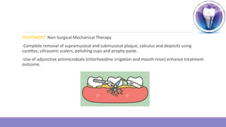
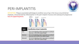
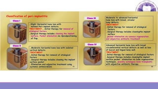
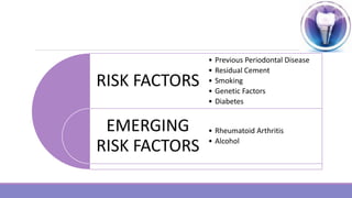
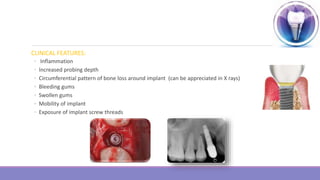
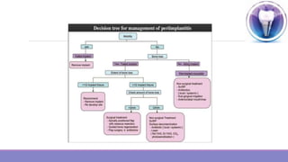
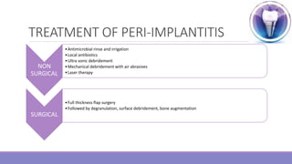
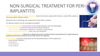
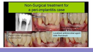
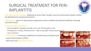
PERI-IMPLANTITIS
JAHNAVI YADAV
FINAL YEAR
DEPT OF PERIODONTOLOGY
CONTENTS
Classification of peri-implant diseases and conditions
Peri-implant Mucositis
Peri-implantitis
Classification
Treatment
Management
IMPLANT
A dental implant (endosseous implant or fixture) is a surgical component that interferes with
bone of jaw or skull to support a dental prosthesis such as crown, denture, facial prosthesis or as
an orthodontic anchor.
A dental implant is a metal post that replaces the root portion of a missing tooth.
CLASSIFICATION OF PER-IMPLANT
DISEASES AND CONDITIONS
New classification was developed by 2017 World Workshop on the Classification of Periodontal
and Peri-implant Disease and Conditions by organizing committee- American Academy of
Periodontology(AAP) and European Federation of Periodontology(EFP)
1. Peri-implant Health
2. Peri-implant Mucositis
3. Peri-implantitis
4. Peri-implant soft and hard tissue deficiencies
PERI-IMPLANT HEALTH: Characterized by –
Absence of clinical signs of inflammation
Absence of bleeding on probing
No increase in probing depth compared to previous examination.
Absence of bone loss beyond crestal bone level changes resulting from initial bone remodeling.
IMPLANT FAILURES
Early Failures (prior to implant placement)
• Hard tissue deficiencies
• Tooth loss
• Trauma from tooth extraction
• Periodontitis
• Endodontic infections
• Root fractures
• Systemic diseases
• Soft tissue deficiencies
• Tooth loss
• Periodontal disease
• Systemic disease
Late Failures (after implant placement)
• Hard tissues deficiencies
• Malpositioning of implants
• Periimplantitis
• Mechanical overload
• Systemic diseases
• Soft tissues deficiencies
• Lack of buccal bone
• Papilla height
• Keratinized tissue
• Migration of teeth and life long skeletal
changes
PERI-IMPLANT MUCOSITIS
DEFINITION: Reversible inflammatory lesion of the soft tissues surrounding an
endosseous implant (functioning implant)
ETIOLOGY: Caused by Plaque accumulation {Smoking, Radiation, Diabetes, Lack of keratinized
mucosa & presence of excess luting cement}
CLINICAL FEATURES: Characterized by bleeding on probing and visual signs of inflammation
(erythema, swelling and/or suppuration)
Increase in probing depth is often observed due to swelling.
TREATMENT: Non-Surgical Mechanical Therapy
-Complete removal of supramucosal and submucosal plaque, calculus and deposits using
curettes, ultrasonic scalers, polishing cups and prophy paste.
-Use of adjunctive antimicrobials (chlorhexidine irrigation and mouth rinse) enhance treatment
outcome.
PERI-IMPLANTITIS
DEFINITION: Plaque associated pathological condition occurring in the tissue around dental
implants, characterized by inflammation in the peri-implant mucosa and subsequent progressive
loss of supporting bone.
RISK FACTORS
EMERGING
RISK FACTORS
• Previous Periodontal Disease
• Residual Cement
• Smoking
• Genetic Factors
• Diabetes
• Rheumatoid Arthritis
• Alcohol
CLINICAL FEATURES:
◦ Inflammation
◦ Increased probing depth
◦ Circumferential pattern of bone loss around implant (can be appreciated in X rays)
◦ Bleeding gums
◦ Swollen gums
◦ Mobility of implant
◦ Exposure of implant screw threads
EXAMINATION AND EVALUATION OF
PERI-IMPLANTITIS
1. Examination of Implants:
•Visual inspection of plaque and calculus accumulation
•Signs of Inflammation
•Peri-implant soft tissue’s color, consistency, contour, quality (Peri implant healthy mucosa is characterized by pink, firm
and well-adapted gingival tissues)
•Palpate to detect: edema, tenderness, exudation
2. Peri-implant Probing:
•Implant Probing should be recorded at the time of final restoration delivery as baseline measurement (3-4mm normal)
•Careful Probing should be done with light force (0.25N)
•Probing around implant may not be same as probing around natural teeth because around teeth, periodontal probe is
restricted by healthy periodontal tissues and fibers BUT there is no such primary source of resistance to probe around
implant, that’s why probing depth differs.
•Probing depth around implants which are presumed to be healthy and w/o BoP has been documented to be about 3mm
around all surfaces.
3. Microbial Testing:
•Greater the probing depths or pockets-higher the levels of microorganisms.
•Usefulness of microbial testing limited to the evaluation of peri-implant sites is to prescribe
proper antibiotics.
4. Mobility:
• Sign of implant failure
•Traditionally examined by finger or instrument pressure
•2 non-invasive ways to evaluate- Periotest
Resonance Frequency Analysis (RFA)
PERIOTEST: Value depends on the dampening characteristics of the periodontium.
RESONANCE FREQUENCY ANALYSIS:
This uses a transducer that is attached to the implant or abutment
A steady signal is applied and response is measured
Increase in value indicates increased implant stability
Whereas decrease in value indicates loss of stability
PERIOTEST
RESONANCE FREQUENCY ANALYSIS
5. Implant Percussion:
oTapping an implant with instrument will produce a sound that can help
to determine osteointegration
•Solid resonating sound indicate implant osteointegration.
•A Dull sound may indicate fibrous encapsulation of the implant.
oHowever, radiographic and other clinical findings are needed for proper diagnosis.
6. Radiographic Examination:
•IOPA should be taken
•Simple, inexpensive, readily available in dental office.
•Can provide details of implant-abutment junction, mesial-distal crestal bone level, peri-implant
radiolucencies.
TREATMENT OF PERI-IMPLANTITIS
NON
SURGICAL
•Antimicrobial rinse and irrigation
•Local antibiotics
•Ultra sonic debridement
•Mechanical debridement with air abrasives
•Laser therapy
SURGICAL
•Full thickness flap surgery
•Followed by degranulation, surface debridement, bone augmentation
NON-SURGICAL TREATMENT FOR PERI-
IMPLANTITIS
Mechanical debridement with air abrasives- Hand instruments coated with titanium, carbon fiber, plastic or
silicon to protect implant surface
Ultrasonic tips or polishing cups coated with carbon fiber or plastic
Air abrasive systems can be used (glycine powder)
Anti-microbial Therapy- Chlorhexidine (CHX) -Broad spectrum anti microbial agent
◦ Systemic antibiotic- Amoxicillin 625mg bid, Metronidazole 200mg tid
◦ Local antibiotics- metronidazole, doxycycline, minocycline, tetracycline etc.
◦ Implant surface decontamination- Saline, citric acid, H2O2,EDTA
Ultrasonic debridement- Ultrasonic scaling instruments oscillate forward and backward at a high‐frequency
producing mechanical vibratory forces that remove the deposits.
Laser Therapy- commonly used lasers for the decontamination of the implant are:
◦ Carbon dioxide (10600nm)
◦ Diode(660nm)
◦ Nd:YAG (1064nm)
◦ Er:YAG (2940nm)
SURGICAL TREATMENT FOR PERI-
IMPLANTITIS
Flap opening and curettage- objective of access flap is to gain access to submucosal implant surface
for debridement and decontamination.
Debridement- goal is to stop disease progression and re-establish periodontal health by removing
necrosed tissues
Decontamination procedure
◦ Chemical Agents: hydrogen peroxide, citric acid, 35% phosphoric acid
◦ Photodynamic Therapy: Photosensitizer + high energy light> destroy bacterial cells
◦ Laser treatment
Implantoplasty if necessary (additional smoothening and polishing of
supracrestal implant surface)
Soft tissue and Bone Augmentation
CIST PROTOCOL
Cumulative Interceptive supportive Therapy
4 components
Proper diagnosis is through probing and periapical radiographs
Each step is used in sequential manner
1. Protocol A (mechanical debridement)-
Initiated when plaque and BOP are present, but pocket depths are 3mm or les, oral hygiene
instructions are reviewed and patients are motivated to initiate and continue maintenance;
mechanical debridement and polishing is done.
2. Protocol B (Antiseptic treatment)-
Indicated when BOP and plaque are present and PD are 4-5mm. Additional mechanical debridement
and polishing is done
Chlorhexidine digluconate can be prescribed as rinse or gels for 30 sec, 2 times a day for 3-4 weeks
3. Protocol C (Antibiotic treatment)-
Involves the addition of systemic or local antibiotic treatment to A and B therapies when PD are
greater than 5 mm
Systemic treatment is metronidazole (250mg tid) for 10 days
Or combination of amoxicillin + metronidazole (500mg/250mg tid) for 10 days
Local antibiotic treatment might include placing a controlled-release device for 10 days
4. Protocol D (Regenerative or resective therapy)-
When bone loss is present and protocols A,B and C have been implemented without resolution, a
surgical approach is necessary.
Regenerative therapy- bone graft, membrane, etc.
Resective therapy- defect osteoplasty/ ostectomy, possible implantoplasty, with an apically positioned
flap.
CIST PROTOCOL
Cumulative Interceptive and Supporting Therapy
Therapy depends on clinical and radiographic diagnosis
MAINTENANCE
IMPLANT SUCCESS
The primary criteria for assessing implant, quality, or health are pain and mobility.
90%–95% has been reported as the success rate of implants over the 10 years.
CONCLUSION
oThe early detection, prevention, and treatment of peri-implant diseases are imperative for
dental implant success. Peri-implant maintenance includes the proper placement of the
dental implant, patient preventive self care, and professional care by the dental team.
oDespite high success, failures do occur therefore patient should be informed about the
possibility of developing inflammation and infection around the implant.
oInformed consent should include need of maintenance therapy.
oOptimal hygiene should be maintained for peri-implantitis therapy.
oCIST Protocol should be followed.
THANK YOU

Periimplantitis

  • 1.
  • 2.
    CONTENTS Classification of peri-implantdiseases and conditions Peri-implant Mucositis Peri-implantitis Classification Treatment Management
  • 3.
    IMPLANT A dental implant(endosseous implant or fixture) is a surgical component that interferes with bone of jaw or skull to support a dental prosthesis such as crown, denture, facial prosthesis or as an orthodontic anchor. A dental implant is a metal post that replaces the root portion of a missing tooth.
  • 4.
    CLASSIFICATION OF PER-IMPLANT DISEASESAND CONDITIONS New classification was developed by 2017 World Workshop on the Classification of Periodontal and Peri-implant Disease and Conditions by organizing committee- American Academy of Periodontology(AAP) and European Federation of Periodontology(EFP) 1. Peri-implant Health 2. Peri-implant Mucositis 3. Peri-implantitis 4. Peri-implant soft and hard tissue deficiencies
  • 5.
    PERI-IMPLANT HEALTH: Characterizedby – Absence of clinical signs of inflammation Absence of bleeding on probing No increase in probing depth compared to previous examination. Absence of bone loss beyond crestal bone level changes resulting from initial bone remodeling.
  • 6.
    IMPLANT FAILURES Early Failures(prior to implant placement) • Hard tissue deficiencies • Tooth loss • Trauma from tooth extraction • Periodontitis • Endodontic infections • Root fractures • Systemic diseases • Soft tissue deficiencies • Tooth loss • Periodontal disease • Systemic disease Late Failures (after implant placement) • Hard tissues deficiencies • Malpositioning of implants • Periimplantitis • Mechanical overload • Systemic diseases • Soft tissues deficiencies • Lack of buccal bone • Papilla height • Keratinized tissue • Migration of teeth and life long skeletal changes
  • 7.
    PERI-IMPLANT MUCOSITIS DEFINITION: Reversibleinflammatory lesion of the soft tissues surrounding an endosseous implant (functioning implant) ETIOLOGY: Caused by Plaque accumulation {Smoking, Radiation, Diabetes, Lack of keratinized mucosa & presence of excess luting cement} CLINICAL FEATURES: Characterized by bleeding on probing and visual signs of inflammation (erythema, swelling and/or suppuration) Increase in probing depth is often observed due to swelling.
  • 8.
    TREATMENT: Non-Surgical MechanicalTherapy -Complete removal of supramucosal and submucosal plaque, calculus and deposits using curettes, ultrasonic scalers, polishing cups and prophy paste. -Use of adjunctive antimicrobials (chlorhexidine irrigation and mouth rinse) enhance treatment outcome.
  • 9.
    PERI-IMPLANTITIS DEFINITION: Plaque associatedpathological condition occurring in the tissue around dental implants, characterized by inflammation in the peri-implant mucosa and subsequent progressive loss of supporting bone.
  • 11.
    RISK FACTORS EMERGING RISK FACTORS •Previous Periodontal Disease • Residual Cement • Smoking • Genetic Factors • Diabetes • Rheumatoid Arthritis • Alcohol
  • 12.
    CLINICAL FEATURES: ◦ Inflammation ◦Increased probing depth ◦ Circumferential pattern of bone loss around implant (can be appreciated in X rays) ◦ Bleeding gums ◦ Swollen gums ◦ Mobility of implant ◦ Exposure of implant screw threads
  • 13.
    EXAMINATION AND EVALUATIONOF PERI-IMPLANTITIS 1. Examination of Implants: •Visual inspection of plaque and calculus accumulation •Signs of Inflammation •Peri-implant soft tissue’s color, consistency, contour, quality (Peri implant healthy mucosa is characterized by pink, firm and well-adapted gingival tissues) •Palpate to detect: edema, tenderness, exudation 2. Peri-implant Probing: •Implant Probing should be recorded at the time of final restoration delivery as baseline measurement (3-4mm normal) •Careful Probing should be done with light force (0.25N) •Probing around implant may not be same as probing around natural teeth because around teeth, periodontal probe is restricted by healthy periodontal tissues and fibers BUT there is no such primary source of resistance to probe around implant, that’s why probing depth differs. •Probing depth around implants which are presumed to be healthy and w/o BoP has been documented to be about 3mm around all surfaces.
  • 14.
    3. Microbial Testing: •Greaterthe probing depths or pockets-higher the levels of microorganisms. •Usefulness of microbial testing limited to the evaluation of peri-implant sites is to prescribe proper antibiotics. 4. Mobility: • Sign of implant failure •Traditionally examined by finger or instrument pressure •2 non-invasive ways to evaluate- Periotest Resonance Frequency Analysis (RFA)
  • 15.
    PERIOTEST: Value dependson the dampening characteristics of the periodontium. RESONANCE FREQUENCY ANALYSIS: This uses a transducer that is attached to the implant or abutment A steady signal is applied and response is measured Increase in value indicates increased implant stability Whereas decrease in value indicates loss of stability PERIOTEST RESONANCE FREQUENCY ANALYSIS
  • 16.
    5. Implant Percussion: oTappingan implant with instrument will produce a sound that can help to determine osteointegration •Solid resonating sound indicate implant osteointegration. •A Dull sound may indicate fibrous encapsulation of the implant. oHowever, radiographic and other clinical findings are needed for proper diagnosis. 6. Radiographic Examination: •IOPA should be taken •Simple, inexpensive, readily available in dental office. •Can provide details of implant-abutment junction, mesial-distal crestal bone level, peri-implant radiolucencies.
  • 18.
    TREATMENT OF PERI-IMPLANTITIS NON SURGICAL •Antimicrobialrinse and irrigation •Local antibiotics •Ultra sonic debridement •Mechanical debridement with air abrasives •Laser therapy SURGICAL •Full thickness flap surgery •Followed by degranulation, surface debridement, bone augmentation
  • 19.
    NON-SURGICAL TREATMENT FORPERI- IMPLANTITIS Mechanical debridement with air abrasives- Hand instruments coated with titanium, carbon fiber, plastic or silicon to protect implant surface Ultrasonic tips or polishing cups coated with carbon fiber or plastic Air abrasive systems can be used (glycine powder) Anti-microbial Therapy- Chlorhexidine (CHX) -Broad spectrum anti microbial agent ◦ Systemic antibiotic- Amoxicillin 625mg bid, Metronidazole 200mg tid ◦ Local antibiotics- metronidazole, doxycycline, minocycline, tetracycline etc. ◦ Implant surface decontamination- Saline, citric acid, H2O2,EDTA Ultrasonic debridement- Ultrasonic scaling instruments oscillate forward and backward at a high‐frequency producing mechanical vibratory forces that remove the deposits. Laser Therapy- commonly used lasers for the decontamination of the implant are: ◦ Carbon dioxide (10600nm) ◦ Diode(660nm) ◦ Nd:YAG (1064nm) ◦ Er:YAG (2940nm)
  • 21.
    SURGICAL TREATMENT FORPERI- IMPLANTITIS Flap opening and curettage- objective of access flap is to gain access to submucosal implant surface for debridement and decontamination. Debridement- goal is to stop disease progression and re-establish periodontal health by removing necrosed tissues Decontamination procedure ◦ Chemical Agents: hydrogen peroxide, citric acid, 35% phosphoric acid ◦ Photodynamic Therapy: Photosensitizer + high energy light> destroy bacterial cells ◦ Laser treatment Implantoplasty if necessary (additional smoothening and polishing of supracrestal implant surface) Soft tissue and Bone Augmentation
  • 22.
    CIST PROTOCOL Cumulative Interceptivesupportive Therapy 4 components Proper diagnosis is through probing and periapical radiographs Each step is used in sequential manner 1. Protocol A (mechanical debridement)- Initiated when plaque and BOP are present, but pocket depths are 3mm or les, oral hygiene instructions are reviewed and patients are motivated to initiate and continue maintenance; mechanical debridement and polishing is done. 2. Protocol B (Antiseptic treatment)- Indicated when BOP and plaque are present and PD are 4-5mm. Additional mechanical debridement and polishing is done Chlorhexidine digluconate can be prescribed as rinse or gels for 30 sec, 2 times a day for 3-4 weeks
  • 23.
    3. Protocol C(Antibiotic treatment)- Involves the addition of systemic or local antibiotic treatment to A and B therapies when PD are greater than 5 mm Systemic treatment is metronidazole (250mg tid) for 10 days Or combination of amoxicillin + metronidazole (500mg/250mg tid) for 10 days Local antibiotic treatment might include placing a controlled-release device for 10 days 4. Protocol D (Regenerative or resective therapy)- When bone loss is present and protocols A,B and C have been implemented without resolution, a surgical approach is necessary. Regenerative therapy- bone graft, membrane, etc. Resective therapy- defect osteoplasty/ ostectomy, possible implantoplasty, with an apically positioned flap.
  • 24.
    CIST PROTOCOL Cumulative Interceptiveand Supporting Therapy Therapy depends on clinical and radiographic diagnosis
  • 25.
  • 26.
    IMPLANT SUCCESS The primarycriteria for assessing implant, quality, or health are pain and mobility. 90%–95% has been reported as the success rate of implants over the 10 years.
  • 27.
    CONCLUSION oThe early detection,prevention, and treatment of peri-implant diseases are imperative for dental implant success. Peri-implant maintenance includes the proper placement of the dental implant, patient preventive self care, and professional care by the dental team. oDespite high success, failures do occur therefore patient should be informed about the possibility of developing inflammation and infection around the implant. oInformed consent should include need of maintenance therapy. oOptimal hygiene should be maintained for peri-implantitis therapy. oCIST Protocol should be followed.
  • 28.