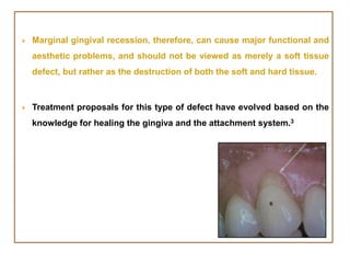
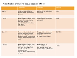
SlideShare a Scribd company logo
PRESENTED BY:
Dr. LYNN JOHNSON
Gingival recession is defined as the displacement of the marginal
tissue apical to the cemento-enamel junction.1
Carranza defines recession as ‘exposure of the tooth surface by
apical migration of the gingiva’.
Histologically, the destruction of gingival tissues, due to mechanical
forces or related to inflammatory periodontal disease, is associated
with loss of periodontal connective tissue fibers and alveolar bone.
As a consequence, exposure of the root surface to oral environment
will occur.
 Gingival recession is a sign of periodontal disease.
 Gingival recession can be localized or generalized and be associated with
one or more surfaces.2
 Gingival recession is a common occurrence and its prevalence increases
with age.
 The recession of the gingiva, either localized or generalized, may be
associated with one or more surfaces, resulting in attachment loss and
root exposure, which can lead to clinical problems such as root surface
hypersensitivity, root caries, cervical root abrasions, difficult plaque
control and diminished cosmetic appeal and aesthetic concern. 3
 Marginal gingival recession, therefore, can cause major functional and
aesthetic problems, and should not be viewed as merely a soft tissue
defect, but rather as the destruction of both the soft and hard tissue.
 Treatment proposals for this type of defect have evolved based on the
knowledge for healing the gingiva and the attachment system.3
Prevalence
According to the US National Survey, 88% of seniors (age 65 and
over) and 50% of adults (18 to 64) present recession in one or more
sites; progressive increase in frequency and extent of recession is
observed with increase in age.5
Multifactorial Gingival Recession Etiology
Periodontal marginal tissue recessions have numerous causes, but there is a
consensus about the gingival recession etiology.
1. An anatomical condition with a pre-existing or acquired alveolar bone
dehiscence combined with localized prominent tooth malposition, inadequate
keratinized gingival dimensions in quality and quantity, high muscle attachment
and frenum pull.
2. Traumatic, overzealous tooth brushing
techniques (ie, forceful, horizontal) frequently
associated with a pre-existing lack of cortical bone,
or acquired bone dehiscence.
3. Occlusal
disturbances and
parafunctional habits
like cervical dental
abrasions etc.
4. Uncontrolled
marginal
inflammation with
accumulation of
dental plaque due to
improper brushing
techniques.
5. Iatrogenic factors related to periodontal,
orthodontic and periodontal/restorative procedures
on thin biotype (eg, gingivectomy, apically positioned
flap, tooth overpreparation violating the biologic
width, incorrect fitting of the restoration with
overcontouring or a gap between the margin of the
crown and the tooth structure).
6. No evident clinical etiology in 17% of gingival recession.3
Treatment Planning Decision Modality 3
If the recession is not progressing
and does not provoke tooth
sensitivity or poor aesthetics, then
tooth-brushing instructions and
regular observation through a
strict maintenance program would
be the optimal treatment.
Progressive gingival recession in the presence of high thermal
sensitivity and/or compromised aesthetic appearance should be treated
with surgical root coverage in Class I and II defects.
Thorough plaque control is the primary condition for the success of
any periodontal surgery.
Smoking is a contraindication for plastic periodontal surgery due to:
• Associated gingival vasoconstriction that often causes necrosis of the
soft tissues;
• Lack of adherence of the fibroblasts; and
• Alteration in immune response.
The ideal surgical objective is
covering the root up to the
cementoenamel junction with
a probing depth of less than 2
mm without probe-induced
bleeding.
The principal challenge lies in
obtaining an excellent blood
supply for the covering
tissues to avoid possible
necrosis and root coverage
failure.
It is always important to select the periodontal procedure that allows the
best aesthetic result, while causing the least amount of trauma.
Miller prescribes complete disclosure at the initial consultation concerning
the root coverage that can realistically be obtained through the selected
form of treatment (Table 1)
Classification of marginal tissue recession (Miller)2
Symptoms Treatment Success
Class I Recession that does not
extend to the mucogingival
junction
Complete root coverage is
achievable
100%
Class II Recession that extends to or
beyond the mucogingival
junction, with no
periodontal attachment loss
(i.e. bone,
soft tissue)
Complete root coverage is
achievable
100%
Class III Recession that extends to or
beyond the mucogingival
junction, with periodontal
attachment loss in interdental
area or malpositioning of the
teeth
Only partial root coverage
possible to the height of
contour
of interproximal tissue.
50-70%
Class IV Recession that extends to or
beyond the mucogingival
junction, with severe bone
or soft-tissue loss in
interdental area
and/or severe malpositioning
of teeth.
Root coverage is
unpredictable
and requires adjunctive
treatment (i.e. orthodontics)
<10%
A number of reports published on recession treatment emphasize the size
of the presurgical defect and its effect on clinical outcomes; in other words,
the deeper and narrower the defect, the greater the achieved root
coverage.
Deeper recessions (ie, 4 mm or more) had greater attachment level gains
than shallow (ie, less than 4 mm) recessions.
The mean percentage of root coverage reported for the subepithelial CT
grafts technique varies between 65% and 98%, while the percentage of
complete root coverage ranges from 0% to 90% depending on the recession
classification.
Pre-requisites of Gingival Recession Surgery3
Indications Contraindications
Small amount of keratinized gingiva
Class I or II recessions
Aesthetic concerns
Single or multiple recessions
Large and deep recessions
Exposed root sensitivities
Insufficient or inefficient patient
hygiene
Smoking patient
Desquamative gingivitis
There are currently 4 common basic categories for root coverage:
In addition, combinations of different procedures are also popular in
many clinical practices and literature reports.
pedicle
grafts
guided tissue
regeneration
techniques with
a membrane
barrier
Treatment of the Exposed Root Surface6
 Before root coverage is attempted, the exposed portion of the root
should be rendered free from bacterial plaque.
 The presence of a filling in the root does not preclude the possibility
for root coverage, but the filling should be removed before the root is
covered with soft tissue.
 Miller advocated the use of root surface demineralization agents as an
important treatment component in the free soft tissue graft procedure.
 This also allows subsequent inter-digitation of these fibrils with those
in the covering connective tissue.
 Flap tension has been reported to be an important factor for the
outcome of the coronally advanced flap procedure.
 The best clinical result is achieved if the flap is passively adapted to
the root surface.
Current muco-gingival plastic surgical techniques for treating gingival
recession - Laterally positioned pedicle graft4
The best-known technique among pedicle grafts is the laterally positioned
pedicle graft introduced by Grupe and Warren and later modified by Grupe.
The success rate of this root coverage procedure was found to be in the
range of 69% ~ 72%.
In this procedure, the adjacent keratinized gingiva is positioned laterally,
and the exposed root surface in the localized gingival recession is
covered.
Guinard and Caffesse reported an average of 1mm postoperative gingival
recession on the adjacent donor site.
The main advantages of the laterally positioned pedicle graft are that it is
relatively easy and not time-consuming, it produces excellent esthetic
results, and a second surgical site is not mandatory.
To reduce the risk for recession on the donor tooth, Grupe suggested that
the marginal soft tissue should not be included in the flap.
INDICATIONS:
 Sufficient width, length, and thickness of keratinized tissue exist
adjacent to the area of gingival recession.
 Coverage of the exposed root is limited to one to two teeth.
 This method is most suitable for root coverage in gingival recession
with narrow mesiodistal dimension ( eg. Mandibular anterior area).
Contraindications
 Insufficient width and thickness of keratinized tissue in the adjacent
donor site.
 Extremely thin bone in the donor site or an osseous defect such as a
dehiscence or fenestration.
 Gingival recession area extremely protrusive.
 Deep periodontal pocket and remarkable loss of interdental alveolar
bone in the adjacent area.
 Narrow oral vestibule.
 Multiple teeth involved.
Pfeifer and Heller advocated the use of a split-thickness flap to minimize
the potential risk for development of dehiscence at the donor tooth.6
The disadvantages, however, include that it is applicable only for
single-site recession, there is a possible danger of gingival recession,
dehiscence, or fenestration at the adjacent donor site, and an
adequate amount of keratinized tissue at a neighboring donor site and
a deep vestibule are needed.
 Step 1: Make incision on gingival margin around exposed roots.
Remove resected soft tissue. Root planing.
 Step 2: A full/ partial thickness flap may be reflected. Make vertical
incision from GM to outline a flap adjacent to recipient site. the flap
should be wider than the recepient site.
 Step 3: Separate flap consisting epithelium and a thin layer of
connective tissue.
 Step 4: Slide the flap laterally onto the adjacent root.
 Step 5:Fix the flap to adjacent root and suture
 Cover the operative field with tin foil and place a periodontal pack.
 There are also other alternative procedures for a laterally positioned
flap, such as a double papilla graft (Cohen and Ross) and an obliquely
rotated graft (Pennel).
The double papilla graft has very limited usefulness due to its poor
predictability, although the esthetic result is excellent.
The obliquely rotated graft has the same disadvantages as the laterally
position pedicle flap, although it can avoid other tension-releasing
incisions as does the laterally positioned pedicle flap.
Coronally Positioned Flap6
 Coronally positioned flap is a technique used to cover exposed roots
with the available gingiva.
 This technique has a major drawback because when significant
recession occurs, there is rarely enough gingival width or
thickness to completely cover the exposed root.
 The ideal case for a coronally positioned flap is a patient who has
adequate thickness and width of the gingiva on the marginal edge
of the flap to be advanced.
 The keratinized gingiva has to be wide enough to secure the
sutures and maintain a stable, tension-free gingival flap during the
healing process .
 The advantages of the coronally positioned flap technique are that only
1 surgical site is involved and it is an excellent color match.
 Case selection is important because if the grafted tissue is thin, only
partial root coverage is achieved, and the tissue is prone to additional
recession.
 In some cases, a split-thickness dissection is performed, while in other
cases, a full-thickness flap is used over the radicular surface of the root
to maintain a sufficient flap thickness.
The pedicle flap is then
advanced in a coronal
direction until it comes to
rest on the recipient bed for
a trial fit.
The fit should be a butt joint
where the flap is inlayed into
or exactly fits into the
recipient site.
Once the tissue lies passively in
place, it should be sutured.
Proper suturing should be
accomplished without any
tension on the flap.
Free gingival graft 4
 The free gingival graft procedure involves a combination of 2 tissue
components (keratinized epithelial and connective tissue) obtained
from the palate or an edentulous ridge and its placement in the
gingival recession area.
 Hattler was the first to utilize keratinized gingiva of the interdental
papillae to cover denuded root surfaces.
 The technique was popularized later by Sullivan and Atkins and was
further refined by other investigators including Sugarman and
Staffileno and Levy.
 Results obtained from different studies indicated that the mean root
coverage treated with a free gingival graft was 88%, with the total root
coverage varying from 70% to 90% of the treated sites.
The promising advantages of this
technique are that it is a relatively
easy technique, it can be applied to
both single and multiple recessions,
it does not depend on adjacent sites
for donor tissue, and its usage is
not relevant to vestibular depth.
The disadvantages is that it creates a
second donor site wound that is prone to
bleeding, pain, and slow healing and it
produces an esthetically less-pleasing result
of the healed tissue, such as pale color and
an irregular surface of the graft site,
especially if the rugae of the palatal mucosa
are included in a large piece of donor tissue.
Sub-epithelial connective tissue graft (sCTG) 4
Because of these drawbacks of free gingival graft mentioned above, the
use of free connective tissue grafts for root coverage was introduced by
Edel in 1974.
The technique was presented by Langer and Calagna as a sub-epithelial
connective tissue graft, and was further described in detail by Langer and
Langer. This method is suitable for covering recessions of both single and
multiple adjacent teeth and is especially indicated when esthetics is a
primary consideration.
The addition of connective tissue under any pedicle flap
yields a mean exposed root coverage of 89.3%, which is
better than other soft tissue grafting tissue techniques.
The harvesting of donor tissue from the
subepithelial connective tissue of the palate
requires a complete knowledge of the anatomy of
the palate.
The best quality connective tissue is
found closest to the teeth rather than
the midline of the palate.
 Individuals with thin periodontal biotypes are susceptible to gingival
recession. They often have thin palatal mucosa that may not be
adequate for grafting.
 In these patients, another periodontal surgical procedure should be
considered. The width of connective tissue needed for most grafts is
determined by the extent of root exposure and the amount of root
coverage anticipated
 Another version of a connective tissue graft was later modified by
Nelson and Harris.
 Nelson modified the original technique by using a pedicle flap to cover
the connective tissue graft and called it a sub-pedicle connective tissue
graft, while Harris further modified this technique by using a bilateral
pedicle flap to cover the connective tissue graft. He called this
technique double pedicle flaps with a connective tissue graft.
The predictability of connective tissue graft procedures is generally
excellent.
For any given site, Nelson reported a mean root coverage of 88%, while
both Levine and Harris reported ~97% root coverage.
Long-term results (27.5 months) of subepithelial connective tissue grafts
have recently been shown to be effective (98.4%) in obtaining root
coverage in 100 patients with 146 Miller class I or II recession defects.
The subepithelial connective tissue graft was reported to be a predictable
method to obtain root coverage (with a mean root coverage of 91.1%) of
recession defects on molars and on other sites (95.8%).
There were an improvement in recession depth (from 4.4 to 0.5 mm), an
increase in the quantity of keratinized tissue (from 0.9 to 3.1 mm), a
decrease in probing depth (from 3.0 to 2.3 mm), and a decrease in
attachment level loss (from 7.4 to 2.8 mm)
The main advantages of this current procedure are that:
- it maintains a blood supply to the graft and therefore has a good
predictability of success;
- it provides good esthetics with preservation of the original flap tissue;
- the donor site wound is less hemorrhagic and painful, and can be healed
by primary closure; and
-it is simultaneously applicable to both single and multiple recessions.
However the critical disadvantage is the fact that this technique is
technically demanding and more time-consuming.
Guided tissue regeneration (GTR) technique4
 In looking for a new attachment or regeneration of tissues at the site of
recession, recent clinical studies have proposed the guided tissue
regeneration (GTR) technique for the treatment of gingival recession.
 Tinti and collaborators are pioneers of this treatment modality. They
have introduced techniques for GTR to obtain root coverage in an
attempt to re-establish a connective tissue attachment on exposed root
surfaces. Pini Prato et al. also exploited guided regeneration techniques
to simultaneously treat osseous defects, exposed roots, and
mucogingival problems.
 The predictability and success rate of the GTR procedures used for
treating gingival recession were addressed in many recent studies and
varied from 45% to 81% with more than 100% improvement in the width
of the keratinized gingiva.
The main advantages of this procedure include
- good esthetics,
- a reasonable potential for true regeneration of the lost periodontal
attachment, and
- the absence of the need for a second donor site.
The disadvantages are that
- it requires 2 surgical stages when non-resorbable membranes are used;
- it is potentially more expensive;
- more effort is required to care for the wound postoperatively;
- and the percentage of root coverage is not usually optimal due to
common membrane exposure and colonization of oral microbiota on
the membrane.
Favorable outcomes for root coverage have recently been reported using
bio-absorbable membranes.
However, the amount of root coverage obtained with a coronally
positioned flap (CPF) was greater than that observed with GTR.
Unfavorable clinical results were reported in a shallow recession study
using a coronally positioned flap in combination with a bio-resorbable
membrane.
The GTR procedure was also reported to produce a mean root coverage of
75.1% in comparison with a mean root coverage of 97.1% in the connective
tissue graft with a partial-thickness double pedicle flap.
The less-favorable clinical outcome with the GTR method was further
confirmed in a recent meta-analysis.
Combination of a CPF with an acellular dermal matrix 4
Due to the presence of many disadvantages associated with CTG, that
procedure combined with an acellular dermal matrix allograft (ADMA) and a
coronally positioned pedicle flap (CPF) has been evaluated as a substitute
for free CTGs in various periodontal procedures, including root coverage.
Root coverage using an acellular dermal matrix graft material and a
coronally positioned flap has thus initially been applied to treat cases with
gingival recession. The long-term stability of the root coverage results
obtained with a graft using an acellular dermal matrix was also
demonstrated by Harris.
Based on evidence shown earlier, the root coverage obtained with an
acellular dermal matrix is predictable, esthetic, and stable over time.
Combination of a CPF with an enamel matrix derivative4
Most of the current literature suggests that the sCTG has the
highest percentage of mean root coverage with the least variability.
Again, due to several unresolved disadvantages with this
technique, an enamel matrix derivative (EMD) has recently been
introduced in the periodontal field to overcome short-comings
associated with this and currently available regenerative
techniques.
EMD is an extract of enamel matrix and contains amelogenins of
various molecular weights. There is evidence to show that
amelogenins are involved not only in enamel formation, but also in
formation of the periodontal attachment during tooth formation
Despite the overall efficacy of EMD regeneration therapy, a
significant variation (similar to the results for GTRs) in clinical
outcomes was observed.
Meanwhile, the current literature also discloses no evidence of
clinically important differences between GTR and Emdogain
treatments in terms of probing attachment level gain and probing
depth reduction.
Preliminary histologic investigations with surgically created defects
and experimental periodontal lesions illustrated the potential of
EMD to induce formation of acellular cementum and promote
significant formation of the supporting periodontal tissues in human
and animal experiments.
However, recent human histologic studies have questioned both the
consistency of the histologic outcomes and the ability of EMD to
predictably stimulate formation of acellular cementum and bone.
Non-functional orientation of the newly formed periodontal
ligament collagen fibers was often observed.
These findings, coupled with the inconsistent bone growth bring
into question both the ability of EMD to predictably induce true
periodontal regeneration in patients and the consistency of the
histologic outcome.
 The periosteum is a highly vascular connective tissue sheath covering
the external surface of all the bones except sites of articulation and
muscle attachment.
 The periosteum comprises of at least two layers, an inner cellular or
cambium layer and an outer fibrous layer.
 The inner layer contains numerous osteoblasts and osteoprogenitor
cells and the outer layer is composed of dense collagen fiber,
fibroblasts and their progenitor cells; osteogenic progenitor cells from
the periosteal cambium layer may work with osteoblasts in initiating
and driving the cell differentiation process of bone repair.
Periosteal pedicle graft7
Research on the structure of periosteum has shown that it is made up of
three discrete zones.
Zone 1 has an
average thickness of 10-20um
consisting predominantly of
osteoblasts representing 90%
of cell population, while
collagen fibrils comprise 15%
of the volume.
Zone 3 has the highest
volume of collagen
fibrils and fibroblasts
among all the three
Zones.
The majority of cells
in zone 2 are
fibroblasts, with
endothelial cells
being most of the
remainder.
Recent papers published have
shown promising results with
the use of periosteum in the
treatment of gingival
recession defects.
After local
anaesthesia, an
intrasulcular
incision is given
with 15c Bard
Parker surgical
blade at the
buccal aspect of
the involved
tooth.
Two horizontal incisions are then made perpendicular to the
adjacent interdental papillae, at the level of the CEJ preserving
the gingival margin of the neighbouring teeth.
Two oblique vertical incisions were extended beyond the
mucogingival junction and a full thickness trapezoidal flap is
raised 3–4 mm apical to the osseous crest which is then
pulled buccally to create tension on the periosteum.
An incision is made through the periosteum where the flap
was still attached to the bone, to create a partial thickness
flap.
The partial thickness flap was extended to expose a sufficient
amount of the periosteum which is then separated from the
underlying bone using a Glickman periosteal elevator .
The process of separating the periosteum is initiated at the
apical extent of the periosteum which is then lifted slowly in a
coronal direction.
The periosteum is not separated completely from the underlying bone,
leaving it attached at its coronal most end .The periosteal pedicle graft
(PPG) thus obtained is then turned over the exposed root surface and
sutured with a synthetic 5-0 bioabsorbable suture.
After stabilizing the periosteal graft, the flap is coronally positioned and
sutured using a sling suture technique with a non-resorbable 4-0 silk
suture .
The releasing incisions are closed with interrupted sutures after which the
operated site is covered with non-eugenol periodontal dressing (coe-Pak)
for protection.
Advantages of Periosteal Pedicle Graft:
When used as a graft for the treatment of gingival recession defects
periosteum has shown promising results. The advantages associated with
PPG are:
• The presence of periosteum adjacent to the defect and in sufficient
quantity.
• Avoiding two surgical sites resulting in less surgical trauma,
postoperative complications.
• Better patient satisfaction.
Similar to connective tissue grafts, the connective tissue component of the
graft may be sufficient to support the documented clinical outcomes.7
Although the exact mechanism involved behind the excellent results
associated with PPG are still not known it is a possibility that the clinical
effectiveness of the periosteal pedicle graft for root coverage may not be
due to the cellular component of the graft.
Tunnel Connective Tissue Graft (TCTG)3
A sulcular incision was designed on both sides, from the first premolar to
the central incisors, and a partial dissection was carefully performed in
order to create a deep pouch beyond the mucogingival junction while
keeping the tip of the interproximal papillae attached to the teeth below
the proximal contact point.
A primary flap on the right and left palatal sites with one line of incision
allows the harvesting of thick, sizable connective tissue. The primary flap
was immediately sutured to prevent bleeding.
The CTG, using 4-0 sutures, was delicately inserted inside the pouch and
was then stabilized with the flap using 5-0 Vicryl sutures. The healing
progressed unevent- fully and the gingival recession was totally covered
with a beautiful aesthetic result on both sides
Advantages Disadvantages
Excellent adaptation on the
recipient site
Highly aesthetic results
High vascularization by the
advanced flap
Increased thickness of the
keratinized gingiva
Harmony in the gingival
color/texture
Traumatic surgery for the
patient
Requires two surgical sites
Delicate harvesting of the graft
Difficult stabilization of the
graft
Palatal graft has limited
quantity and thickness
Lengthy surgery/healing
Conclusion
Gingival recession is one of the main esthetic complaints of patients.
This also exposes patients to sensitivity and greater risk for root
caries. Mucogingival surgery endeavors’ to reestablish the
periodontium to a healthy circumstance Periodontal plastic surgery
strives to restore the periodon- tium to a healthy, efficient, and
aesthetic state. For coverage of exposed roots, there is a vast range
of mucogingival grafting procedures available in the present epoch.
These procedures are quite predictable and produce satisfactory
solutions to the problems presented by gingival recessions. Choice
of appropriate procedure and surgical technique will recommend
successful and exceedingly predictable results in the management of
gingival recession .5
1. Leonardo Trombel. Periodontal regeneration in gingival recession defects.
Periodontology 2000, Vol. 19, 1998, 138-150
2. Suchetha A, Darshan B Mundinamane, Bharwani Ashit G, Soorya KV. Classification
of gingival recession - The dilemma continues. SRM University Journal of Dental
Sciences Volume 2, Issue 1, January - March 2011
3. Andre P. Saadoun,. Current trends in gingival recession coverage—part I the
tunnel connective tissue graft. Continuing Education 2006:14. Vol. 18, No. 7
4. Lein-tuan Hou, Ji-jong Yan, Cheng-mei Liu, Jiann-feng Huang, S-m Jehing
Man-ying Wong, Paul Paoying Lin. Treatment of the gingival recession ──
literature review of current progress . Chin Dent J 2005 ‧ Vol 24 ‧ No 2;71-78
5. Koppolu Pradeep, Palaparthy Rajababu, Durvasula Satyanarayana, and Vidya
Sagar. Gingival Recession: Review and Strategies in Treatment of Recession.
Hindawi Publishing Corporation. Case Reports in Dentistry Volume 2012, 1- 6
pages
6. Louis F. Rose. Surgical Therapies for the Treatment of Gingival Recession. Inside
Dentistry, May 2006, Volume 2, Issue 4
7. Ajay Mahajan et al. Effective management of gingival recession defects using
periosteal pedicle grafts. e-Journal of Dentistry July - Sep 2012 Vol 2 Issue 3

More Related Content

What's hot

030.furcation involvement and its therapy
030.furcation involvement and its therapy030.furcation involvement and its therapy
030.furcation involvement and its therapy
Dr.Jaffar Raza BDS
 
bone loss patterns
   bone loss patterns    bone loss patterns
bone loss patterns
neeti shinde
 
ROOT COVERAGE PROCEDURES
ROOT COVERAGE PROCEDURESROOT COVERAGE PROCEDURES
ROOT COVERAGE PROCEDURES
Dr Ripunjay Tripathi
 
Resective osseous surgery
Resective osseous surgeryResective osseous surgery
Resective osseous surgery
Shilpa Shiv
 
Regenerative techniques for periodontal therapy
Regenerative  techniques for periodontal therapyRegenerative  techniques for periodontal therapy
Regenerative techniques for periodontal therapy
Enas Elgendy
 
Gingival recession part 2
Gingival recession part 2Gingival recession part 2
Gingival recession part 2
ManishaSinha17
 
Periodontal bone defects
Periodontal bone defectsPeriodontal bone defects
Periodontal bone defects
Heenal Adhyaru
 
Retainer in FPD
Retainer in FPD Retainer in FPD
Retainer in FPD
Hind Tabbal
 
Furcation involvement and management
Furcation involvement and managementFurcation involvement and management
Furcation involvement and management
Aishwarya Hajare
 
Radiographic aids in the diagnosis of periodontal disease
Radiographic aids in the diagnosis of periodontal diseaseRadiographic aids in the diagnosis of periodontal disease
Radiographic aids in the diagnosis of periodontal disease
Dara Ghaznavi
 
Periodontal flap
Periodontal flapPeriodontal flap
Periodontal flap
Rinisha Sinha
 
Furcation involvements and its treatments
Furcation  involvements and its treatmentsFurcation  involvements and its treatments
Furcation involvements and its treatments
Diana Abo el Ola
 
Reconstructive osseous surgeries
Reconstructive osseous surgeriesReconstructive osseous surgeries
Reconstructive osseous surgeries
Achi Joshi
 
bite planes
bite planesbite planes
Gingival Recession
Gingival RecessionGingival Recession
Gingival Recession
Salam Jawad
 
RADIOGRAPHIC AIDS IN THE DIAGNOSIS OF PERIODONTAL DISEASE
RADIOGRAPHIC AIDS IN THE DIAGNOSIS OF PERIODONTAL DISEASERADIOGRAPHIC AIDS IN THE DIAGNOSIS OF PERIODONTAL DISEASE
RADIOGRAPHIC AIDS IN THE DIAGNOSIS OF PERIODONTAL DISEASERupal Patle
 
Furcation involvement
Furcation involvement Furcation involvement
Furcation involvement
Syed Dhasthaheer
 
Failures in FPD
Failures in FPDFailures in FPD
Failures in FPD
Dr.Sachin Sunny Otta
 

What's hot (20)

030.furcation involvement and its therapy
030.furcation involvement and its therapy030.furcation involvement and its therapy
030.furcation involvement and its therapy
 
bone loss patterns
   bone loss patterns    bone loss patterns
bone loss patterns
 
ROOT COVERAGE PROCEDURES
ROOT COVERAGE PROCEDURESROOT COVERAGE PROCEDURES
ROOT COVERAGE PROCEDURES
 
Resective osseous surgery
Resective osseous surgeryResective osseous surgery
Resective osseous surgery
 
Regenerative techniques for periodontal therapy
Regenerative  techniques for periodontal therapyRegenerative  techniques for periodontal therapy
Regenerative techniques for periodontal therapy
 
Gingival recession part 2
Gingival recession part 2Gingival recession part 2
Gingival recession part 2
 
Periodontal bone defects
Periodontal bone defectsPeriodontal bone defects
Periodontal bone defects
 
Retainer in FPD
Retainer in FPD Retainer in FPD
Retainer in FPD
 
Furcation involvement and management
Furcation involvement and managementFurcation involvement and management
Furcation involvement and management
 
Radiographic aids in the diagnosis of periodontal disease
Radiographic aids in the diagnosis of periodontal diseaseRadiographic aids in the diagnosis of periodontal disease
Radiographic aids in the diagnosis of periodontal disease
 
Periodontal flap
Periodontal flapPeriodontal flap
Periodontal flap
 
Furcation involvements and its treatments
Furcation  involvements and its treatmentsFurcation  involvements and its treatments
Furcation involvements and its treatments
 
Reconstructive osseous surgeries
Reconstructive osseous surgeriesReconstructive osseous surgeries
Reconstructive osseous surgeries
 
Bone loss
Bone loss Bone loss
Bone loss
 
bite planes
bite planesbite planes
bite planes
 
Gingival recession
Gingival recession Gingival recession
Gingival recession
 
Gingival Recession
Gingival RecessionGingival Recession
Gingival Recession
 
RADIOGRAPHIC AIDS IN THE DIAGNOSIS OF PERIODONTAL DISEASE
RADIOGRAPHIC AIDS IN THE DIAGNOSIS OF PERIODONTAL DISEASERADIOGRAPHIC AIDS IN THE DIAGNOSIS OF PERIODONTAL DISEASE
RADIOGRAPHIC AIDS IN THE DIAGNOSIS OF PERIODONTAL DISEASE
 
Furcation involvement
Furcation involvement Furcation involvement
Furcation involvement
 
Failures in FPD
Failures in FPDFailures in FPD
Failures in FPD
 

Similar to Lynn-GINGIVAL RECESSION.ppt

fenestration.docx
fenestration.docxfenestration.docx
fenestration.docx
Dr.Mohammed Alruby
 
Iatrogenic factors in periodontal disease
Iatrogenic factors  in periodontal diseaseIatrogenic factors  in periodontal disease
Iatrogenic factors in periodontal disease
lobna elsaadawy
 
Restorative interrelationships(carranza 2012)
Restorative interrelationships(carranza 2012) Restorative interrelationships(carranza 2012)
Restorative interrelationships(carranza 2012)
Meysam Aryam
 
Biological width by Dr.Ali Mohammed AbuTrab
Biological width by Dr.Ali Mohammed AbuTrabBiological width by Dr.Ali Mohammed AbuTrab
Biological width by Dr.Ali Mohammed AbuTrab
Ali Mohammed AbuTrab
 
periodontal consideration.pptx
periodontal consideration.pptxperiodontal consideration.pptx
periodontal consideration.pptx
raiesahashem
 
NON-SURGICAL THERAPY.ppt
NON-SURGICAL THERAPY.pptNON-SURGICAL THERAPY.ppt
NON-SURGICAL THERAPY.ppt
malti19
 
621636012-4Phase-II-Periodontal-Therapy-1.pdf
621636012-4Phase-II-Periodontal-Therapy-1.pdf621636012-4Phase-II-Periodontal-Therapy-1.pdf
621636012-4Phase-II-Periodontal-Therapy-1.pdf
mb3861240
 
Furcation.pptx
Furcation.pptxFurcation.pptx
Furcation.pptx
MohammadEissaAhmadi
 
Sequelae caused by wearing dentures
Sequelae caused by wearing denturesSequelae caused by wearing dentures
Sequelae caused by wearing dentures
Priyam Javed
 
Root Resection – A Dark Horse in Management Offurcation Involved Maxillary Mo...
Root Resection – A Dark Horse in Management Offurcation Involved Maxillary Mo...Root Resection – A Dark Horse in Management Offurcation Involved Maxillary Mo...
Root Resection – A Dark Horse in Management Offurcation Involved Maxillary Mo...
iosrjce
 
Effects of restorative procedure on periodontium
Effects of restorative procedure on periodontiumEffects of restorative procedure on periodontium
Effects of restorative procedure on periodontiumParth Thakkar
 
32338285 effects-of-restorative-procedure-on-um-perio
32338285 effects-of-restorative-procedure-on-um-perio32338285 effects-of-restorative-procedure-on-um-perio
32338285 effects-of-restorative-procedure-on-um-periohaneenoo
 
furcation involvmentt dental disease.pptx
furcation involvmentt dental disease.pptxfurcation involvmentt dental disease.pptx
furcation involvmentt dental disease.pptx
MohamedYElZahar
 
Iatroenic factors in periodontits
Iatroenic factors in periodontitsIatroenic factors in periodontits
Iatroenic factors in periodontits
Swati Gupta
 
Non surgical management of gingival recession- Dr Harshavardhan Patwal
Non surgical management of   gingival  recession- Dr Harshavardhan PatwalNon surgical management of   gingival  recession- Dr Harshavardhan Patwal
Non surgical management of gingival recession- Dr Harshavardhan Patwal
Dr Harshavardhan Patwal
 
prevention of peri implant disease 8.pdf
prevention of peri implant disease 8.pdfprevention of peri implant disease 8.pdf
prevention of peri implant disease 8.pdf
mlhdakafera
 
Treatment of gingival recession using coronally advanced flap
Treatment of gingival recession using coronally advanced flapTreatment of gingival recession using coronally advanced flap
Treatment of gingival recession using coronally advanced flap
Shruti Maroo
 
Periodontium and prosthodontics
Periodontium and prosthodonticsPeriodontium and prosthodontics
Periodontium and prosthodontics
Indian dental academy
 
Mucogingival surgery in periodontics
Mucogingival surgery in periodonticsMucogingival surgery in periodontics
Mucogingival surgery in periodontics
Binaya Subedi
 

Similar to Lynn-GINGIVAL RECESSION.ppt (20)

fenestration.docx
fenestration.docxfenestration.docx
fenestration.docx
 
Iatrogenic factors in periodontal disease
Iatrogenic factors  in periodontal diseaseIatrogenic factors  in periodontal disease
Iatrogenic factors in periodontal disease
 
Restorative interrelationships(carranza 2012)
Restorative interrelationships(carranza 2012) Restorative interrelationships(carranza 2012)
Restorative interrelationships(carranza 2012)
 
Biological width by Dr.Ali Mohammed AbuTrab
Biological width by Dr.Ali Mohammed AbuTrabBiological width by Dr.Ali Mohammed AbuTrab
Biological width by Dr.Ali Mohammed AbuTrab
 
periodontal consideration.pptx
periodontal consideration.pptxperiodontal consideration.pptx
periodontal consideration.pptx
 
NON-SURGICAL THERAPY.ppt
NON-SURGICAL THERAPY.pptNON-SURGICAL THERAPY.ppt
NON-SURGICAL THERAPY.ppt
 
621636012-4Phase-II-Periodontal-Therapy-1.pdf
621636012-4Phase-II-Periodontal-Therapy-1.pdf621636012-4Phase-II-Periodontal-Therapy-1.pdf
621636012-4Phase-II-Periodontal-Therapy-1.pdf
 
Furcation.pptx
Furcation.pptxFurcation.pptx
Furcation.pptx
 
Sequelae caused by wearing dentures
Sequelae caused by wearing denturesSequelae caused by wearing dentures
Sequelae caused by wearing dentures
 
Root Resection – A Dark Horse in Management Offurcation Involved Maxillary Mo...
Root Resection – A Dark Horse in Management Offurcation Involved Maxillary Mo...Root Resection – A Dark Horse in Management Offurcation Involved Maxillary Mo...
Root Resection – A Dark Horse in Management Offurcation Involved Maxillary Mo...
 
Effects of restorative procedure on periodontium
Effects of restorative procedure on periodontiumEffects of restorative procedure on periodontium
Effects of restorative procedure on periodontium
 
32338285 effects-of-restorative-procedure-on-um-perio
32338285 effects-of-restorative-procedure-on-um-perio32338285 effects-of-restorative-procedure-on-um-perio
32338285 effects-of-restorative-procedure-on-um-perio
 
furcation involvmentt dental disease.pptx
furcation involvmentt dental disease.pptxfurcation involvmentt dental disease.pptx
furcation involvmentt dental disease.pptx
 
Iatroenic factors in periodontits
Iatroenic factors in periodontitsIatroenic factors in periodontits
Iatroenic factors in periodontits
 
Non surgical management of gingival recession- Dr Harshavardhan Patwal
Non surgical management of   gingival  recession- Dr Harshavardhan PatwalNon surgical management of   gingival  recession- Dr Harshavardhan Patwal
Non surgical management of gingival recession- Dr Harshavardhan Patwal
 
prevention of peri implant disease 8.pdf
prevention of peri implant disease 8.pdfprevention of peri implant disease 8.pdf
prevention of peri implant disease 8.pdf
 
Treatment of gingival recession using coronally advanced flap
Treatment of gingival recession using coronally advanced flapTreatment of gingival recession using coronally advanced flap
Treatment of gingival recession using coronally advanced flap
 
Root Caries
Root CariesRoot Caries
Root Caries
 
Periodontium and prosthodontics
Periodontium and prosthodonticsPeriodontium and prosthodontics
Periodontium and prosthodontics
 
Mucogingival surgery in periodontics
Mucogingival surgery in periodonticsMucogingival surgery in periodontics
Mucogingival surgery in periodontics
 

More from Rama Dental College Hospital and Research Center

AI in Periodontics by Dr. Neelam Das.pptx
AI in Periodontics by Dr. Neelam Das.pptxAI in Periodontics by Dr. Neelam Das.pptx
AI in Periodontics by Dr. Neelam Das.pptx
Rama Dental College Hospital and Research Center
 
History of Periodontics by Dr. Neelam Das.pptx
History of Periodontics by Dr. Neelam Das.pptxHistory of Periodontics by Dr. Neelam Das.pptx
History of Periodontics by Dr. Neelam Das.pptx
Rama Dental College Hospital and Research Center
 
Nanotechnology in Periodontics by Dr. Neelam Das.pptx
Nanotechnology in Periodontics by Dr. Neelam Das.pptxNanotechnology in Periodontics by Dr. Neelam Das.pptx
Nanotechnology in Periodontics by Dr. Neelam Das.pptx
Rama Dental College Hospital and Research Center
 
chronic periodontitis.pptx
chronic periodontitis.pptxchronic periodontitis.pptx
clinical diagnosis.pptx
clinical diagnosis.pptxclinical diagnosis.pptx
Dental plaque.ppt
Dental plaque.pptDental plaque.ppt
Controversies in Periodontal Practice - CDD.ppt
Controversies in Periodontal Practice - CDD.pptControversies in Periodontal Practice - CDD.ppt
Controversies in Periodontal Practice - CDD.ppt
Rama Dental College Hospital and Research Center
 
Periodontal Indices by Dr. Neelam Das .pptx
Periodontal Indices by Dr. Neelam Das .pptxPeriodontal Indices by Dr. Neelam Das .pptx
Periodontal Indices by Dr. Neelam Das .pptx
Rama Dental College Hospital and Research Center
 
crown lengthening.pptx
crown lengthening.pptxcrown lengthening.pptx
mechanical plaque control.pptx
mechanical plaque control.pptxmechanical plaque control.pptx
Interdisciplinary Periodontics ppt.pptx
Interdisciplinary Periodontics ppt.pptxInterdisciplinary Periodontics ppt.pptx
Interdisciplinary Periodontics ppt.pptx
Rama Dental College Hospital and Research Center
 
Endo – Perio lesions.ppt
Endo – Perio lesions.pptEndo – Perio lesions.ppt
Periodontal ligament.pptx by Dr. Ira Gupta
Periodontal ligament.pptx by Dr. Ira GuptaPeriodontal ligament.pptx by Dr. Ira Gupta
Periodontal ligament.pptx by Dr. Ira Gupta
Rama Dental College Hospital and Research Center
 
STRESS & PERIODONTAL DISEASE.pptx by Dr. Neelam Das
STRESS & PERIODONTAL DISEASE.pptx  by Dr. Neelam DasSTRESS & PERIODONTAL DISEASE.pptx  by Dr. Neelam Das
STRESS & PERIODONTAL DISEASE.pptx by Dr. Neelam Das
Rama Dental College Hospital and Research Center
 
Antimicrobials in periodontics.pptx by Dr. Shruti Gupta
Antimicrobials in periodontics.pptx  by Dr. Shruti GuptaAntimicrobials in periodontics.pptx  by Dr. Shruti Gupta
Antimicrobials in periodontics.pptx by Dr. Shruti Gupta
Rama Dental College Hospital and Research Center
 
Periodontal therapy in female patients.pdf
Periodontal therapy in female patients.pdfPeriodontal therapy in female patients.pdf
Periodontal therapy in female patients.pdf
Rama Dental College Hospital and Research Center
 
agressive periodontitis.pptx
agressive periodontitis.pptxagressive periodontitis.pptx
SURGERY TO INCREASE WIDTH OF ATTACHED GINGIVA.pptx
SURGERY TO INCREASE WIDTH OF ATTACHED GINGIVA.pptxSURGERY TO INCREASE WIDTH OF ATTACHED GINGIVA.pptx
SURGERY TO INCREASE WIDTH OF ATTACHED GINGIVA.pptx
Rama Dental College Hospital and Research Center
 
NON-SURGICAL PERIODONTAL INSTRUMENTS.pptx
NON-SURGICAL PERIODONTAL INSTRUMENTS.pptxNON-SURGICAL PERIODONTAL INSTRUMENTS.pptx
NON-SURGICAL PERIODONTAL INSTRUMENTS.pptx
Rama Dental College Hospital and Research Center
 
Nanotechnology.pdf
Nanotechnology.pdfNanotechnology.pdf

More from Rama Dental College Hospital and Research Center (20)

AI in Periodontics by Dr. Neelam Das.pptx
AI in Periodontics by Dr. Neelam Das.pptxAI in Periodontics by Dr. Neelam Das.pptx
AI in Periodontics by Dr. Neelam Das.pptx
 
History of Periodontics by Dr. Neelam Das.pptx
History of Periodontics by Dr. Neelam Das.pptxHistory of Periodontics by Dr. Neelam Das.pptx
History of Periodontics by Dr. Neelam Das.pptx
 
Nanotechnology in Periodontics by Dr. Neelam Das.pptx
Nanotechnology in Periodontics by Dr. Neelam Das.pptxNanotechnology in Periodontics by Dr. Neelam Das.pptx
Nanotechnology in Periodontics by Dr. Neelam Das.pptx
 
chronic periodontitis.pptx
chronic periodontitis.pptxchronic periodontitis.pptx
chronic periodontitis.pptx
 
clinical diagnosis.pptx
clinical diagnosis.pptxclinical diagnosis.pptx
clinical diagnosis.pptx
 
Dental plaque.ppt
Dental plaque.pptDental plaque.ppt
Dental plaque.ppt
 
Controversies in Periodontal Practice - CDD.ppt
Controversies in Periodontal Practice - CDD.pptControversies in Periodontal Practice - CDD.ppt
Controversies in Periodontal Practice - CDD.ppt
 
Periodontal Indices by Dr. Neelam Das .pptx
Periodontal Indices by Dr. Neelam Das .pptxPeriodontal Indices by Dr. Neelam Das .pptx
Periodontal Indices by Dr. Neelam Das .pptx
 
crown lengthening.pptx
crown lengthening.pptxcrown lengthening.pptx
crown lengthening.pptx
 
mechanical plaque control.pptx
mechanical plaque control.pptxmechanical plaque control.pptx
mechanical plaque control.pptx
 
Interdisciplinary Periodontics ppt.pptx
Interdisciplinary Periodontics ppt.pptxInterdisciplinary Periodontics ppt.pptx
Interdisciplinary Periodontics ppt.pptx
 
Endo – Perio lesions.ppt
Endo – Perio lesions.pptEndo – Perio lesions.ppt
Endo – Perio lesions.ppt
 
Periodontal ligament.pptx by Dr. Ira Gupta
Periodontal ligament.pptx by Dr. Ira GuptaPeriodontal ligament.pptx by Dr. Ira Gupta
Periodontal ligament.pptx by Dr. Ira Gupta
 
STRESS & PERIODONTAL DISEASE.pptx by Dr. Neelam Das
STRESS & PERIODONTAL DISEASE.pptx  by Dr. Neelam DasSTRESS & PERIODONTAL DISEASE.pptx  by Dr. Neelam Das
STRESS & PERIODONTAL DISEASE.pptx by Dr. Neelam Das
 
Antimicrobials in periodontics.pptx by Dr. Shruti Gupta
Antimicrobials in periodontics.pptx  by Dr. Shruti GuptaAntimicrobials in periodontics.pptx  by Dr. Shruti Gupta
Antimicrobials in periodontics.pptx by Dr. Shruti Gupta
 
Periodontal therapy in female patients.pdf
Periodontal therapy in female patients.pdfPeriodontal therapy in female patients.pdf
Periodontal therapy in female patients.pdf
 
agressive periodontitis.pptx
agressive periodontitis.pptxagressive periodontitis.pptx
agressive periodontitis.pptx
 
SURGERY TO INCREASE WIDTH OF ATTACHED GINGIVA.pptx
SURGERY TO INCREASE WIDTH OF ATTACHED GINGIVA.pptxSURGERY TO INCREASE WIDTH OF ATTACHED GINGIVA.pptx
SURGERY TO INCREASE WIDTH OF ATTACHED GINGIVA.pptx
 
NON-SURGICAL PERIODONTAL INSTRUMENTS.pptx
NON-SURGICAL PERIODONTAL INSTRUMENTS.pptxNON-SURGICAL PERIODONTAL INSTRUMENTS.pptx
NON-SURGICAL PERIODONTAL INSTRUMENTS.pptx
 
Nanotechnology.pdf
Nanotechnology.pdfNanotechnology.pdf
Nanotechnology.pdf
 

Recently uploaded

New Directions in Targeted Therapeutic Approaches for Older Adults With Mantl...
New Directions in Targeted Therapeutic Approaches for Older Adults With Mantl...New Directions in Targeted Therapeutic Approaches for Older Adults With Mantl...
New Directions in Targeted Therapeutic Approaches for Older Adults With Mantl...
i3 Health
 
Phone Us ❤85270-49040❤ #ℂall #gIRLS In Surat By Surat @ℂall @Girls Hotel With...
Phone Us ❤85270-49040❤ #ℂall #gIRLS In Surat By Surat @ℂall @Girls Hotel With...Phone Us ❤85270-49040❤ #ℂall #gIRLS In Surat By Surat @ℂall @Girls Hotel With...
Phone Us ❤85270-49040❤ #ℂall #gIRLS In Surat By Surat @ℂall @Girls Hotel With...
Savita Shen $i11
 
Triangles of Neck and Clinical Correlation by Dr. RIG.pptx
Triangles of Neck and Clinical Correlation by Dr. RIG.pptxTriangles of Neck and Clinical Correlation by Dr. RIG.pptx
Triangles of Neck and Clinical Correlation by Dr. RIG.pptx
Dr. Rabia Inam Gandapore
 
POST OPERATIVE OLIGURIA and its management
POST OPERATIVE OLIGURIA and its managementPOST OPERATIVE OLIGURIA and its management
POST OPERATIVE OLIGURIA and its management
touseefaziz1
 
Antiulcer drugs Advance Pharmacology .pptx
Antiulcer drugs Advance Pharmacology .pptxAntiulcer drugs Advance Pharmacology .pptx
Antiulcer drugs Advance Pharmacology .pptx
Rohit chaurpagar
 
Non-respiratory Functions of the Lungs.pdf
Non-respiratory Functions of the Lungs.pdfNon-respiratory Functions of the Lungs.pdf
Non-respiratory Functions of the Lungs.pdf
MedicoseAcademics
 
Ocular injury ppt Upendra pal optometrist upums saifai etawah
Ocular injury  ppt  Upendra pal  optometrist upums saifai etawahOcular injury  ppt  Upendra pal  optometrist upums saifai etawah
Ocular injury ppt Upendra pal optometrist upums saifai etawah
pal078100
 
Factory Supply Best Quality Pmk Oil CAS 28578–16–7 PMK Powder in Stock
Factory Supply Best Quality Pmk Oil CAS 28578–16–7 PMK Powder in StockFactory Supply Best Quality Pmk Oil CAS 28578–16–7 PMK Powder in Stock
Factory Supply Best Quality Pmk Oil CAS 28578–16–7 PMK Powder in Stock
rebeccabio
 
Flu Vaccine Alert in Bangalore Karnataka
Flu Vaccine Alert in Bangalore KarnatakaFlu Vaccine Alert in Bangalore Karnataka
Flu Vaccine Alert in Bangalore Karnataka
addon Scans
 
Tom Selleck Health: A Comprehensive Look at the Iconic Actor’s Wellness Journey
Tom Selleck Health: A Comprehensive Look at the Iconic Actor’s Wellness JourneyTom Selleck Health: A Comprehensive Look at the Iconic Actor’s Wellness Journey
Tom Selleck Health: A Comprehensive Look at the Iconic Actor’s Wellness Journey
greendigital
 
ARTHROLOGY PPT NCISM SYLLABUS AYURVEDA STUDENTS
ARTHROLOGY PPT NCISM SYLLABUS AYURVEDA STUDENTSARTHROLOGY PPT NCISM SYLLABUS AYURVEDA STUDENTS
ARTHROLOGY PPT NCISM SYLLABUS AYURVEDA STUDENTS
Dr. Vinay Pareek
 
Charaka Samhita Sutra Sthana 9 Chapter khuddakachatuspadadhyaya
Charaka Samhita Sutra Sthana 9 Chapter khuddakachatuspadadhyayaCharaka Samhita Sutra Sthana 9 Chapter khuddakachatuspadadhyaya
Charaka Samhita Sutra Sthana 9 Chapter khuddakachatuspadadhyaya
Dr KHALID B.M
 
Hemodialysis: Chapter 3, Dialysis Water Unit - Dr.Gawad
Hemodialysis: Chapter 3, Dialysis Water Unit - Dr.GawadHemodialysis: Chapter 3, Dialysis Water Unit - Dr.Gawad
Hemodialysis: Chapter 3, Dialysis Water Unit - Dr.Gawad
NephroTube - Dr.Gawad
 
The hemodynamic and autonomic determinants of elevated blood pressure in obes...
The hemodynamic and autonomic determinants of elevated blood pressure in obes...The hemodynamic and autonomic determinants of elevated blood pressure in obes...
The hemodynamic and autonomic determinants of elevated blood pressure in obes...
Catherine Liao
 
Cervical & Brachial Plexus By Dr. RIG.pptx
Cervical & Brachial Plexus By Dr. RIG.pptxCervical & Brachial Plexus By Dr. RIG.pptx
Cervical & Brachial Plexus By Dr. RIG.pptx
Dr. Rabia Inam Gandapore
 
Lung Cancer: Artificial Intelligence, Synergetics, Complex System Analysis, S...
Lung Cancer: Artificial Intelligence, Synergetics, Complex System Analysis, S...Lung Cancer: Artificial Intelligence, Synergetics, Complex System Analysis, S...
Lung Cancer: Artificial Intelligence, Synergetics, Complex System Analysis, S...
Oleg Kshivets
 
ANATOMY AND PHYSIOLOGY OF URINARY SYSTEM.pptx
ANATOMY AND PHYSIOLOGY OF URINARY SYSTEM.pptxANATOMY AND PHYSIOLOGY OF URINARY SYSTEM.pptx
ANATOMY AND PHYSIOLOGY OF URINARY SYSTEM.pptx
Swetaba Besh
 
Charaka Samhita Sutra sthana Chapter 15 Upakalpaniyaadhyaya
Charaka Samhita Sutra sthana Chapter 15 UpakalpaniyaadhyayaCharaka Samhita Sutra sthana Chapter 15 Upakalpaniyaadhyaya
Charaka Samhita Sutra sthana Chapter 15 Upakalpaniyaadhyaya
Dr KHALID B.M
 
Prix Galien International 2024 Forum Program
Prix Galien International 2024 Forum ProgramPrix Galien International 2024 Forum Program
Prix Galien International 2024 Forum Program
Levi Shapiro
 
Evaluation of antidepressant activity of clitoris ternatea in animals
Evaluation of antidepressant activity of clitoris ternatea in animalsEvaluation of antidepressant activity of clitoris ternatea in animals
Evaluation of antidepressant activity of clitoris ternatea in animals
Shweta
 

Recently uploaded (20)

New Directions in Targeted Therapeutic Approaches for Older Adults With Mantl...
New Directions in Targeted Therapeutic Approaches for Older Adults With Mantl...New Directions in Targeted Therapeutic Approaches for Older Adults With Mantl...
New Directions in Targeted Therapeutic Approaches for Older Adults With Mantl...
 
Phone Us ❤85270-49040❤ #ℂall #gIRLS In Surat By Surat @ℂall @Girls Hotel With...
Phone Us ❤85270-49040❤ #ℂall #gIRLS In Surat By Surat @ℂall @Girls Hotel With...Phone Us ❤85270-49040❤ #ℂall #gIRLS In Surat By Surat @ℂall @Girls Hotel With...
Phone Us ❤85270-49040❤ #ℂall #gIRLS In Surat By Surat @ℂall @Girls Hotel With...
 
Triangles of Neck and Clinical Correlation by Dr. RIG.pptx
Triangles of Neck and Clinical Correlation by Dr. RIG.pptxTriangles of Neck and Clinical Correlation by Dr. RIG.pptx
Triangles of Neck and Clinical Correlation by Dr. RIG.pptx
 
POST OPERATIVE OLIGURIA and its management
POST OPERATIVE OLIGURIA and its managementPOST OPERATIVE OLIGURIA and its management
POST OPERATIVE OLIGURIA and its management
 
Antiulcer drugs Advance Pharmacology .pptx
Antiulcer drugs Advance Pharmacology .pptxAntiulcer drugs Advance Pharmacology .pptx
Antiulcer drugs Advance Pharmacology .pptx
 
Non-respiratory Functions of the Lungs.pdf
Non-respiratory Functions of the Lungs.pdfNon-respiratory Functions of the Lungs.pdf
Non-respiratory Functions of the Lungs.pdf
 
Ocular injury ppt Upendra pal optometrist upums saifai etawah
Ocular injury  ppt  Upendra pal  optometrist upums saifai etawahOcular injury  ppt  Upendra pal  optometrist upums saifai etawah
Ocular injury ppt Upendra pal optometrist upums saifai etawah
 
Factory Supply Best Quality Pmk Oil CAS 28578–16–7 PMK Powder in Stock
Factory Supply Best Quality Pmk Oil CAS 28578–16–7 PMK Powder in StockFactory Supply Best Quality Pmk Oil CAS 28578–16–7 PMK Powder in Stock
Factory Supply Best Quality Pmk Oil CAS 28578–16–7 PMK Powder in Stock
 
Flu Vaccine Alert in Bangalore Karnataka
Flu Vaccine Alert in Bangalore KarnatakaFlu Vaccine Alert in Bangalore Karnataka
Flu Vaccine Alert in Bangalore Karnataka
 
Tom Selleck Health: A Comprehensive Look at the Iconic Actor’s Wellness Journey
Tom Selleck Health: A Comprehensive Look at the Iconic Actor’s Wellness JourneyTom Selleck Health: A Comprehensive Look at the Iconic Actor’s Wellness Journey
Tom Selleck Health: A Comprehensive Look at the Iconic Actor’s Wellness Journey
 
ARTHROLOGY PPT NCISM SYLLABUS AYURVEDA STUDENTS
ARTHROLOGY PPT NCISM SYLLABUS AYURVEDA STUDENTSARTHROLOGY PPT NCISM SYLLABUS AYURVEDA STUDENTS
ARTHROLOGY PPT NCISM SYLLABUS AYURVEDA STUDENTS
 
Charaka Samhita Sutra Sthana 9 Chapter khuddakachatuspadadhyaya
Charaka Samhita Sutra Sthana 9 Chapter khuddakachatuspadadhyayaCharaka Samhita Sutra Sthana 9 Chapter khuddakachatuspadadhyaya
Charaka Samhita Sutra Sthana 9 Chapter khuddakachatuspadadhyaya
 
Hemodialysis: Chapter 3, Dialysis Water Unit - Dr.Gawad
Hemodialysis: Chapter 3, Dialysis Water Unit - Dr.GawadHemodialysis: Chapter 3, Dialysis Water Unit - Dr.Gawad
Hemodialysis: Chapter 3, Dialysis Water Unit - Dr.Gawad
 
The hemodynamic and autonomic determinants of elevated blood pressure in obes...
The hemodynamic and autonomic determinants of elevated blood pressure in obes...The hemodynamic and autonomic determinants of elevated blood pressure in obes...
The hemodynamic and autonomic determinants of elevated blood pressure in obes...
 
Cervical & Brachial Plexus By Dr. RIG.pptx
Cervical & Brachial Plexus By Dr. RIG.pptxCervical & Brachial Plexus By Dr. RIG.pptx
Cervical & Brachial Plexus By Dr. RIG.pptx
 
Lung Cancer: Artificial Intelligence, Synergetics, Complex System Analysis, S...
Lung Cancer: Artificial Intelligence, Synergetics, Complex System Analysis, S...Lung Cancer: Artificial Intelligence, Synergetics, Complex System Analysis, S...
Lung Cancer: Artificial Intelligence, Synergetics, Complex System Analysis, S...
 
ANATOMY AND PHYSIOLOGY OF URINARY SYSTEM.pptx
ANATOMY AND PHYSIOLOGY OF URINARY SYSTEM.pptxANATOMY AND PHYSIOLOGY OF URINARY SYSTEM.pptx
ANATOMY AND PHYSIOLOGY OF URINARY SYSTEM.pptx
 
Charaka Samhita Sutra sthana Chapter 15 Upakalpaniyaadhyaya
Charaka Samhita Sutra sthana Chapter 15 UpakalpaniyaadhyayaCharaka Samhita Sutra sthana Chapter 15 Upakalpaniyaadhyaya
Charaka Samhita Sutra sthana Chapter 15 Upakalpaniyaadhyaya
 
Prix Galien International 2024 Forum Program
Prix Galien International 2024 Forum ProgramPrix Galien International 2024 Forum Program
Prix Galien International 2024 Forum Program
 
Evaluation of antidepressant activity of clitoris ternatea in animals
Evaluation of antidepressant activity of clitoris ternatea in animalsEvaluation of antidepressant activity of clitoris ternatea in animals
Evaluation of antidepressant activity of clitoris ternatea in animals
 

Lynn-GINGIVAL RECESSION.ppt

  • 2. Gingival recession is defined as the displacement of the marginal tissue apical to the cemento-enamel junction.1 Carranza defines recession as ‘exposure of the tooth surface by apical migration of the gingiva’. Histologically, the destruction of gingival tissues, due to mechanical forces or related to inflammatory periodontal disease, is associated with loss of periodontal connective tissue fibers and alveolar bone. As a consequence, exposure of the root surface to oral environment will occur.
  • 3.  Gingival recession is a sign of periodontal disease.  Gingival recession can be localized or generalized and be associated with one or more surfaces.2  Gingival recession is a common occurrence and its prevalence increases with age.  The recession of the gingiva, either localized or generalized, may be associated with one or more surfaces, resulting in attachment loss and root exposure, which can lead to clinical problems such as root surface hypersensitivity, root caries, cervical root abrasions, difficult plaque control and diminished cosmetic appeal and aesthetic concern. 3
  • 4.  Marginal gingival recession, therefore, can cause major functional and aesthetic problems, and should not be viewed as merely a soft tissue defect, but rather as the destruction of both the soft and hard tissue.  Treatment proposals for this type of defect have evolved based on the knowledge for healing the gingiva and the attachment system.3
  • 5. Prevalence According to the US National Survey, 88% of seniors (age 65 and over) and 50% of adults (18 to 64) present recession in one or more sites; progressive increase in frequency and extent of recession is observed with increase in age.5
  • 6. Multifactorial Gingival Recession Etiology Periodontal marginal tissue recessions have numerous causes, but there is a consensus about the gingival recession etiology. 1. An anatomical condition with a pre-existing or acquired alveolar bone dehiscence combined with localized prominent tooth malposition, inadequate keratinized gingival dimensions in quality and quantity, high muscle attachment and frenum pull. 2. Traumatic, overzealous tooth brushing techniques (ie, forceful, horizontal) frequently associated with a pre-existing lack of cortical bone, or acquired bone dehiscence.
  • 7. 3. Occlusal disturbances and parafunctional habits like cervical dental abrasions etc. 4. Uncontrolled marginal inflammation with accumulation of dental plaque due to improper brushing techniques. 5. Iatrogenic factors related to periodontal, orthodontic and periodontal/restorative procedures on thin biotype (eg, gingivectomy, apically positioned flap, tooth overpreparation violating the biologic width, incorrect fitting of the restoration with overcontouring or a gap between the margin of the crown and the tooth structure).
  • 8. 6. No evident clinical etiology in 17% of gingival recession.3 Treatment Planning Decision Modality 3 If the recession is not progressing and does not provoke tooth sensitivity or poor aesthetics, then tooth-brushing instructions and regular observation through a strict maintenance program would be the optimal treatment.
  • 9. Progressive gingival recession in the presence of high thermal sensitivity and/or compromised aesthetic appearance should be treated with surgical root coverage in Class I and II defects. Thorough plaque control is the primary condition for the success of any periodontal surgery.
  • 10. Smoking is a contraindication for plastic periodontal surgery due to: • Associated gingival vasoconstriction that often causes necrosis of the soft tissues; • Lack of adherence of the fibroblasts; and • Alteration in immune response. The ideal surgical objective is covering the root up to the cementoenamel junction with a probing depth of less than 2 mm without probe-induced bleeding. The principal challenge lies in obtaining an excellent blood supply for the covering tissues to avoid possible necrosis and root coverage failure.
  • 11. It is always important to select the periodontal procedure that allows the best aesthetic result, while causing the least amount of trauma. Miller prescribes complete disclosure at the initial consultation concerning the root coverage that can realistically be obtained through the selected form of treatment (Table 1)
  • 12. Classification of marginal tissue recession (Miller)2 Symptoms Treatment Success Class I Recession that does not extend to the mucogingival junction Complete root coverage is achievable 100% Class II Recession that extends to or beyond the mucogingival junction, with no periodontal attachment loss (i.e. bone, soft tissue) Complete root coverage is achievable 100% Class III Recession that extends to or beyond the mucogingival junction, with periodontal attachment loss in interdental area or malpositioning of the teeth Only partial root coverage possible to the height of contour of interproximal tissue. 50-70% Class IV Recession that extends to or beyond the mucogingival junction, with severe bone or soft-tissue loss in interdental area and/or severe malpositioning of teeth. Root coverage is unpredictable and requires adjunctive treatment (i.e. orthodontics) <10%
  • 13. A number of reports published on recession treatment emphasize the size of the presurgical defect and its effect on clinical outcomes; in other words, the deeper and narrower the defect, the greater the achieved root coverage. Deeper recessions (ie, 4 mm or more) had greater attachment level gains than shallow (ie, less than 4 mm) recessions. The mean percentage of root coverage reported for the subepithelial CT grafts technique varies between 65% and 98%, while the percentage of complete root coverage ranges from 0% to 90% depending on the recession classification.
  • 14. Pre-requisites of Gingival Recession Surgery3 Indications Contraindications Small amount of keratinized gingiva Class I or II recessions Aesthetic concerns Single or multiple recessions Large and deep recessions Exposed root sensitivities Insufficient or inefficient patient hygiene Smoking patient Desquamative gingivitis
  • 15. There are currently 4 common basic categories for root coverage: In addition, combinations of different procedures are also popular in many clinical practices and literature reports. pedicle grafts guided tissue regeneration techniques with a membrane barrier
  • 16. Treatment of the Exposed Root Surface6  Before root coverage is attempted, the exposed portion of the root should be rendered free from bacterial plaque.  The presence of a filling in the root does not preclude the possibility for root coverage, but the filling should be removed before the root is covered with soft tissue.  Miller advocated the use of root surface demineralization agents as an important treatment component in the free soft tissue graft procedure.
  • 17.  This also allows subsequent inter-digitation of these fibrils with those in the covering connective tissue.  Flap tension has been reported to be an important factor for the outcome of the coronally advanced flap procedure.  The best clinical result is achieved if the flap is passively adapted to the root surface.
  • 18. Current muco-gingival plastic surgical techniques for treating gingival recession - Laterally positioned pedicle graft4 The best-known technique among pedicle grafts is the laterally positioned pedicle graft introduced by Grupe and Warren and later modified by Grupe. The success rate of this root coverage procedure was found to be in the range of 69% ~ 72%. In this procedure, the adjacent keratinized gingiva is positioned laterally, and the exposed root surface in the localized gingival recession is covered. Guinard and Caffesse reported an average of 1mm postoperative gingival recession on the adjacent donor site.
  • 19. The main advantages of the laterally positioned pedicle graft are that it is relatively easy and not time-consuming, it produces excellent esthetic results, and a second surgical site is not mandatory. To reduce the risk for recession on the donor tooth, Grupe suggested that the marginal soft tissue should not be included in the flap.
  • 20. INDICATIONS:  Sufficient width, length, and thickness of keratinized tissue exist adjacent to the area of gingival recession.  Coverage of the exposed root is limited to one to two teeth.  This method is most suitable for root coverage in gingival recession with narrow mesiodistal dimension ( eg. Mandibular anterior area).
  • 21. Contraindications  Insufficient width and thickness of keratinized tissue in the adjacent donor site.  Extremely thin bone in the donor site or an osseous defect such as a dehiscence or fenestration.  Gingival recession area extremely protrusive.  Deep periodontal pocket and remarkable loss of interdental alveolar bone in the adjacent area.  Narrow oral vestibule.  Multiple teeth involved.
  • 22. Pfeifer and Heller advocated the use of a split-thickness flap to minimize the potential risk for development of dehiscence at the donor tooth.6 The disadvantages, however, include that it is applicable only for single-site recession, there is a possible danger of gingival recession, dehiscence, or fenestration at the adjacent donor site, and an adequate amount of keratinized tissue at a neighboring donor site and a deep vestibule are needed.
  • 23.  Step 1: Make incision on gingival margin around exposed roots. Remove resected soft tissue. Root planing.  Step 2: A full/ partial thickness flap may be reflected. Make vertical incision from GM to outline a flap adjacent to recipient site. the flap should be wider than the recepient site.
  • 24.  Step 3: Separate flap consisting epithelium and a thin layer of connective tissue.  Step 4: Slide the flap laterally onto the adjacent root.  Step 5:Fix the flap to adjacent root and suture  Cover the operative field with tin foil and place a periodontal pack.
  • 25.  There are also other alternative procedures for a laterally positioned flap, such as a double papilla graft (Cohen and Ross) and an obliquely rotated graft (Pennel). The double papilla graft has very limited usefulness due to its poor predictability, although the esthetic result is excellent. The obliquely rotated graft has the same disadvantages as the laterally position pedicle flap, although it can avoid other tension-releasing incisions as does the laterally positioned pedicle flap.
  • 26. Coronally Positioned Flap6  Coronally positioned flap is a technique used to cover exposed roots with the available gingiva.  This technique has a major drawback because when significant recession occurs, there is rarely enough gingival width or thickness to completely cover the exposed root.  The ideal case for a coronally positioned flap is a patient who has adequate thickness and width of the gingiva on the marginal edge of the flap to be advanced.  The keratinized gingiva has to be wide enough to secure the sutures and maintain a stable, tension-free gingival flap during the healing process .
  • 27.  The advantages of the coronally positioned flap technique are that only 1 surgical site is involved and it is an excellent color match.  Case selection is important because if the grafted tissue is thin, only partial root coverage is achieved, and the tissue is prone to additional recession.  In some cases, a split-thickness dissection is performed, while in other cases, a full-thickness flap is used over the radicular surface of the root to maintain a sufficient flap thickness.
  • 28. The pedicle flap is then advanced in a coronal direction until it comes to rest on the recipient bed for a trial fit. The fit should be a butt joint where the flap is inlayed into or exactly fits into the recipient site. Once the tissue lies passively in place, it should be sutured. Proper suturing should be accomplished without any tension on the flap.
  • 29. Free gingival graft 4  The free gingival graft procedure involves a combination of 2 tissue components (keratinized epithelial and connective tissue) obtained from the palate or an edentulous ridge and its placement in the gingival recession area.  Hattler was the first to utilize keratinized gingiva of the interdental papillae to cover denuded root surfaces.  The technique was popularized later by Sullivan and Atkins and was further refined by other investigators including Sugarman and Staffileno and Levy.  Results obtained from different studies indicated that the mean root coverage treated with a free gingival graft was 88%, with the total root coverage varying from 70% to 90% of the treated sites.
  • 30. The promising advantages of this technique are that it is a relatively easy technique, it can be applied to both single and multiple recessions, it does not depend on adjacent sites for donor tissue, and its usage is not relevant to vestibular depth. The disadvantages is that it creates a second donor site wound that is prone to bleeding, pain, and slow healing and it produces an esthetically less-pleasing result of the healed tissue, such as pale color and an irregular surface of the graft site, especially if the rugae of the palatal mucosa are included in a large piece of donor tissue.
  • 31. Sub-epithelial connective tissue graft (sCTG) 4 Because of these drawbacks of free gingival graft mentioned above, the use of free connective tissue grafts for root coverage was introduced by Edel in 1974. The technique was presented by Langer and Calagna as a sub-epithelial connective tissue graft, and was further described in detail by Langer and Langer. This method is suitable for covering recessions of both single and multiple adjacent teeth and is especially indicated when esthetics is a primary consideration.
  • 32. The addition of connective tissue under any pedicle flap yields a mean exposed root coverage of 89.3%, which is better than other soft tissue grafting tissue techniques. The harvesting of donor tissue from the subepithelial connective tissue of the palate requires a complete knowledge of the anatomy of the palate. The best quality connective tissue is found closest to the teeth rather than the midline of the palate.
  • 33.  Individuals with thin periodontal biotypes are susceptible to gingival recession. They often have thin palatal mucosa that may not be adequate for grafting.  In these patients, another periodontal surgical procedure should be considered. The width of connective tissue needed for most grafts is determined by the extent of root exposure and the amount of root coverage anticipated
  • 34.
  • 35.
  • 36.  Another version of a connective tissue graft was later modified by Nelson and Harris.  Nelson modified the original technique by using a pedicle flap to cover the connective tissue graft and called it a sub-pedicle connective tissue graft, while Harris further modified this technique by using a bilateral pedicle flap to cover the connective tissue graft. He called this technique double pedicle flaps with a connective tissue graft. The predictability of connective tissue graft procedures is generally excellent. For any given site, Nelson reported a mean root coverage of 88%, while both Levine and Harris reported ~97% root coverage.
  • 37. Long-term results (27.5 months) of subepithelial connective tissue grafts have recently been shown to be effective (98.4%) in obtaining root coverage in 100 patients with 146 Miller class I or II recession defects. The subepithelial connective tissue graft was reported to be a predictable method to obtain root coverage (with a mean root coverage of 91.1%) of recession defects on molars and on other sites (95.8%). There were an improvement in recession depth (from 4.4 to 0.5 mm), an increase in the quantity of keratinized tissue (from 0.9 to 3.1 mm), a decrease in probing depth (from 3.0 to 2.3 mm), and a decrease in attachment level loss (from 7.4 to 2.8 mm)
  • 38. The main advantages of this current procedure are that: - it maintains a blood supply to the graft and therefore has a good predictability of success; - it provides good esthetics with preservation of the original flap tissue; - the donor site wound is less hemorrhagic and painful, and can be healed by primary closure; and -it is simultaneously applicable to both single and multiple recessions. However the critical disadvantage is the fact that this technique is technically demanding and more time-consuming.
  • 39. Guided tissue regeneration (GTR) technique4  In looking for a new attachment or regeneration of tissues at the site of recession, recent clinical studies have proposed the guided tissue regeneration (GTR) technique for the treatment of gingival recession.  Tinti and collaborators are pioneers of this treatment modality. They have introduced techniques for GTR to obtain root coverage in an attempt to re-establish a connective tissue attachment on exposed root surfaces. Pini Prato et al. also exploited guided regeneration techniques to simultaneously treat osseous defects, exposed roots, and mucogingival problems.  The predictability and success rate of the GTR procedures used for treating gingival recession were addressed in many recent studies and varied from 45% to 81% with more than 100% improvement in the width of the keratinized gingiva.
  • 40.
  • 41.
  • 42. The main advantages of this procedure include - good esthetics, - a reasonable potential for true regeneration of the lost periodontal attachment, and - the absence of the need for a second donor site. The disadvantages are that - it requires 2 surgical stages when non-resorbable membranes are used; - it is potentially more expensive; - more effort is required to care for the wound postoperatively; - and the percentage of root coverage is not usually optimal due to common membrane exposure and colonization of oral microbiota on the membrane.
  • 43. Favorable outcomes for root coverage have recently been reported using bio-absorbable membranes. However, the amount of root coverage obtained with a coronally positioned flap (CPF) was greater than that observed with GTR. Unfavorable clinical results were reported in a shallow recession study using a coronally positioned flap in combination with a bio-resorbable membrane. The GTR procedure was also reported to produce a mean root coverage of 75.1% in comparison with a mean root coverage of 97.1% in the connective tissue graft with a partial-thickness double pedicle flap. The less-favorable clinical outcome with the GTR method was further confirmed in a recent meta-analysis.
  • 44. Combination of a CPF with an acellular dermal matrix 4 Due to the presence of many disadvantages associated with CTG, that procedure combined with an acellular dermal matrix allograft (ADMA) and a coronally positioned pedicle flap (CPF) has been evaluated as a substitute for free CTGs in various periodontal procedures, including root coverage. Root coverage using an acellular dermal matrix graft material and a coronally positioned flap has thus initially been applied to treat cases with gingival recession. The long-term stability of the root coverage results obtained with a graft using an acellular dermal matrix was also demonstrated by Harris. Based on evidence shown earlier, the root coverage obtained with an acellular dermal matrix is predictable, esthetic, and stable over time.
  • 45. Combination of a CPF with an enamel matrix derivative4 Most of the current literature suggests that the sCTG has the highest percentage of mean root coverage with the least variability. Again, due to several unresolved disadvantages with this technique, an enamel matrix derivative (EMD) has recently been introduced in the periodontal field to overcome short-comings associated with this and currently available regenerative techniques. EMD is an extract of enamel matrix and contains amelogenins of various molecular weights. There is evidence to show that amelogenins are involved not only in enamel formation, but also in formation of the periodontal attachment during tooth formation
  • 46. Despite the overall efficacy of EMD regeneration therapy, a significant variation (similar to the results for GTRs) in clinical outcomes was observed. Meanwhile, the current literature also discloses no evidence of clinically important differences between GTR and Emdogain treatments in terms of probing attachment level gain and probing depth reduction. Preliminary histologic investigations with surgically created defects and experimental periodontal lesions illustrated the potential of EMD to induce formation of acellular cementum and promote significant formation of the supporting periodontal tissues in human and animal experiments.
  • 47. However, recent human histologic studies have questioned both the consistency of the histologic outcomes and the ability of EMD to predictably stimulate formation of acellular cementum and bone. Non-functional orientation of the newly formed periodontal ligament collagen fibers was often observed. These findings, coupled with the inconsistent bone growth bring into question both the ability of EMD to predictably induce true periodontal regeneration in patients and the consistency of the histologic outcome.
  • 48.  The periosteum is a highly vascular connective tissue sheath covering the external surface of all the bones except sites of articulation and muscle attachment.  The periosteum comprises of at least two layers, an inner cellular or cambium layer and an outer fibrous layer.  The inner layer contains numerous osteoblasts and osteoprogenitor cells and the outer layer is composed of dense collagen fiber, fibroblasts and their progenitor cells; osteogenic progenitor cells from the periosteal cambium layer may work with osteoblasts in initiating and driving the cell differentiation process of bone repair. Periosteal pedicle graft7
  • 49. Research on the structure of periosteum has shown that it is made up of three discrete zones. Zone 1 has an average thickness of 10-20um consisting predominantly of osteoblasts representing 90% of cell population, while collagen fibrils comprise 15% of the volume. Zone 3 has the highest volume of collagen fibrils and fibroblasts among all the three Zones. The majority of cells in zone 2 are fibroblasts, with endothelial cells being most of the remainder.
  • 50. Recent papers published have shown promising results with the use of periosteum in the treatment of gingival recession defects. After local anaesthesia, an intrasulcular incision is given with 15c Bard Parker surgical blade at the buccal aspect of the involved tooth. Two horizontal incisions are then made perpendicular to the adjacent interdental papillae, at the level of the CEJ preserving the gingival margin of the neighbouring teeth.
  • 51. Two oblique vertical incisions were extended beyond the mucogingival junction and a full thickness trapezoidal flap is raised 3–4 mm apical to the osseous crest which is then pulled buccally to create tension on the periosteum. An incision is made through the periosteum where the flap was still attached to the bone, to create a partial thickness flap. The partial thickness flap was extended to expose a sufficient amount of the periosteum which is then separated from the underlying bone using a Glickman periosteal elevator . The process of separating the periosteum is initiated at the apical extent of the periosteum which is then lifted slowly in a coronal direction.
  • 52. The periosteum is not separated completely from the underlying bone, leaving it attached at its coronal most end .The periosteal pedicle graft (PPG) thus obtained is then turned over the exposed root surface and sutured with a synthetic 5-0 bioabsorbable suture. After stabilizing the periosteal graft, the flap is coronally positioned and sutured using a sling suture technique with a non-resorbable 4-0 silk suture . The releasing incisions are closed with interrupted sutures after which the operated site is covered with non-eugenol periodontal dressing (coe-Pak) for protection.
  • 53. Advantages of Periosteal Pedicle Graft: When used as a graft for the treatment of gingival recession defects periosteum has shown promising results. The advantages associated with PPG are: • The presence of periosteum adjacent to the defect and in sufficient quantity. • Avoiding two surgical sites resulting in less surgical trauma, postoperative complications. • Better patient satisfaction.
  • 54. Similar to connective tissue grafts, the connective tissue component of the graft may be sufficient to support the documented clinical outcomes.7 Although the exact mechanism involved behind the excellent results associated with PPG are still not known it is a possibility that the clinical effectiveness of the periosteal pedicle graft for root coverage may not be due to the cellular component of the graft.
  • 55. Tunnel Connective Tissue Graft (TCTG)3 A sulcular incision was designed on both sides, from the first premolar to the central incisors, and a partial dissection was carefully performed in order to create a deep pouch beyond the mucogingival junction while keeping the tip of the interproximal papillae attached to the teeth below the proximal contact point. A primary flap on the right and left palatal sites with one line of incision allows the harvesting of thick, sizable connective tissue. The primary flap was immediately sutured to prevent bleeding. The CTG, using 4-0 sutures, was delicately inserted inside the pouch and was then stabilized with the flap using 5-0 Vicryl sutures. The healing progressed unevent- fully and the gingival recession was totally covered with a beautiful aesthetic result on both sides
  • 56. Advantages Disadvantages Excellent adaptation on the recipient site Highly aesthetic results High vascularization by the advanced flap Increased thickness of the keratinized gingiva Harmony in the gingival color/texture Traumatic surgery for the patient Requires two surgical sites Delicate harvesting of the graft Difficult stabilization of the graft Palatal graft has limited quantity and thickness Lengthy surgery/healing
  • 57. Conclusion Gingival recession is one of the main esthetic complaints of patients. This also exposes patients to sensitivity and greater risk for root caries. Mucogingival surgery endeavors’ to reestablish the periodontium to a healthy circumstance Periodontal plastic surgery strives to restore the periodon- tium to a healthy, efficient, and aesthetic state. For coverage of exposed roots, there is a vast range of mucogingival grafting procedures available in the present epoch. These procedures are quite predictable and produce satisfactory solutions to the problems presented by gingival recessions. Choice of appropriate procedure and surgical technique will recommend successful and exceedingly predictable results in the management of gingival recession .5
  • 58. 1. Leonardo Trombel. Periodontal regeneration in gingival recession defects. Periodontology 2000, Vol. 19, 1998, 138-150 2. Suchetha A, Darshan B Mundinamane, Bharwani Ashit G, Soorya KV. Classification of gingival recession - The dilemma continues. SRM University Journal of Dental Sciences Volume 2, Issue 1, January - March 2011 3. Andre P. Saadoun,. Current trends in gingival recession coverage—part I the tunnel connective tissue graft. Continuing Education 2006:14. Vol. 18, No. 7 4. Lein-tuan Hou, Ji-jong Yan, Cheng-mei Liu, Jiann-feng Huang, S-m Jehing Man-ying Wong, Paul Paoying Lin. Treatment of the gingival recession ── literature review of current progress . Chin Dent J 2005 ‧ Vol 24 ‧ No 2;71-78
  • 59. 5. Koppolu Pradeep, Palaparthy Rajababu, Durvasula Satyanarayana, and Vidya Sagar. Gingival Recession: Review and Strategies in Treatment of Recession. Hindawi Publishing Corporation. Case Reports in Dentistry Volume 2012, 1- 6 pages 6. Louis F. Rose. Surgical Therapies for the Treatment of Gingival Recession. Inside Dentistry, May 2006, Volume 2, Issue 4 7. Ajay Mahajan et al. Effective management of gingival recession defects using periosteal pedicle grafts. e-Journal of Dentistry July - Sep 2012 Vol 2 Issue 3