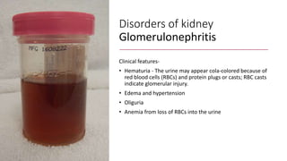
Nursing management of
patient with Genito-urinary
system disorders
Mr. Sushil Sudarshan Humane
MSN, RN
Review of
urinary
system
Functions of urinary system
• Filter. fluid from the bloodstream.
• Waste processing. filtrate, allowing wastes and excess ions to leave the body, returning needed
substances to the blood in just the right proportions.
• Elimination. eliminating nitrogenous wastes, toxins, and drugs from the body.
• Regulation. the proper balance between water and salts and between acids and bases is
maintained.
• Other regulatory functions. By producing the enzyme renin, they help regulate blood pressure,
and their hormone erythropoietin stimulates red blood cell production in the bone marrow.
• Conversion. Kidney cells also convert vitamin D to its active form.
Renin-angiotensin
mechanism
Review of urinary system
• Location. These small, dark red, kidney-bean shape, retroperitoneal position (beneath the parietal peritoneum) in the superior
lumbar region, T12 to the L3 vertebra.
• Positioning. right kidney is positioned slightly lower than the left.
• Size. 12 cm (5 inches) long, 6 cm (2.5 inches) wide, and 3 cm (1 inch) thick,
• Adrenal gland.
• Fibrous capsule. glistening appearance.
• Perirenal fat capsule. acts to cushion
• Renal fascia. outermost capsule, anchors the kidney, hold it in place
• Renal cortex. The outer region, light in color
• Renal medulla. Deep, darker, reddish-brown area
• Renal pyramids. triangular regions with a striped appearance, the renal, or medullary pyramid
• Renal columns. The pyramids are separated by extensions of cortex-like tissue
Nephron
• Nephrons. million tiny structures called nephrons
• Glomerulus. a glomerulus is a knot of capillaries.
• Renal tubule.
• Bowman’s capsule. cup-shaped, the
glomerulus,Podocytes- octopus-like cells- foot
processes that intertwine with one another and cling to
the glomerulus.
• Collecting duct.
• Proximal convoluted tubule.
• Loop of Henle.- hairpin loop following the proximal
convoluted tubule.
• Afferent arteriole. is the “feeder vessel”.
• Efferent arteriole.
• Peritubular capillaries.
Ureters
• Size.- 25 to 30 cm (10 to 12 inches) long and 6 mm (1/4
inch) in diameter.
• Location. Each ureter runs behind the peritoneum from the
renal hilum to the posterior aspect of the bladder, which it
enters at a slight angle.
• Function. carry urine from the kidneys to the bladder,
peristalsis, valve-like folds of bladder mucosa that flap over
the ureter openings.
Urinary bladder
A smooth, collapsible, muscular sac that stores urine
temporarily.
• Location. retroperitoneally in the pelvis just posterior
to the symphysis pubis.
• Function. The detrusor muscles and the transitional
epithelium both make the bladder uniquely suited for
its function of urine storage.
• Trigone. The smooth triangular region of the bladder
base outlined by these three openings is called the
trigone, where infections tend to persist.
• Detrusor muscles. The bladder wall contains three
layers of smooth muscle, collectively called the
detrusor muscle, and its mucosa is a special type of
epithelium, transitional epithelium.
Urethra
• Internal urethral sphincter. At the bladder-urethral
junction,an involuntary sphincter
• External urethral sphincter. the external urethral sphincter,
skeletal muscle, voluntarily controlled.
• Female urethra. 3 to 4 cm (1 1/2 inches) long, and its external
orifice, or opening, lies anteriorly to the vaginal opening.
• Male urethra. 20 cm (8 inches) long and has three named regions:
the prostatic, membranous, and spongy (penile) urethrae; it
opens at the tip of the penis after traveling down its length.
Characteristics of urine
• In 24 hours, 150 to 180 liters of blood plasma filters
• Daily volume. In 24 hours, 1.0 to 1.8 liters of urine are produced.
• Components. Urine contains nitrogenous wastes and unneeded
substances.
• Color. clear and pale to deep yellow.
• Odor. sterile and slightly aromatic, but if allowed to stand, it takes on
an ammonia odor caused by the action of bacteria on the urine solutes.
• pH. slightly acidic (around 6),
• Specific gravity. ranges from 1.001 to 1.035.
• Solutes. sodium and potassium ions, urea, uric acid, creatinine,
ammonia, bicarbonate ions, and various other ions.
Nursing
assessment
History, and physical examination
• During physical examination for
genitourinary dysfunction areas of
emphasis include the abdomen, suprapubic
region, genitalia and lower back, and lower
extremities.
• Direct palpation of the kidneys may help
determine their size and mobility
• The right kidney is easier to feel because it
is somewhat lower than the left one
Nursing assessment
History, and physical examination
• Renal dysfunction may produce
tenderness over the costovertebral
angle, which is the angle formed by
the lower border of the 12th, or
bottom, rib and the spine.
• The abdomen is auscultated to
assess for bruits (low- pitched
murmurs that indicate renal artery
stenosis or an aortic aneurysm).
• The abdomen is also assessed for
the presence of peritoneal fluid,
which may occur with kidney
dysfunction.
Nursing assessment
• The bladder should be percussed after the patient
voids to check for residual urine
• Percussion of the bladder begins at the midline just
above the umbilicus and proceeds downward.
• The sound changes from tympanic to dull when
percussing over the bladder.
• The bladder, which can be palpated only if it is
moderately distended, feels like a smooth, firm, round
mass rising out of the abdomen, usually at midline
• Dullness to percussion of the bladder following voiding
indicates incomplete bladder emptying.
Nursing assessment
• The prostate gland is palpated by
digital rectal examination (DRE)
• Blood is drawn for PSA before the DRE
because manipulation of the prostate
can cause the PSA level to rise
temporarily.
• The inguinal area is examined for
enlarged nodes, an inguinal or femoral
hernia, or varicocele (varicose veins of
the spermatic cord)
Nursing assessment
• In female, the vulva, urethral meatus, and vagina are
examined
• The patient is assessed for edema and changes in body
weight.
• Edema may be observed, particularly in the face and
dependent parts of the body, such as the ankles and
sacral areas
• An increase in body weight commonly accompanies
edema. A 1-kg weight gain equals approximately 1,000
mL of fluid.
Disorders of kidney
Mr. Sushil S. Humane
MSN, RN
Disorders of kidney
Glomerulonephritis
Acute Nephritic Syndrome
• Definition – Acute
glomerulonephritis refers to a group
of kidney diseases in which there is
an inflammatory reaction in the
glomeruli.
• It is not an infection of the kidney,
but rather the result of the immune
mechanisms of the body.
Disorders of kidney
Glomerulonephritis
Risk factors
• Group A beta- hemolytic streptococcal infection of
the throat
• Impetigo (infection of the skin)
• Acute viral infections- upper respiratory tract
infections, mumps, varicella zoster virus, Epstein-
Barr virus, hepatitis B, and human
immunodeficiency virus [HIV] infection).
• Antigens outside the body (eg, medications, foreign
serum)
• In other patients, the kidney tissue itself serves as
the inciting antigen.
Disorders of kidney
Glomerulonephritis
Categories –
• Primary: Disease is mainly in glomeruli
• Secondary: Glomerular diseases that are
the consequence of systemic disease
• Idiopathic: Cause is unknown
• Acute: Occurs over days or weeks
• Chronic: Occurs over months or years
• Rapidly progressing: Constant loss of renal
function with minimal chance of recovery
Disorders of kidney
Glomerulonephritis
Categories –
• Diffuse: Involves all glomeruli
• Focal: Involves some glomeruli
• Segmental: Involves portions of
individual glomeruli
• Membranous: Evidence of thickened
glomerular capillary walls
• Proliferative: Number of glomerular
cells involved
Disorders of kidney
Glomerulonephritis
Clinical features-
• Hematuria - The urine may appear cola-colored because of
red blood cells (RBCs) and protein plugs or casts; RBC casts
indicate glomerular injury.
• Edema and hypertension
• Oliguria
• Anemia from loss of RBCs into the urine
Pathophysiology
Clinical features-
• In the more severe form of the disease, patients also complain of
headache, malaise, and flank pain.
• Elderly patients may experience circulatory overload with dyspnea,
engorged neck veins, cardiomegaly, and pulmonary edema.
• Atypical symptoms include confusion, somnolence, and seizures,
which are often confused with the symptoms of a primary neurologic
disorder
Disorders of kidney
Glomerulonephritis
Diagnostic evaluation- History
• On examination- kidney is large, tender, edematous and congested
• Urinanalysis- protienuria, hematuria , oliguria
• Blood studies-
• Serum creatinine, BUN increased
• Hypoalbuminemia, hyperlipidemia
• Elevated serum IgA level
• Antistreptolysin O titers are usually elevated in post streptococcal
glomerulonephritis
• Electron microscopy and immunofluorescent analysis help identify the nature of
the lesion
• Kidney biopsy may be needed for definitive diagnosis.
Disorders of kidney
Glomerulonephritis
Complications-
• Hypertensive Encephalopathy
• Heart Failure
• Pulmonary Edema
• ESRD
Disorders of kidney
Glomerulonephritis
Management- Goal
• To conserve renal function
• To treat complication adequately
• Types of management-
• Non pharmacological management
• Dietary management
• Pharmacological management
• Nursing management
Disorders of kidney
Glomerulonephritis
Non-pharmacological management-
• Complete bed rest – As excessive activity may increase the
proteinuria and hematuria. It should be encouraged until the urine
clears and BUN, creatinine and BP return to normal.
• Strict intake output charting
• Fluid restrictions
• Plasmapheresis to decrease the serum anti body level
• Dialysis if, uremic symptoms are severe.
Disorders of kidney
Glomerulonephritis
Dietary management-
• Protein restricted diet as the level of BUN and creatinine is high in
blood
• Low fat diet due to hyperlipidemia
• Sodium restriction if hypertension, edema or congestive heart failure
are present.
• Increased carbohydrate diet to provide energy and to prevent the
catabolism of protein.
Disorders of kidney
Glomerulonephritis
Pharmacological management-
• Residual streptococcal infection is suspected, penicillin is the agent of
choice.
• Diuretics and antihypertensive agents may be given to control
hypertension.
• Corticosteroids and cytotoxic agents are used to reduce the
inflammation.
• H2 blockers (to prevent stress ulcers)
• Phosphate binding agents (to reduce phosphate and elevate calcium).
Disorders of kidney
Glomerulonephritis
Nursing management-
• Monitor vital signs, intake and output, and maintain dietary
restrictions during acute phase.
• Encourage rest during the acute phase as directed until the urine
clears and BUN, creatinine, and blood pressure normalize. (Rest also
facilitates diuresis.)
• Administer medications as ordered, and evaluate patient's response
to antihypertensives, diuretics, H2 blockers, phosphate-binding
agents, and antibiotics (if indicated).
Disorders of kidney
Glomerulonephritis
Nursing management-
• Carefully monitor fluid balance
• Replace fluids according to the patient's fluid losses (urine,
respiration, feces)
• Daily body weight as prescribed.
• Monitor pulmonary artery pressure and CVP, if indicated.
• Monitor for signs and symptoms of heart failure: distended neck
veins, tachycardia, gallop rhythm, enlarged and tender liver, crackles
at bases of lungs. Observe for hypertensive encephalopathy, any
evidence of seizure activity.
Disorders of kidney
Glomerulonephritis
Nursing management-
• Regular monitoring of blood pressure, urinary protein, and BUN
concentrations to determine if there is exacerbation of disease
activity.
• Encourage patient to treat any infection promptly.
• Tell patient to report any signs of decreasing renal function and to
obtain treatment immediately.
Disorders of kidney
Glomerulonephritis
Mr Sushil S. Humane MSN, RN
Nephrotic syndrome
• Definition- Nephrotic syndrome is a clinical disorder
characterized by marked increase of protein in the
urine (proteinuria), decrease in albumin in the blood
(hypoalbuminemia), edema, and excess lipids in the
blood (hyperlipidemia).
• These occur because of increased permeability of the
glomerular capillary membrane.
Nephrotic syndrome
Classification of nephrotic syndrome-
ETOLOGICAL CLASSIFICATION
• Primary NEPHROTIC syndrome. Disease limited to kidney
• Secondary NEPHROTIC syndrome. Other systems involved
HISTOLOGICAL CLASISIFICATION
• MCD (Minimal change disease )
• FSGN (Focal segmental glomerulosclerosis )
• MN (Membranous nephropathy)
• MPGN (membranous proliferative glomerulonephrosclerosis)
Nephrotic syndrome
Etiology-
• Membranous nephropathy (MN)
• Hepatitis B
• Sjogren's syndrome
• Systemic lupus erythematosus (SLE)
• Diabetes mellitus
• Sarcoidosis
• Syphilis
• Drugs
• Malignancy (cancer)
Nephrotic syndrome
Etiology-
• Focal segmental glomerulosclerosis (FSGS)
• Hypertensive Nephrosclerosis
• Human immunodeficiency virus (HIV)
• Diabetes mellitus
• Obesity
• Kidney loss
• Minimal change disease (MCD)
• Drugs
• Malignancy, especially Hodgkin's lymphoma
Nephrotic syndrome
Clinical features-
• The major manifestation of nephrotic
syndrome is edema.
• It is usually soft and pitting and commonly
occurs around the eyes (periorbital), in
dependent areas (sacrum, ankles, and
hands), and in the abdomen (ascites).
• Patients may also exhibit irritability,
headache, and malaise.
Nephrotic syndrome
Diagnostic evaluation-
• Urinalysis- marked proteinuria, microscopic hematuria,
• 24-hour urine for protein (increased) and creatinine
clearance (decreased)
• Protein electrophoresis and immunoelectrophoresis of
the urine to categorize the proteinuria
• Needle biopsy of kidney for histologic examination of
renal tissue to confirm diagnosis
• Serum chemistry- decreased total protein and albumin,
normal or increased creatinine, increased triglycerides,
Nephrotic syndrome
Complications-
• Infection (due to a deficient
immune response)
• Thromboembolism (especially
of the renal vein)
• Pulmonary emboli
• ARF(due to hypovolemia)
• Accelerated atherosclerosis
(due to hyperlipidemia)
Nephrotic syndrome
Management-
• Treatment of causative glomerular disease
• Diuretics (used cautiously) and angiotensin
converting enzyme inhibitors to control
proteinuria
• Corticosteroids or immunosuppressant
agents to decrease proteinuria
• General management of edema
• Sodium and fluid restriction; liberal
potassium
• Infusion of salt-poor albumin
• Dietary protein supplements
• Low-saturated-fat diet
Nephrotic syndrome
Nursing Management-
• Monitor daily weight, intake and output, and urine specific
gravity.
• Monitor CVP (if indicated), vital signs, orthostatic blood
pressure, and heart rate to detect hypovolemia.
• Monitor serum BUN and creatinine to assess renal
function.
• Administer diuretics or immunosuppressants as prescribed,
and evaluate patient's response.
• Infuse I.V. albumin as ordered.
• Encourage bed rest for a few days to help mobilize edema;
however, some ambulation is necessary to reduce risk of
thromboembolic complications.
Renal failure
Mr Sushil S. Humane
MSN, RN
Renal failure
• Definition- Acute renal failure is a
sudden and almost complete loss
of kidney function caused by
failure of renal circulation or by
glomerular or tubular
dysfunction.
Renal failure
Etiology-
• Pre – renal (hypoperfusion
of kidney)
• Intra – renal (actual
damage to the kidney
tissue)
• Post – renal (obstruction to
urine flow)
Renal failure
Renal failure
Renal failure
Renal failure
RISK FACTORS
• Advanced age
• Blockages in the blood vessels in your arms or legs
• Diabetes
• High blood pressure
• Heart failure
• Kidney diseases
• Liver disease
Renal failure
PHASES OF ARF
• Initiating phase
• Oliguric phase
• Diuretic phase
• Recovery phase
Renal
failure
PHASES OF ARF
• Initiating phase Begins with the initial insult and
ends when oliguria develops
• Oliguric phase Urine output less than 400 ml/day
• Diuretic phase Urine output become normal but
nitrogenous waste products still remain elevated in
blood
• Recovery phase It signifies the improvement of
renal function It takes 3-12 months to return
normal
Renal failure
Clinical features-
• Vomiting and/or diarrhea, which
may lead to dehydration.
• Nausea.
• Weight loss.
• Nocturnal urination.
• pale urine.
• Less frequent urination, or in
smaller amounts than usual, with
dark coloured urine
• Haematuria.
• Pressure, or difficulty urinating.
• Itching.
Renal
failure
Clinical features-
• Bone damage.
• Non-union in broken bones.
• Muscle cramps (caused by low levels of calcium
which can cause hypocalcaemia)
• Abnormal heart rhythms.
• Muscle paralysis.
• Swelling of the legs, ankles, feet, face and/or
hands.
• Shortness of breath due to extra fluid on the lungs
• Pain in the back or side
• Feeling tired and/or weak.
Renal
failure
Clinical features-
• Memory problems.
• Difficulty concentrating.
• Dizziness.
• Low blood pressure.
• Anorexia
• Pruritus
• Seizures (if blood urea nitrogen level is very high)
Renal
failure
Diagnostic evaluation-
• History regarding the etiological factors and risk
factors.
• Physical symptoms
• Urine output – scanty, bloody, and low specific
gravity
• Increased BUN and creatinine level in blood
• Hyperkalemia
• Metabolic acidosis
• Hyperphoshatemia
• Hypocalcemia
• Anemia
Renal
failure
Prevention-
• Provide adequate hydration to patient at high risk
for dehydration
• Prevent and treat shock with blood and fluid
replacement therapy
• Manage hypotension
• Monitor critically ill patient for central venous and
arterial pressures and hourly urine output to
detect the onset of renal failure as early as
possible.
• Continuously assess the renal function
Renal
failure
Prevention-
• Prevent and treat infections
• Cautiously administer the blood
• Closely monitor the all medications that
metabolized and excreted by the kidney for dosage
and blood levels for the toxic effects.
• Pay special attention to wound, burns and other
precursors of sepsis.
Renal
failure
COMPLICATIONS ARF can affect the entire body in
the form of –
• Infection
• Hyperkalaemia, Hyperphosphataemia,
Hyponatraemia
• Water overload
• Pericarditis
• Pulmonary oedema.
• Reduced level of consciousness.
• Immune deficiency
Renal
failure
Management-
• To correct fluid and electrolyte balance.
• To correct dehydration.
• To Keep other body systems working properly
Renal calculi
Mr Sushil S Humane
MSN, RN
Renal calculi
• Urolithiasis refers to stones (calculi) in the urinary
tract.
• Stones are formed in the urinary tract when
urinary concentrations of substances such as
calcium oxalate, calcium phosphate, and uric acid
increase.
• This is referred to as supersaturation and is depen-
dent on the amount of the substance, ionic
strength, and pH of the urine.
Renal calculi
Incidence-
• The occurrence of urinary stones occurs predomi-
nantly in the third to fifth decades of life and
• Affects men more than women.
• About half of patients with a single renal stone
have another episode within 5 years.
• Most stones contain calcium or magnesium in
combination with phosphorus or oxalate.
• Most stones are radiopaque and can be detected
by x-ray studies
Renal calculi
Types of stone-
• Calcium stone
• Oxalate stone
• Cystiene stone
• Struvite stone
Renal calculi
Types of stone-
• Calcium stone
• Most stones (75%) are composed mainly of
calcium oxalate crystals.
• Increased calcium concentrations in blood and
urine promote precipitation of calcium and
formation of stones.
• Causes of hypercalcemia (high serum calcium) and
hypercalciuria (high urine calcium) include the
following:
Renal calculi
Types of stone-
• Calcium stone
• Hyperparathyroidism
• Renal tubular acidosis
• Cancers
• Granulomatous diseases (sarcoidosis,
tuberculosis), which may cause increased vitamin
D production by the granulomatous tissue
• Excessive intake of vitamin D
• Excessive intake of milk and alkali
• Myeloproliferative diseases (leukemia,
polycythemia vera, multiple myeloma), which
produce an unusual proliferation of blood cells
from the bone marrow
Renal calculi
Types of stone-
• Uric acid stones
• 5% to 10% of all stones
• gout
• myeloproliferative disorders
• Diet high in purines and abnormal purine
metabolism
Renal calculi
Types of stone-
• Struvite stones
• 15% of urinary calculi
• form in persistently alkaline, ammonia-rich urine
• caused by the presence of urease splitting bacteria
such as Proteus, Pseudomonas, Klebsiella, Staphy-
lococcus, or Mycoplasma species.
• Predisposing factors for struvite stones (commonly
called infection stones) include neurogenic bladder,
foreign bodies, and recurrent UTIs
Renal calculi
Types of stone-
• Cystine stones
• 1% to 2% of all stones occur in patients with a rare
inherited defect in renal absorption of cystine (an
amino acid).
Renal calculi
Causes and predisposing factors:
• Chronic dehydration, poor fluid intake, and
immobility
• Living in mountainous, desert, or tropical areas
• Infection, urinary stasis, and periods of immobility
• Inflammatory bowel disease and in patients with
an ileostomy or bowel resection because these
patients absorb more oxalate.
• Medications- antacids, acetazolamide (Diamox),
vitamin D, laxatives, and high doses of aspirin
Renal calculi
Location of stones-
• Kidney
• Ureter
• Bladder
• Urethra
Renal calculi
Clinical features-
• Pain
• Heamturia
• Dysuria
• Oedema
• Pyuria
Associated symptoms-
• Nausea, vomiting, diarrhea, abdominal discomfort
• Chills and fever (may)
Renal calculi
Clinical features-
Pain-
• Stones in the renal pelvis may be associated with
an intense, deep ache in the costovertebral region
• Pain originating in the renal area radiates
anteriorly and downward toward the bladder in
the female and toward the testis in the male.
• If the pain suddenly becomes acute, with
tenderness over the costovertebral area, and
nausea and vomiting appear termed as renal colic
Renal calculi
Clinical features-
• Pain-
• Stones lodged in the ureter (ureteral obstruction)
cause acute, excruciating, colicky, wavelike pain,
radiating down the thigh and to the genitalia
• It is called ureteral colic
• Colic is mediated by prostaglandin E, a substance
that increases ureteral contractility and renal blood
flow and that leads to increased intraureteral
pressure and pain
• If the stone present in the bladder and obstruct he
urine flow, produces the pain at suprapubic region
along with bladder distension
Renal calculi
Clinical features-
• Hematuria- Hematuria is often present because of
the abrasive action of the stone.
• Dysuria- Painful micturition is termed as dysuria.
• Obstruction in urine flow tend to cause the
dysuria.
Renal calculi
Clinical features-
• Oedema- When the stones block the flow of urine,
obstruction develops, producing an increase in
hydrostatic pressure and distending the renal
pelvis and proximal ureter.
• Thereby GFR decreases leads to sodium and water
retetion and gives rise to oedema.
• Pyuria- Obstruction in urine flow, urinary retention
and urinary stasis may cause the UTI and featured
as pyuria.
Renal calculi
Clinical features-
• Associated symptoms- Nausea, vomiting, diarrhea,
abdominal discomfort due to renointestinal
reflexes and shared nerve supply (celiac ganglion)
between the ureters and intestineand the
anatomic proximity of the kidneys to the stomach,
pancreas, and large intestine.
• Features of infection- Due to UTI. These features
may be chill, high grade fever dysuria etc.
Renal calculi
Diagnostic evaluation-
• History
• Physical examination
• Urinanalysis
• Blood studies
• Stone chemistry
• Radiographic studies
Renal calculi
Diagnostic evaluation
• History -
• Diet
• Water
• Occupation
• medication
• Past and recent medical history
• Collect the informations regarding the reasons for
seeking health care services
Renal calculi
Diagnostic evaluation
• Physical examination - Locate, nature and
characteristics of pain
• Assess the level of pain ,tenderness etc.
• Observe for the associated symptoms.
Renal calculi
Diagnostic evaluation
• Urinanalysis- hematuria and pyuria
• pH < 5.5 indicates uric acid stone
• pH > 7.5 indicates struvite stone
• urine culture and drug sensitivity studies to detect
infection.
• 24-hour urine test for measurement of calcium,
uric acid, creatinine, sodium,citrate and oxalate
Renal calculi
Diagnostic evaluation
Blood studies-
• Hyperuracemia
• Hypercalcemia
• Neutrophilia
• Elevated serum parathyroid hormone
Renal calculi
Diagnostic evaluation
• Stone chemistry-
• Collection of stone through a strainer is useful.
• Analyze the stone chemically to find out the
composition which helps in therapeutic
management.
Renal calculi
Diagnostic evaluation
• Radiographic studies-
• Kidney, ureters, and bladder radiography may show
stone.
• Intra venous urogram (intravenous pyelogram) to
determine site and evaluate degree of obstruction
• Retrograde pyelography
• Ultrasound
• Helical or axial CAT Scan
Renal calculi
Management General Principles
• If small stone (< 4 mm) and able to treat as
outpatient, 80% will pass stone spontaneously with
hydration, pain control, and reassurance.
• Hospitalized for intractable pain, persistent
vomiting, high-grade fever, obstruction with
infection, and solitary kidney with obstruction.
• Medical management
• Surgical management
• Nursing management
Renal calculi
Management
• Medical management Goal- Immediate goal-
• To relieve the pain until its causes can be
eliminated. Long term goal (basic goal)- To
eradicate the stone
• To determine the stone type
• To prevent nephron destruction
• To control infection
• To relieve any obstruction
Renal calculi
Management
• Medical management
• Opioid analgesics or NSAIDs are administered to
prevent shock and syncope that may result from
the excruciating pain.
• NSAIDs provide specific pain relief because they
inhibit the synthesis of prostaglandin E.
• Hot baths or moist heat to the flank areas may also
be useful.
Renal calculi
Management
• Medical management
• Fluids are encouraged. This increases the
hydrostatic pressure behind the stone, assisting it
in its downward passage.
• A high, around-the-clock fluid intake reduces the
concentration of urinary crystalloids, dilutes the
urine, and ensures a high urine output.
Renal calculi
Management
• Medical management
• Fluids are encouraged. This increases the hydrostatic pressure behind
the stone, assisting it in its downward passage.
• A high, around-the-clock fluid intake reduces the concentration of
urinary crystalloids, dilutes the urine, and ensures a high urine
output.
Renal calculi
Management
• Medical management Calcium stone- Cellulose sodium phosphate
(Calcibind) may be effective in preventing calcium stones. It binds calcium
from food in the intestinal tract, reducing the amount of calcium absorbed
into the circulation.
• restrict calcium in diet
• Therapy with thiazide diuretics may be beneficial in reducing the calcium
loss in the urine and lowering the elevated paratharmone levels.
• The urine may be acidified by use of medications such as ammonium
chloride or acetohydroxamic acid
• Sodium and protein restriction diet
Renal calculi
Management
• Medical management Uric acid stone-
• low-purine diet such as shellfish, anchovies,
asparagus, mushrooms, and organ meats are
avoided
• Allopurinol may be prescribed to reduce serum
uric acid levels and urinary uric acid excretion.
• Proteins may be limited in diet
Renal calculi
Management
• Medical management Cystine stone -
• Low-protein diet
• Penicillamine is administered to reduce the
amount of cystine in the urine
• urine is alkalinized.
Renal calculi
Management
• Medical management Oxalate stone -
• Encourage the increased fluid intake
• Avoid the food contains oxalate such as- spinach,
strawberries, tea, peanuts, wheat bran
Renal calculi
Management Non surgical management-
• Ureteroscopy
• ESWL (Extra Corporeal Shock wave lithotripsy)
• Endoscopic procedures
• Electrohydrolic lithotripsy
• Chemolysis
Renal calculi
Management
• Non surgical management- Ureteroscopy
• Ureteroscopy involves visualizing the stone and
then destroying it.
• Access to the stone is accomplished by inserting a
ureteroscope into the ureter and then inserting a
laser, electrohydraulic lithotriptor, or ultrasound
device through the ureteroscope to fragment and
remove the stones.
• A stent may be inserted and left in place for 48
hours or more after the procedure to keep the
ureter patent
Renal calculi
Management
• Non surgical management- ESWL- ESWL is a
noninvasive procedure used to break up stones in
the calyx of the kidney
• In ESWL, a high-energy amplitude of pressure, or
shock wave, is generated by the abrupt release of
energy and transmitted through water and soft
tissues.
• When the shock wave encounters a substance of
different intensity (a renal stone), a compression
wave causes the surface of the stone to fragment.
• Repeated shock waves focused on the stone
eventually reduce it to many small pieces. These
small pieces are excreted in the urine, usually
without difficulty.
Renal calculi
Management
• Non-surgical
management- ESWL
Renal calculi
Management
• Non surgical management-
• Endoscopic procedures-
• A percutaneous nephrostomy or a
percutaneous nephrolithotomy may be
performed, and a nephroscope is introduced
through the dilated percutaneous tract into the
renal parenchyma.
• Depending on its size, the stone may be
extracted with forceps or by a stone retrieval
basket. Alternatively, an ultrasound probe may
be introduced through the nephrostomy tube.
Renal calculi Management-Non surgical
management-
• Electrohydraulic lithotripsy-
• an electrical discharge is used
to create a hydraulic shock
wave to break up the stone.
• A probe is passed through the
cystoscope, and the tip of the
lithotriptor is placed near the
stone
• This procedure is performed
under topical anesthesia.
Renal calculi
Management
• Non-surgical management-
• Chemolysis- Chemolysis, stone
dissolution using infusions of chemical
solutions (eg, alkylating agents,
acidifying agents)
• A percutaneous nephrostomy is
performed, and the warm irrigating
solution is allowed to flow
continuously onto the stone.
Renal calculi
Management
• Surgical management- Nephrolithotomy - Incision
into the kidney with removal of the stone
• Nephrectomy – removal of kidney
• Pyelolithotomy - removal of stone from renal pelvis
• Ureterolithotomy - removal of stone from ureter
• Cystostomy – removal of stone from bladder
• Cystolitholapaxy - an instrument is inserted
through the urethra into the bladder, and the stone
is crushed in the jaws of this instrument
Thank you

Genito-urinary system disorders-1.pptx

  • 1.
    Nursing management of patientwith Genito-urinary system disorders Mr. Sushil Sudarshan Humane MSN, RN
  • 2.
  • 3.
    Functions of urinarysystem • Filter. fluid from the bloodstream. • Waste processing. filtrate, allowing wastes and excess ions to leave the body, returning needed substances to the blood in just the right proportions. • Elimination. eliminating nitrogenous wastes, toxins, and drugs from the body. • Regulation. the proper balance between water and salts and between acids and bases is maintained. • Other regulatory functions. By producing the enzyme renin, they help regulate blood pressure, and their hormone erythropoietin stimulates red blood cell production in the bone marrow. • Conversion. Kidney cells also convert vitamin D to its active form.
  • 4.
  • 5.
    Review of urinarysystem • Location. These small, dark red, kidney-bean shape, retroperitoneal position (beneath the parietal peritoneum) in the superior lumbar region, T12 to the L3 vertebra. • Positioning. right kidney is positioned slightly lower than the left. • Size. 12 cm (5 inches) long, 6 cm (2.5 inches) wide, and 3 cm (1 inch) thick, • Adrenal gland. • Fibrous capsule. glistening appearance. • Perirenal fat capsule. acts to cushion • Renal fascia. outermost capsule, anchors the kidney, hold it in place • Renal cortex. The outer region, light in color • Renal medulla. Deep, darker, reddish-brown area • Renal pyramids. triangular regions with a striped appearance, the renal, or medullary pyramid • Renal columns. The pyramids are separated by extensions of cortex-like tissue
  • 7.
    Nephron • Nephrons. milliontiny structures called nephrons • Glomerulus. a glomerulus is a knot of capillaries. • Renal tubule. • Bowman’s capsule. cup-shaped, the glomerulus,Podocytes- octopus-like cells- foot processes that intertwine with one another and cling to the glomerulus. • Collecting duct. • Proximal convoluted tubule. • Loop of Henle.- hairpin loop following the proximal convoluted tubule. • Afferent arteriole. is the “feeder vessel”. • Efferent arteriole. • Peritubular capillaries.
  • 8.
    Ureters • Size.- 25to 30 cm (10 to 12 inches) long and 6 mm (1/4 inch) in diameter. • Location. Each ureter runs behind the peritoneum from the renal hilum to the posterior aspect of the bladder, which it enters at a slight angle. • Function. carry urine from the kidneys to the bladder, peristalsis, valve-like folds of bladder mucosa that flap over the ureter openings.
  • 9.
    Urinary bladder A smooth,collapsible, muscular sac that stores urine temporarily. • Location. retroperitoneally in the pelvis just posterior to the symphysis pubis. • Function. The detrusor muscles and the transitional epithelium both make the bladder uniquely suited for its function of urine storage. • Trigone. The smooth triangular region of the bladder base outlined by these three openings is called the trigone, where infections tend to persist. • Detrusor muscles. The bladder wall contains three layers of smooth muscle, collectively called the detrusor muscle, and its mucosa is a special type of epithelium, transitional epithelium.
  • 10.
    Urethra • Internal urethralsphincter. At the bladder-urethral junction,an involuntary sphincter • External urethral sphincter. the external urethral sphincter, skeletal muscle, voluntarily controlled. • Female urethra. 3 to 4 cm (1 1/2 inches) long, and its external orifice, or opening, lies anteriorly to the vaginal opening. • Male urethra. 20 cm (8 inches) long and has three named regions: the prostatic, membranous, and spongy (penile) urethrae; it opens at the tip of the penis after traveling down its length.
  • 13.
    Characteristics of urine •In 24 hours, 150 to 180 liters of blood plasma filters • Daily volume. In 24 hours, 1.0 to 1.8 liters of urine are produced. • Components. Urine contains nitrogenous wastes and unneeded substances. • Color. clear and pale to deep yellow. • Odor. sterile and slightly aromatic, but if allowed to stand, it takes on an ammonia odor caused by the action of bacteria on the urine solutes. • pH. slightly acidic (around 6), • Specific gravity. ranges from 1.001 to 1.035. • Solutes. sodium and potassium ions, urea, uric acid, creatinine, ammonia, bicarbonate ions, and various other ions.
  • 14.
    Nursing assessment History, and physicalexamination • During physical examination for genitourinary dysfunction areas of emphasis include the abdomen, suprapubic region, genitalia and lower back, and lower extremities. • Direct palpation of the kidneys may help determine their size and mobility • The right kidney is easier to feel because it is somewhat lower than the left one
  • 15.
    Nursing assessment History, andphysical examination • Renal dysfunction may produce tenderness over the costovertebral angle, which is the angle formed by the lower border of the 12th, or bottom, rib and the spine. • The abdomen is auscultated to assess for bruits (low- pitched murmurs that indicate renal artery stenosis or an aortic aneurysm). • The abdomen is also assessed for the presence of peritoneal fluid, which may occur with kidney dysfunction.
  • 16.
    Nursing assessment • Thebladder should be percussed after the patient voids to check for residual urine • Percussion of the bladder begins at the midline just above the umbilicus and proceeds downward. • The sound changes from tympanic to dull when percussing over the bladder. • The bladder, which can be palpated only if it is moderately distended, feels like a smooth, firm, round mass rising out of the abdomen, usually at midline • Dullness to percussion of the bladder following voiding indicates incomplete bladder emptying.
  • 17.
    Nursing assessment • Theprostate gland is palpated by digital rectal examination (DRE) • Blood is drawn for PSA before the DRE because manipulation of the prostate can cause the PSA level to rise temporarily. • The inguinal area is examined for enlarged nodes, an inguinal or femoral hernia, or varicocele (varicose veins of the spermatic cord)
  • 18.
    Nursing assessment • Infemale, the vulva, urethral meatus, and vagina are examined • The patient is assessed for edema and changes in body weight. • Edema may be observed, particularly in the face and dependent parts of the body, such as the ankles and sacral areas • An increase in body weight commonly accompanies edema. A 1-kg weight gain equals approximately 1,000 mL of fluid.
  • 19.
    Disorders of kidney Mr.Sushil S. Humane MSN, RN
  • 20.
    Disorders of kidney Glomerulonephritis AcuteNephritic Syndrome • Definition – Acute glomerulonephritis refers to a group of kidney diseases in which there is an inflammatory reaction in the glomeruli. • It is not an infection of the kidney, but rather the result of the immune mechanisms of the body.
  • 21.
    Disorders of kidney Glomerulonephritis Riskfactors • Group A beta- hemolytic streptococcal infection of the throat • Impetigo (infection of the skin) • Acute viral infections- upper respiratory tract infections, mumps, varicella zoster virus, Epstein- Barr virus, hepatitis B, and human immunodeficiency virus [HIV] infection). • Antigens outside the body (eg, medications, foreign serum) • In other patients, the kidney tissue itself serves as the inciting antigen.
  • 22.
    Disorders of kidney Glomerulonephritis Categories– • Primary: Disease is mainly in glomeruli • Secondary: Glomerular diseases that are the consequence of systemic disease • Idiopathic: Cause is unknown • Acute: Occurs over days or weeks • Chronic: Occurs over months or years • Rapidly progressing: Constant loss of renal function with minimal chance of recovery
  • 23.
    Disorders of kidney Glomerulonephritis Categories– • Diffuse: Involves all glomeruli • Focal: Involves some glomeruli • Segmental: Involves portions of individual glomeruli • Membranous: Evidence of thickened glomerular capillary walls • Proliferative: Number of glomerular cells involved
  • 24.
    Disorders of kidney Glomerulonephritis Clinicalfeatures- • Hematuria - The urine may appear cola-colored because of red blood cells (RBCs) and protein plugs or casts; RBC casts indicate glomerular injury. • Edema and hypertension • Oliguria • Anemia from loss of RBCs into the urine
  • 25.
  • 26.
    Clinical features- • Inthe more severe form of the disease, patients also complain of headache, malaise, and flank pain. • Elderly patients may experience circulatory overload with dyspnea, engorged neck veins, cardiomegaly, and pulmonary edema. • Atypical symptoms include confusion, somnolence, and seizures, which are often confused with the symptoms of a primary neurologic disorder Disorders of kidney Glomerulonephritis
  • 27.
    Diagnostic evaluation- History •On examination- kidney is large, tender, edematous and congested • Urinanalysis- protienuria, hematuria , oliguria • Blood studies- • Serum creatinine, BUN increased • Hypoalbuminemia, hyperlipidemia • Elevated serum IgA level • Antistreptolysin O titers are usually elevated in post streptococcal glomerulonephritis • Electron microscopy and immunofluorescent analysis help identify the nature of the lesion • Kidney biopsy may be needed for definitive diagnosis. Disorders of kidney Glomerulonephritis
  • 28.
    Complications- • Hypertensive Encephalopathy •Heart Failure • Pulmonary Edema • ESRD Disorders of kidney Glomerulonephritis
  • 29.
    Management- Goal • Toconserve renal function • To treat complication adequately • Types of management- • Non pharmacological management • Dietary management • Pharmacological management • Nursing management Disorders of kidney Glomerulonephritis
  • 30.
    Non-pharmacological management- • Completebed rest – As excessive activity may increase the proteinuria and hematuria. It should be encouraged until the urine clears and BUN, creatinine and BP return to normal. • Strict intake output charting • Fluid restrictions • Plasmapheresis to decrease the serum anti body level • Dialysis if, uremic symptoms are severe. Disorders of kidney Glomerulonephritis
  • 31.
    Dietary management- • Proteinrestricted diet as the level of BUN and creatinine is high in blood • Low fat diet due to hyperlipidemia • Sodium restriction if hypertension, edema or congestive heart failure are present. • Increased carbohydrate diet to provide energy and to prevent the catabolism of protein. Disorders of kidney Glomerulonephritis
  • 32.
    Pharmacological management- • Residualstreptococcal infection is suspected, penicillin is the agent of choice. • Diuretics and antihypertensive agents may be given to control hypertension. • Corticosteroids and cytotoxic agents are used to reduce the inflammation. • H2 blockers (to prevent stress ulcers) • Phosphate binding agents (to reduce phosphate and elevate calcium). Disorders of kidney Glomerulonephritis
  • 33.
    Nursing management- • Monitorvital signs, intake and output, and maintain dietary restrictions during acute phase. • Encourage rest during the acute phase as directed until the urine clears and BUN, creatinine, and blood pressure normalize. (Rest also facilitates diuresis.) • Administer medications as ordered, and evaluate patient's response to antihypertensives, diuretics, H2 blockers, phosphate-binding agents, and antibiotics (if indicated). Disorders of kidney Glomerulonephritis
  • 34.
    Nursing management- • Carefullymonitor fluid balance • Replace fluids according to the patient's fluid losses (urine, respiration, feces) • Daily body weight as prescribed. • Monitor pulmonary artery pressure and CVP, if indicated. • Monitor for signs and symptoms of heart failure: distended neck veins, tachycardia, gallop rhythm, enlarged and tender liver, crackles at bases of lungs. Observe for hypertensive encephalopathy, any evidence of seizure activity. Disorders of kidney Glomerulonephritis
  • 35.
    Nursing management- • Regularmonitoring of blood pressure, urinary protein, and BUN concentrations to determine if there is exacerbation of disease activity. • Encourage patient to treat any infection promptly. • Tell patient to report any signs of decreasing renal function and to obtain treatment immediately. Disorders of kidney Glomerulonephritis
  • 36.
    Mr Sushil S.Humane MSN, RN
  • 37.
    Nephrotic syndrome • Definition-Nephrotic syndrome is a clinical disorder characterized by marked increase of protein in the urine (proteinuria), decrease in albumin in the blood (hypoalbuminemia), edema, and excess lipids in the blood (hyperlipidemia). • These occur because of increased permeability of the glomerular capillary membrane.
  • 38.
    Nephrotic syndrome Classification ofnephrotic syndrome- ETOLOGICAL CLASSIFICATION • Primary NEPHROTIC syndrome. Disease limited to kidney • Secondary NEPHROTIC syndrome. Other systems involved HISTOLOGICAL CLASISIFICATION • MCD (Minimal change disease ) • FSGN (Focal segmental glomerulosclerosis ) • MN (Membranous nephropathy) • MPGN (membranous proliferative glomerulonephrosclerosis)
  • 39.
    Nephrotic syndrome Etiology- • Membranousnephropathy (MN) • Hepatitis B • Sjogren's syndrome • Systemic lupus erythematosus (SLE) • Diabetes mellitus • Sarcoidosis • Syphilis • Drugs • Malignancy (cancer)
  • 40.
    Nephrotic syndrome Etiology- • Focalsegmental glomerulosclerosis (FSGS) • Hypertensive Nephrosclerosis • Human immunodeficiency virus (HIV) • Diabetes mellitus • Obesity • Kidney loss • Minimal change disease (MCD) • Drugs • Malignancy, especially Hodgkin's lymphoma
  • 42.
    Nephrotic syndrome Clinical features- •The major manifestation of nephrotic syndrome is edema. • It is usually soft and pitting and commonly occurs around the eyes (periorbital), in dependent areas (sacrum, ankles, and hands), and in the abdomen (ascites). • Patients may also exhibit irritability, headache, and malaise.
  • 43.
    Nephrotic syndrome Diagnostic evaluation- •Urinalysis- marked proteinuria, microscopic hematuria, • 24-hour urine for protein (increased) and creatinine clearance (decreased) • Protein electrophoresis and immunoelectrophoresis of the urine to categorize the proteinuria • Needle biopsy of kidney for histologic examination of renal tissue to confirm diagnosis • Serum chemistry- decreased total protein and albumin, normal or increased creatinine, increased triglycerides,
  • 44.
    Nephrotic syndrome Complications- • Infection(due to a deficient immune response) • Thromboembolism (especially of the renal vein) • Pulmonary emboli • ARF(due to hypovolemia) • Accelerated atherosclerosis (due to hyperlipidemia)
  • 45.
    Nephrotic syndrome Management- • Treatmentof causative glomerular disease • Diuretics (used cautiously) and angiotensin converting enzyme inhibitors to control proteinuria • Corticosteroids or immunosuppressant agents to decrease proteinuria • General management of edema • Sodium and fluid restriction; liberal potassium • Infusion of salt-poor albumin • Dietary protein supplements • Low-saturated-fat diet
  • 46.
    Nephrotic syndrome Nursing Management- •Monitor daily weight, intake and output, and urine specific gravity. • Monitor CVP (if indicated), vital signs, orthostatic blood pressure, and heart rate to detect hypovolemia. • Monitor serum BUN and creatinine to assess renal function. • Administer diuretics or immunosuppressants as prescribed, and evaluate patient's response. • Infuse I.V. albumin as ordered. • Encourage bed rest for a few days to help mobilize edema; however, some ambulation is necessary to reduce risk of thromboembolic complications.
  • 47.
    Renal failure Mr SushilS. Humane MSN, RN
  • 48.
    Renal failure • Definition-Acute renal failure is a sudden and almost complete loss of kidney function caused by failure of renal circulation or by glomerular or tubular dysfunction.
  • 49.
    Renal failure Etiology- • Pre– renal (hypoperfusion of kidney) • Intra – renal (actual damage to the kidney tissue) • Post – renal (obstruction to urine flow)
  • 50.
  • 51.
  • 52.
  • 53.
    Renal failure RISK FACTORS •Advanced age • Blockages in the blood vessels in your arms or legs • Diabetes • High blood pressure • Heart failure • Kidney diseases • Liver disease
  • 54.
    Renal failure PHASES OFARF • Initiating phase • Oliguric phase • Diuretic phase • Recovery phase
  • 55.
    Renal failure PHASES OF ARF •Initiating phase Begins with the initial insult and ends when oliguria develops • Oliguric phase Urine output less than 400 ml/day • Diuretic phase Urine output become normal but nitrogenous waste products still remain elevated in blood • Recovery phase It signifies the improvement of renal function It takes 3-12 months to return normal
  • 56.
    Renal failure Clinical features- •Vomiting and/or diarrhea, which may lead to dehydration. • Nausea. • Weight loss. • Nocturnal urination. • pale urine. • Less frequent urination, or in smaller amounts than usual, with dark coloured urine • Haematuria. • Pressure, or difficulty urinating. • Itching.
  • 57.
    Renal failure Clinical features- • Bonedamage. • Non-union in broken bones. • Muscle cramps (caused by low levels of calcium which can cause hypocalcaemia) • Abnormal heart rhythms. • Muscle paralysis. • Swelling of the legs, ankles, feet, face and/or hands. • Shortness of breath due to extra fluid on the lungs • Pain in the back or side • Feeling tired and/or weak.
  • 58.
    Renal failure Clinical features- • Memoryproblems. • Difficulty concentrating. • Dizziness. • Low blood pressure. • Anorexia • Pruritus • Seizures (if blood urea nitrogen level is very high)
  • 59.
    Renal failure Diagnostic evaluation- • Historyregarding the etiological factors and risk factors. • Physical symptoms • Urine output – scanty, bloody, and low specific gravity • Increased BUN and creatinine level in blood • Hyperkalemia • Metabolic acidosis • Hyperphoshatemia • Hypocalcemia • Anemia
  • 60.
    Renal failure Prevention- • Provide adequatehydration to patient at high risk for dehydration • Prevent and treat shock with blood and fluid replacement therapy • Manage hypotension • Monitor critically ill patient for central venous and arterial pressures and hourly urine output to detect the onset of renal failure as early as possible. • Continuously assess the renal function
  • 61.
    Renal failure Prevention- • Prevent andtreat infections • Cautiously administer the blood • Closely monitor the all medications that metabolized and excreted by the kidney for dosage and blood levels for the toxic effects. • Pay special attention to wound, burns and other precursors of sepsis.
  • 62.
    Renal failure COMPLICATIONS ARF canaffect the entire body in the form of – • Infection • Hyperkalaemia, Hyperphosphataemia, Hyponatraemia • Water overload • Pericarditis • Pulmonary oedema. • Reduced level of consciousness. • Immune deficiency
  • 63.
    Renal failure Management- • To correctfluid and electrolyte balance. • To correct dehydration. • To Keep other body systems working properly
  • 64.
    Renal calculi Mr SushilS Humane MSN, RN
  • 65.
    Renal calculi • Urolithiasisrefers to stones (calculi) in the urinary tract. • Stones are formed in the urinary tract when urinary concentrations of substances such as calcium oxalate, calcium phosphate, and uric acid increase. • This is referred to as supersaturation and is depen- dent on the amount of the substance, ionic strength, and pH of the urine.
  • 66.
    Renal calculi Incidence- • Theoccurrence of urinary stones occurs predomi- nantly in the third to fifth decades of life and • Affects men more than women. • About half of patients with a single renal stone have another episode within 5 years. • Most stones contain calcium or magnesium in combination with phosphorus or oxalate. • Most stones are radiopaque and can be detected by x-ray studies
  • 67.
    Renal calculi Types ofstone- • Calcium stone • Oxalate stone • Cystiene stone • Struvite stone
  • 68.
    Renal calculi Types ofstone- • Calcium stone • Most stones (75%) are composed mainly of calcium oxalate crystals. • Increased calcium concentrations in blood and urine promote precipitation of calcium and formation of stones. • Causes of hypercalcemia (high serum calcium) and hypercalciuria (high urine calcium) include the following:
  • 69.
    Renal calculi Types ofstone- • Calcium stone • Hyperparathyroidism • Renal tubular acidosis • Cancers • Granulomatous diseases (sarcoidosis, tuberculosis), which may cause increased vitamin D production by the granulomatous tissue • Excessive intake of vitamin D • Excessive intake of milk and alkali • Myeloproliferative diseases (leukemia, polycythemia vera, multiple myeloma), which produce an unusual proliferation of blood cells from the bone marrow
  • 70.
    Renal calculi Types ofstone- • Uric acid stones • 5% to 10% of all stones • gout • myeloproliferative disorders • Diet high in purines and abnormal purine metabolism
  • 71.
    Renal calculi Types ofstone- • Struvite stones • 15% of urinary calculi • form in persistently alkaline, ammonia-rich urine • caused by the presence of urease splitting bacteria such as Proteus, Pseudomonas, Klebsiella, Staphy- lococcus, or Mycoplasma species. • Predisposing factors for struvite stones (commonly called infection stones) include neurogenic bladder, foreign bodies, and recurrent UTIs
  • 72.
    Renal calculi Types ofstone- • Cystine stones • 1% to 2% of all stones occur in patients with a rare inherited defect in renal absorption of cystine (an amino acid).
  • 73.
    Renal calculi Causes andpredisposing factors: • Chronic dehydration, poor fluid intake, and immobility • Living in mountainous, desert, or tropical areas • Infection, urinary stasis, and periods of immobility • Inflammatory bowel disease and in patients with an ileostomy or bowel resection because these patients absorb more oxalate. • Medications- antacids, acetazolamide (Diamox), vitamin D, laxatives, and high doses of aspirin
  • 74.
    Renal calculi Location ofstones- • Kidney • Ureter • Bladder • Urethra
  • 75.
    Renal calculi Clinical features- •Pain • Heamturia • Dysuria • Oedema • Pyuria Associated symptoms- • Nausea, vomiting, diarrhea, abdominal discomfort • Chills and fever (may)
  • 76.
    Renal calculi Clinical features- Pain- •Stones in the renal pelvis may be associated with an intense, deep ache in the costovertebral region • Pain originating in the renal area radiates anteriorly and downward toward the bladder in the female and toward the testis in the male. • If the pain suddenly becomes acute, with tenderness over the costovertebral area, and nausea and vomiting appear termed as renal colic
  • 77.
    Renal calculi Clinical features- •Pain- • Stones lodged in the ureter (ureteral obstruction) cause acute, excruciating, colicky, wavelike pain, radiating down the thigh and to the genitalia • It is called ureteral colic • Colic is mediated by prostaglandin E, a substance that increases ureteral contractility and renal blood flow and that leads to increased intraureteral pressure and pain • If the stone present in the bladder and obstruct he urine flow, produces the pain at suprapubic region along with bladder distension
  • 78.
    Renal calculi Clinical features- •Hematuria- Hematuria is often present because of the abrasive action of the stone. • Dysuria- Painful micturition is termed as dysuria. • Obstruction in urine flow tend to cause the dysuria.
  • 79.
    Renal calculi Clinical features- •Oedema- When the stones block the flow of urine, obstruction develops, producing an increase in hydrostatic pressure and distending the renal pelvis and proximal ureter. • Thereby GFR decreases leads to sodium and water retetion and gives rise to oedema. • Pyuria- Obstruction in urine flow, urinary retention and urinary stasis may cause the UTI and featured as pyuria.
  • 80.
    Renal calculi Clinical features- •Associated symptoms- Nausea, vomiting, diarrhea, abdominal discomfort due to renointestinal reflexes and shared nerve supply (celiac ganglion) between the ureters and intestineand the anatomic proximity of the kidneys to the stomach, pancreas, and large intestine. • Features of infection- Due to UTI. These features may be chill, high grade fever dysuria etc.
  • 81.
    Renal calculi Diagnostic evaluation- •History • Physical examination • Urinanalysis • Blood studies • Stone chemistry • Radiographic studies
  • 82.
    Renal calculi Diagnostic evaluation •History - • Diet • Water • Occupation • medication • Past and recent medical history • Collect the informations regarding the reasons for seeking health care services
  • 83.
    Renal calculi Diagnostic evaluation •Physical examination - Locate, nature and characteristics of pain • Assess the level of pain ,tenderness etc. • Observe for the associated symptoms.
  • 84.
    Renal calculi Diagnostic evaluation •Urinanalysis- hematuria and pyuria • pH < 5.5 indicates uric acid stone • pH > 7.5 indicates struvite stone • urine culture and drug sensitivity studies to detect infection. • 24-hour urine test for measurement of calcium, uric acid, creatinine, sodium,citrate and oxalate
  • 85.
    Renal calculi Diagnostic evaluation Bloodstudies- • Hyperuracemia • Hypercalcemia • Neutrophilia • Elevated serum parathyroid hormone
  • 86.
    Renal calculi Diagnostic evaluation •Stone chemistry- • Collection of stone through a strainer is useful. • Analyze the stone chemically to find out the composition which helps in therapeutic management.
  • 87.
    Renal calculi Diagnostic evaluation •Radiographic studies- • Kidney, ureters, and bladder radiography may show stone. • Intra venous urogram (intravenous pyelogram) to determine site and evaluate degree of obstruction • Retrograde pyelography • Ultrasound • Helical or axial CAT Scan
  • 88.
    Renal calculi Management GeneralPrinciples • If small stone (< 4 mm) and able to treat as outpatient, 80% will pass stone spontaneously with hydration, pain control, and reassurance. • Hospitalized for intractable pain, persistent vomiting, high-grade fever, obstruction with infection, and solitary kidney with obstruction. • Medical management • Surgical management • Nursing management
  • 89.
    Renal calculi Management • Medicalmanagement Goal- Immediate goal- • To relieve the pain until its causes can be eliminated. Long term goal (basic goal)- To eradicate the stone • To determine the stone type • To prevent nephron destruction • To control infection • To relieve any obstruction
  • 90.
    Renal calculi Management • Medicalmanagement • Opioid analgesics or NSAIDs are administered to prevent shock and syncope that may result from the excruciating pain. • NSAIDs provide specific pain relief because they inhibit the synthesis of prostaglandin E. • Hot baths or moist heat to the flank areas may also be useful.
  • 91.
    Renal calculi Management • Medicalmanagement • Fluids are encouraged. This increases the hydrostatic pressure behind the stone, assisting it in its downward passage. • A high, around-the-clock fluid intake reduces the concentration of urinary crystalloids, dilutes the urine, and ensures a high urine output.
  • 92.
    Renal calculi Management • Medicalmanagement • Fluids are encouraged. This increases the hydrostatic pressure behind the stone, assisting it in its downward passage. • A high, around-the-clock fluid intake reduces the concentration of urinary crystalloids, dilutes the urine, and ensures a high urine output.
  • 93.
    Renal calculi Management • Medicalmanagement Calcium stone- Cellulose sodium phosphate (Calcibind) may be effective in preventing calcium stones. It binds calcium from food in the intestinal tract, reducing the amount of calcium absorbed into the circulation. • restrict calcium in diet • Therapy with thiazide diuretics may be beneficial in reducing the calcium loss in the urine and lowering the elevated paratharmone levels. • The urine may be acidified by use of medications such as ammonium chloride or acetohydroxamic acid • Sodium and protein restriction diet
  • 94.
    Renal calculi Management • Medicalmanagement Uric acid stone- • low-purine diet such as shellfish, anchovies, asparagus, mushrooms, and organ meats are avoided • Allopurinol may be prescribed to reduce serum uric acid levels and urinary uric acid excretion. • Proteins may be limited in diet
  • 95.
    Renal calculi Management • Medicalmanagement Cystine stone - • Low-protein diet • Penicillamine is administered to reduce the amount of cystine in the urine • urine is alkalinized.
  • 96.
    Renal calculi Management • Medicalmanagement Oxalate stone - • Encourage the increased fluid intake • Avoid the food contains oxalate such as- spinach, strawberries, tea, peanuts, wheat bran
  • 97.
    Renal calculi Management Nonsurgical management- • Ureteroscopy • ESWL (Extra Corporeal Shock wave lithotripsy) • Endoscopic procedures • Electrohydrolic lithotripsy • Chemolysis
  • 98.
    Renal calculi Management • Nonsurgical management- Ureteroscopy • Ureteroscopy involves visualizing the stone and then destroying it. • Access to the stone is accomplished by inserting a ureteroscope into the ureter and then inserting a laser, electrohydraulic lithotriptor, or ultrasound device through the ureteroscope to fragment and remove the stones. • A stent may be inserted and left in place for 48 hours or more after the procedure to keep the ureter patent
  • 99.
    Renal calculi Management • Nonsurgical management- ESWL- ESWL is a noninvasive procedure used to break up stones in the calyx of the kidney • In ESWL, a high-energy amplitude of pressure, or shock wave, is generated by the abrupt release of energy and transmitted through water and soft tissues. • When the shock wave encounters a substance of different intensity (a renal stone), a compression wave causes the surface of the stone to fragment. • Repeated shock waves focused on the stone eventually reduce it to many small pieces. These small pieces are excreted in the urine, usually without difficulty.
  • 100.
  • 101.
    Renal calculi Management • Nonsurgical management- • Endoscopic procedures- • A percutaneous nephrostomy or a percutaneous nephrolithotomy may be performed, and a nephroscope is introduced through the dilated percutaneous tract into the renal parenchyma. • Depending on its size, the stone may be extracted with forceps or by a stone retrieval basket. Alternatively, an ultrasound probe may be introduced through the nephrostomy tube.
  • 102.
    Renal calculi Management-Nonsurgical management- • Electrohydraulic lithotripsy- • an electrical discharge is used to create a hydraulic shock wave to break up the stone. • A probe is passed through the cystoscope, and the tip of the lithotriptor is placed near the stone • This procedure is performed under topical anesthesia.
  • 103.
    Renal calculi Management • Non-surgicalmanagement- • Chemolysis- Chemolysis, stone dissolution using infusions of chemical solutions (eg, alkylating agents, acidifying agents) • A percutaneous nephrostomy is performed, and the warm irrigating solution is allowed to flow continuously onto the stone.
  • 104.
    Renal calculi Management • Surgicalmanagement- Nephrolithotomy - Incision into the kidney with removal of the stone • Nephrectomy – removal of kidney • Pyelolithotomy - removal of stone from renal pelvis • Ureterolithotomy - removal of stone from ureter • Cystostomy – removal of stone from bladder • Cystolitholapaxy - an instrument is inserted through the urethra into the bladder, and the stone is crushed in the jaws of this instrument
  • 105.