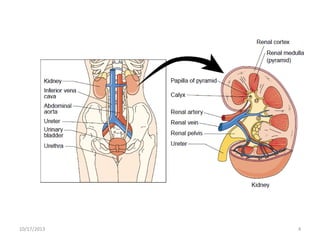
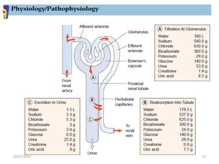
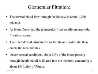
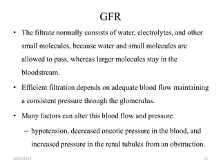
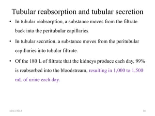
This document provides an overview of genito-urinary disorders and the anatomy and physiology of the urinary system. It describes the key components of the urinary system including the kidneys, ureters, bladder, and urethra. It then focuses on the anatomy and functions of the kidneys, nephrons, glomerulus, and the three step process of urine formation through glomerular filtration, tubular reabsorption, and tubular secretion. Common genito-urinary disorders like urinary tract infections, cystitis, pyelonephritis, prostatitis, urethritis, and nephrotic syndrome are also summarized.
At the end of this lecture the student will be able to understand the following:
Anatomy and physiology of renal & urology system
Assessment of renal & urology system
Introduction to renal & urology system disorders
Definition of UTI
Etiology/Pathophysiology of UTI
Risk factors of UTI
Clinical manifestation UTI
Complications of UTI
Diagnostic test of UTI
Medical management UTI
Nursing management UTI
This presentation comprises of congenital anomalies of kidney and urinary tract made concise and in depth for PG preparation. It contains all important topics of the regarding subject covered in detail.
Urolithiasis is a common disease that is estimated to
produce medical costs of $2.1 billion per year in the United States alone.
Renal colic affects approximately 1.2 million people
each year in USA and accounts for approximately 1% of
all hospital admissions.
Most active emergency departments (EDs) manage
patients with acute renal colic every day.
Pyelonephritis
It is the inflammation of the kidney & upper urinary tract that usually results from the bacterial infection of the bladder.
Pyelonephritis can be classified in several different catagories:
-acute pyelonephritis
-chronic pyelonephritis
-xanthogranulomatous pyelonephritis
Symptomatic presence of micro-organisms within the urinary tract i.e., kidney, ureters, bladder and urethra.
• Associated with inflammation of urinary tract.
Kidney stones (also called renal calculi, nephrolithiasis or urolithiasis) are hard deposits made of minerals and salts that form inside your kidneys. Diet, excess body weight, some medical conditions, and certain supplements and medications are among the many causes of kidney stones.
Nephrotic syndrome is a kidney disorder that causes your body to pass too much protein in your urine. Nephrotic syndrome is usually caused by damage to the clusters of small blood vessels in your kidneys that filter waste and excess water from your blood.
Gallstones are hardened deposits of bile that can form in your gallbladder. Bile is a digestive fluid produced in your liver and stored in your gallbladder. When you eat, your gallbladder contracts and empties bile into your small intestine (duodenum)
definition of hydronephrosis,
causes and types of hydronephrosis
pathophysiology of hydronephrosis
clinical manifestation and diagnostic test for hydronephrosis
management
Brief description of genitourinary system-related disorders with their nursing management. This presentation involves glomerulonephritis, nephrotic syndrome, acute renal failure, and renal calculi.
continuation on the urinary tract disorders. congenital and acquired disorders well covered. pyelonephritis also forms part of the text. thanks for reading. remeber to like and follow
Cirrhosis is a late stage of scarring (fibrosis) of the liver caused by many forms of liver diseases and conditions, such as hepatitis and chronic alcoholism
Recomendações da OMS sobre cuidados maternos e neonatais para uma experiência pós-natal positiva.
Em consonância com os ODS – Objetivos do Desenvolvimento Sustentável e a Estratégia Global para a Saúde das Mulheres, Crianças e Adolescentes, e aplicando uma abordagem baseada nos direitos humanos, os esforços de cuidados pós-natais devem expandir-se para além da cobertura e da simples sobrevivência, de modo a incluir cuidados de qualidade.
Estas diretrizes visam melhorar a qualidade dos cuidados pós-natais essenciais e de rotina prestados às mulheres e aos recém-nascidos, com o objetivo final de melhorar a saúde e o bem-estar materno e neonatal.
Uma “experiência pós-natal positiva” é um resultado importante para todas as mulheres que dão à luz e para os seus recém-nascidos, estabelecendo as bases para a melhoria da saúde e do bem-estar a curto e longo prazo. Uma experiência pós-natal positiva é definida como aquela em que as mulheres, pessoas que gestam, os recém-nascidos, os casais, os pais, os cuidadores e as famílias recebem informação consistente, garantia e apoio de profissionais de saúde motivados; e onde um sistema de saúde flexível e com recursos reconheça as necessidades das mulheres e dos bebês e respeite o seu contexto cultural.
Estas diretrizes consolidadas apresentam algumas recomendações novas e já bem fundamentadas sobre cuidados pós-natais de rotina para mulheres e neonatos que recebem cuidados no pós-parto em unidades de saúde ou na comunidade, independentemente dos recursos disponíveis.
É fornecido um conjunto abrangente de recomendações para cuidados durante o período puerperal, com ênfase nos cuidados essenciais que todas as mulheres e recém-nascidos devem receber, e com a devida atenção à qualidade dos cuidados; isto é, a entrega e a experiência do cuidado recebido. Estas diretrizes atualizam e ampliam as recomendações da OMS de 2014 sobre cuidados pós-natais da mãe e do recém-nascido e complementam as atuais diretrizes da OMS sobre a gestão de complicações pós-natais.
O estabelecimento da amamentação e o manejo das principais intercorrências é contemplada.
Recomendamos muito.
Vamos discutir essas recomendações no nosso curso de pós-graduação em Aleitamento no Instituto Ciclos.
Esta publicação só está disponível em inglês até o momento.
Prof. Marcus Renato de Carvalho
www.agostodourado.com
These simplified slides by Dr. Sidra Arshad present an overview of the non-respiratory functions of the respiratory tract.
Learning objectives:
1. Enlist the non-respiratory functions of the respiratory tract
2. Briefly explain how these functions are carried out
3. Discuss the significance of dead space
4. Differentiate between minute ventilation and alveolar ventilation
5. Describe the cough and sneeze reflexes
Study Resources:
1. Chapter 39, Guyton and Hall Textbook of Medical Physiology, 14th edition
2. Chapter 34, Ganong’s Review of Medical Physiology, 26th edition
3. Chapter 17, Human Physiology by Lauralee Sherwood, 9th edition
4. Non-respiratory functions of the lungs https://academic.oup.com/bjaed/article/13/3/98/278874
ARTIFICIAL INTELLIGENCE IN HEALTHCARE.pdfAnujkumaranit
Artificial intelligence (AI) refers to the simulation of human intelligence processes by machines, especially computer systems. It encompasses tasks such as learning, reasoning, problem-solving, perception, and language understanding. AI technologies are revolutionizing various fields, from healthcare to finance, by enabling machines to perform tasks that typically require human intelligence.
Ethanol (CH3CH2OH), or beverage alcohol, is a two-carbon alcohol
that is rapidly distributed in the body and brain. Ethanol alters many
neurochemical systems and has rewarding and addictive properties. It
is the oldest recreational drug and likely contributes to more morbidity,
mortality, and public health costs than all illicit drugs combined. The
5th edition of the Diagnostic and Statistical Manual of Mental Disorders
(DSM-5) integrates alcohol abuse and alcohol dependence into a single
disorder called alcohol use disorder (AUD), with mild, moderate,
and severe subclassifications (American Psychiatric Association, 2013).
In the DSM-5, all types of substance abuse and dependence have been
combined into a single substance use disorder (SUD) on a continuum
from mild to severe. A diagnosis of AUD requires that at least two of
the 11 DSM-5 behaviors be present within a 12-month period (mild
AUD: 2–3 criteria; moderate AUD: 4–5 criteria; severe AUD: 6–11 criteria).
The four main behavioral effects of AUD are impaired control over
drinking, negative social consequences, risky use, and altered physiological
effects (tolerance, withdrawal). This chapter presents an overview
of the prevalence and harmful consequences of AUD in the U.S.,
the systemic nature of the disease, neurocircuitry and stages of AUD,
comorbidities, fetal alcohol spectrum disorders, genetic risk factors, and
pharmacotherapies for AUD.
Acute scrotum is a general term referring to an emergency condition affecting the contents or the wall of the scrotum.
There are a number of conditions that present acutely, predominantly with pain and/or swelling
A careful and detailed history and examination, and in some cases, investigations allow differentiation between these diagnoses. A prompt diagnosis is essential as the patient may require urgent surgical intervention
Testicular torsion refers to twisting of the spermatic cord, causing ischaemia of the testicle.
Testicular torsion results from inadequate fixation of the testis to the tunica vaginalis producing ischemia from reduced arterial inflow and venous outflow obstruction.
The prevalence of testicular torsion in adult patients hospitalized with acute scrotal pain is approximately 25 to 50 percent
Title: Sense of Smell
Presenter: Dr. Faiza, Assistant Professor of Physiology
Qualifications:
MBBS (Best Graduate, AIMC Lahore)
FCPS Physiology
ICMT, CHPE, DHPE (STMU)
MPH (GC University, Faisalabad)
MBA (Virtual University of Pakistan)
Learning Objectives:
Describe the primary categories of smells and the concept of odor blindness.
Explain the structure and location of the olfactory membrane and mucosa, including the types and roles of cells involved in olfaction.
Describe the pathway and mechanisms of olfactory signal transmission from the olfactory receptors to the brain.
Illustrate the biochemical cascade triggered by odorant binding to olfactory receptors, including the role of G-proteins and second messengers in generating an action potential.
Identify different types of olfactory disorders such as anosmia, hyposmia, hyperosmia, and dysosmia, including their potential causes.
Key Topics:
Olfactory Genes:
3% of the human genome accounts for olfactory genes.
400 genes for odorant receptors.
Olfactory Membrane:
Located in the superior part of the nasal cavity.
Medially: Folds downward along the superior septum.
Laterally: Folds over the superior turbinate and upper surface of the middle turbinate.
Total surface area: 5-10 square centimeters.
Olfactory Mucosa:
Olfactory Cells: Bipolar nerve cells derived from the CNS (100 million), with 4-25 olfactory cilia per cell.
Sustentacular Cells: Produce mucus and maintain ionic and molecular environment.
Basal Cells: Replace worn-out olfactory cells with an average lifespan of 1-2 months.
Bowman’s Gland: Secretes mucus.
Stimulation of Olfactory Cells:
Odorant dissolves in mucus and attaches to receptors on olfactory cilia.
Involves a cascade effect through G-proteins and second messengers, leading to depolarization and action potential generation in the olfactory nerve.
Quality of a Good Odorant:
Small (3-20 Carbon atoms), volatile, water-soluble, and lipid-soluble.
Facilitated by odorant-binding proteins in mucus.
Membrane Potential and Action Potential:
Resting membrane potential: -55mV.
Action potential frequency in the olfactory nerve increases with odorant strength.
Adaptation Towards the Sense of Smell:
Rapid adaptation within the first second, with further slow adaptation.
Psychological adaptation greater than receptor adaptation, involving feedback inhibition from the central nervous system.
Primary Sensations of Smell:
Camphoraceous, Musky, Floral, Pepperminty, Ethereal, Pungent, Putrid.
Odor Detection Threshold:
Examples: Hydrogen sulfide (0.0005 ppm), Methyl-mercaptan (0.002 ppm).
Some toxic substances are odorless at lethal concentrations.
Characteristics of Smell:
Odor blindness for single substances due to lack of appropriate receptor protein.
Behavioral and emotional influences of smell.
Transmission of Olfactory Signals:
From olfactory cells to glomeruli in the olfactory bulb, involving lateral inhibition.
Primitive, less old, and new olfactory systems with different path
micro teaching on communication m.sc nursing.pdfAnurag Sharma
Microteaching is a unique model of practice teaching. It is a viable instrument for the. desired change in the teaching behavior or the behavior potential which, in specified types of real. classroom situations, tends to facilitate the achievement of specified types of objectives.
Title: Sense of Taste
Presenter: Dr. Faiza, Assistant Professor of Physiology
Qualifications:
MBBS (Best Graduate, AIMC Lahore)
FCPS Physiology
ICMT, CHPE, DHPE (STMU)
MPH (GC University, Faisalabad)
MBA (Virtual University of Pakistan)
Learning Objectives:
Describe the structure and function of taste buds.
Describe the relationship between the taste threshold and taste index of common substances.
Explain the chemical basis and signal transduction of taste perception for each type of primary taste sensation.
Recognize different abnormalities of taste perception and their causes.
Key Topics:
Significance of Taste Sensation:
Differentiation between pleasant and harmful food
Influence on behavior
Selection of food based on metabolic needs
Receptors of Taste:
Taste buds on the tongue
Influence of sense of smell, texture of food, and pain stimulation (e.g., by pepper)
Primary and Secondary Taste Sensations:
Primary taste sensations: Sweet, Sour, Salty, Bitter, Umami
Chemical basis and signal transduction mechanisms for each taste
Taste Threshold and Index:
Taste threshold values for Sweet (sucrose), Salty (NaCl), Sour (HCl), and Bitter (Quinine)
Taste index relationship: Inversely proportional to taste threshold
Taste Blindness:
Inability to taste certain substances, particularly thiourea compounds
Example: Phenylthiocarbamide
Structure and Function of Taste Buds:
Composition: Epithelial cells, Sustentacular/Supporting cells, Taste cells, Basal cells
Features: Taste pores, Taste hairs/microvilli, and Taste nerve fibers
Location of Taste Buds:
Found in papillae of the tongue (Fungiform, Circumvallate, Foliate)
Also present on the palate, tonsillar pillars, epiglottis, and proximal esophagus
Mechanism of Taste Stimulation:
Interaction of taste substances with receptors on microvilli
Signal transduction pathways for Umami, Sweet, Bitter, Sour, and Salty tastes
Taste Sensitivity and Adaptation:
Decrease in sensitivity with age
Rapid adaptation of taste sensation
Role of Saliva in Taste:
Dissolution of tastants to reach receptors
Washing away the stimulus
Taste Preferences and Aversions:
Mechanisms behind taste preference and aversion
Influence of receptors and neural pathways
Impact of Sensory Nerve Damage:
Degeneration of taste buds if the sensory nerve fiber is cut
Abnormalities of Taste Detection:
Conditions: Ageusia, Hypogeusia, Dysgeusia (parageusia)
Causes: Nerve damage, neurological disorders, infections, poor oral hygiene, adverse drug effects, deficiencies, aging, tobacco use, altered neurotransmitter levels
Neurotransmitters and Taste Threshold:
Effects of serotonin (5-HT) and norepinephrine (NE) on taste sensitivity
Supertasters:
25% of the population with heightened sensitivity to taste, especially bitterness
Increased number of fungiform papillae
NVBDCP.pptx Nation vector borne disease control programSapna Thakur
NVBDCP was launched in 2003-2004 . Vector-Borne Disease: Disease that results from an infection transmitted to humans and other animals by blood-feeding arthropods, such as mosquitoes, ticks, and fleas. Examples of vector-borne diseases include Dengue fever, West Nile Virus, Lyme disease, and malaria.
TEST BANK for Operations Management, 14th Edition by William J. Stevenson, Ve...kevinkariuki227
TEST BANK for Operations Management, 14th Edition by William J. Stevenson, Verified Chapters 1 - 19, Complete Newest Version.pdf
Flu Vaccine Alert in Bangalore Karnatakaaddon Scans
As flu season approaches, health officials in Bangalore, Karnataka, are urging residents to get their flu vaccinations. The seasonal flu, while common, can lead to severe health complications, particularly for vulnerable populations such as young children, the elderly, and those with underlying health conditions.
Dr. Vidisha Kumari, a leading epidemiologist in Bangalore, emphasizes the importance of getting vaccinated. "The flu vaccine is our best defense against the influenza virus. It not only protects individuals but also helps prevent the spread of the virus in our communities," he says.
This year, the flu season is expected to coincide with a potential increase in other respiratory illnesses. The Karnataka Health Department has launched an awareness campaign highlighting the significance of flu vaccinations. They have set up multiple vaccination centers across Bangalore, making it convenient for residents to receive their shots.
To encourage widespread vaccination, the government is also collaborating with local schools, workplaces, and community centers to facilitate vaccination drives. Special attention is being given to ensuring that the vaccine is accessible to all, including marginalized communities who may have limited access to healthcare.
Residents are reminded that the flu vaccine is safe and effective. Common side effects are mild and may include soreness at the injection site, mild fever, or muscle aches. These side effects are generally short-lived and far less severe than the flu itself.
Healthcare providers are also stressing the importance of continuing COVID-19 precautions. Wearing masks, practicing good hand hygiene, and maintaining social distancing are still crucial, especially in crowded places.
Protect yourself and your loved ones by getting vaccinated. Together, we can help keep Bangalore healthy and safe this flu season. For more information on vaccination centers and schedules, residents can visit the Karnataka Health Department’s official website or follow their social media pages.
Stay informed, stay safe, and get your flu shot today!
Prix Galien International 2024 Forum ProgramLevi Shapiro
June 20, 2024, Prix Galien International and Jerusalem Ethics Forum in ROME. Detailed agenda including panels:
- ADVANCES IN CARDIOLOGY: A NEW PARADIGM IS COMING
- WOMEN’S HEALTH: FERTILITY PRESERVATION
- WHAT’S NEW IN THE TREATMENT OF INFECTIOUS,
ONCOLOGICAL AND INFLAMMATORY SKIN DISEASES?
- ARTIFICIAL INTELLIGENCE AND ETHICS
- GENE THERAPY
- BEYOND BORDERS: GLOBAL INITIATIVES FOR DEMOCRATIZING LIFE SCIENCE TECHNOLOGIES AND PROMOTING ACCESS TO HEALTHCARE
- ETHICAL CHALLENGES IN LIFE SCIENCES
- Prix Galien International Awards Ceremony
2. Anatomic and Physiologic Overview
• The urinary system comprises the kidneys, ureters, bladder,
and urethra.
10/17/2013
2
3. Kidneys
• The kidneys are a pair of brownish-red structures located
retroperitoneally
• The kidney consists of two distinct regions, the renal
parenchyma and the renal pelvis.
• The renal parenchyma is divided into the cortex and the
medulla.
• The cortex contains the glomeruli, proximal and distal tubules,
and cortical collecting ducts and their adjacent peritubular
capillaries.
10/17/2013
3
5. Kidneys
• The afferent arteriole branches to form the glomerulus, which
is the capillary bed responsible for glomerular filtration.
• Blood leaves the glomerulus through the efferent arteriole and
flows back to the inferior vena cava.
• Each kidney contains about 1 million nephrons, the functional
units of the kidney.
• Each kidney is capable of providing adequate renal function if
the opposite kidney is damaged or becomes nonfunctional.
10/17/2013
5
7. Glomerulus
• The glomerulus is composed of three filtering layers: the
capillary endothelium, the basement membrane, and the
epithelium.
• The glomerular membrane normally allows filtration of fluid
and small molecules yet limits passage of larger molecules,
such as blood cells and albumin.
• Kidney function begins to decrease at a rate of approximately
1% each year beginning at approximately age 30.
10/17/2013
7
9. Urine Formation
• Urine is formed in the nephrons through a complex three-step
process: glomerular filtration, tubular reabsorption, and
tubular secretion.
• The various substances normally filtered by the glomerulus,
reabsorbed by the tubules, and excreted in the urine include
sodium, chloride, bicarbonate, potassium, glucose, urea,
creatinine, and uric acid.
• Within the tubule, some of these substances are selectively
reabsorbed into the blood.
• Some substances, such as glucose, are completely reabsorbed in
10/17/2013
the tubule and normally do not appear in the urine.
9
10. Urine Formation
• Amino acids and glucose are usually filtered at the level of the
glomerulus and reabsorbed so that neither is excreted in the
urine.
• Glucose, however, appears in the urine (glycosuria) if the
amount of glucose in the blood and glomerular filtrate exceeds
the amount that the tubules are able to reabsorb.
10/17/2013
10
11. Urine Formation
• Normally, glucose is completely reabsorbed when the blood
glucose level is less than 200 mg/dL (11 mmol/L).
• In diabetes, when the blood glucose level exceeds the kidneys’
reabsorption capacity, glucose appears in the urine.
• Glycosuria is also common in pregnancy.
• Protein molecules are also generally not found in the urine;
10/17/2013
11
12. Urine Formation
• however, low-molecular-weight proteins (globulins and
albumin) may periodically be excreted in small amounts.
• Transient proteinuria in amounts less than 150 mg/dL is
considered normal and does not require further evaluation.
• Persistent proteinuria usually signifies damage to the
glomeruli.
10/17/2013
12
14. Glomerular filtration:
• The normal blood flow through the kidneys is about 1,200
mL/min.
• As blood flows into the glomerulus from an afferent arteriole,
filtration occurs.
• The filtered fluid, also known as filtrate or ultrafiltrate, then
enters the renal tubules.
• Under normal conditions, about 20% of the blood passing
through the glomeruli is filtered into the nephron, amounting to
about 180 L/day of filtrate.
10/17/2013
14
15. GFR
• The filtrate normally consists of water, electrolytes, and other
small molecules, because water and small molecules are
allowed to pass, whereas larger molecules stay in the
bloodstream.
• Efficient filtration depends on adequate blood flow maintaining
a consistent pressure through the glomerulus.
• Many factors can alter this blood flow and pressure
– hypotension, decreased oncotic pressure in the blood, and
increased pressure in the renal tubules from an obstruction.
10/17/2013
15
16. Tubular reabsorption and tubular secretion
• In tubular reabsorption, a substance moves from the filtrate
back into the peritubular capillaries.
• In tubular secretion, a substance moves from the peritubular
capillaries into tubular filtrate.
• Of the 180 L of filtrate that the kidneys produce each day, 99%
is reabsorbed into the bloodstream, resulting in 1,000 to 1,500
mL of urine each day.
10/17/2013
16
17. Cont’d
• Although most reabsorption occurs in the proximal tubule,
reabsorption occurs along the entire tubule.
•
Filtrate becomes concentrated under the influence of
antidiuretic hormone (ADH) and becomes urine
10/17/2013
17
19. GFR
• The normal adult GFR is about 100 to 120 mL/min .
• Creatinine clearance is an excellent measure of renal function;
as renal function declines, creatinine clearance decreases.
• The average person voids 1,200 to 1,500 mL of urine in 24
hours
• This amount varies depending on fluid intake,
sweating, environmental temperature, vomiting, or
diarrhea.
10/17/2013
19
23. Reading assignment
• What are the types of diagnostic tests used to identify
genito-urinary problems?
10/17/2013
23
24. Urinary tract infections
• Caused by pathogenic microorganisms in the urinary tract
• UTIs are generally classified as infections involving the upper
or lower urinary tract
• Lower UTIs include bacterial cystitis, bacterial prostatitis
and bacterial urethritis
10/17/2013
24
25. Urinary tract infections
• There can be acute or chronic nonbacterial causes of
inflammation in any of these areas that can be misdiagnosed as
bacterial infections.
• Upper UTIs are much less common and include acute or
chronic pyelonephritis, interstitial nephritis, and renal
abscesses.
10/17/2013
25
26. Findings on Exam in UTI
• Physical Exam:
– CVA tenderness (pyelonephritis)
– Urethral discharge (urethritis)
– Tender prostate on DRE (prostatitis)
• Labs: Urinalysis
– + leukocyte esterase
– + nitrites
• More likely gram-negative rods
– + WBCs
– + RBCs
10/17/2013
26
27. Culture in UTI
• Positive Urine Culture = >105 CFU/mL
• Most common pathogen for cystitis, prostatitis,
pyelonephritis:
– Escherichia coli
– Staphylococcus saprophyticus
– Proteus mirabilis
– Klebsiella
– Enterococcus
• Most common pathogen for urethritis
• Chlamydia trachomatis
• Neisseria Gonorrhea
10/17/2013
27
28. Lower Urinary Tract Infection - Cystitis
• Uncomplicated (Simple) cystitis
– In healthy woman, with no signs of systemic disease
• Complicated cystitis
– In men, or woman with comorbid medical problems.
• Recurrent cystitis
10/17/2013
28
29. Uncomplicated (simple) Cystitis
• Definition
– Healthy adult woman (over age 12)
– Non-pregnant
– No fever, nausea, vomiting, flank pain
• Diagnosis
– Dipstick urinalysis (no culture or lab tests needed)
• Treatment
– Trimethroprim/Sulfamethoxazole for 3 days
– May use fluoroquinolone (ciprofoxacin or levofloxacin)
• Risk factors:
– Sexual intercourse
• May recommend post-coital voiding
10/17/2013
29
30. Complicated Cystitis
– Females with comorbid medical conditions
– All male patients
– Indwelling foley catheters
– Urosepsis/hospitalization
• Diagnosis
– Urinalysis, Urine culture
– Further labs, if appropriate.
• Treatment
– Fluoroquinolone (or other broad spectrum antibiotic)
– 7-14 days of treatment (depending on severity)
– May treat even longer (2-4 weeks) in males with UTI
10/17/2013
30
31. Recurrent Cystitis
• Want to make sure urine culture and sensitivity obtained.
• May consider urologic work-up to evaluate for anatomical
abnormality.
• Treat for 7-14 days.
10/17/2013
31
32. Pyelonephritis
• Infection of the kidney
• Associated with constitutional symptoms – fever, nausea,
vomiting, headache
• Diagnosis:
• Urinalysis, urine culture, CBC, Chemistry
• Treatment:
• 2-weeks of Trimethroprim/sulfamethoxazole or
fluoroquinolone
• Complications:
– Perinephric/Renal abscess:
• Diagnosis: CT with contrast, renal ultrasound
• May need surgical drainage.
– Nephrolithiasis with UTI
• Suspect in patient with severe flank pain
• Need urology consult for treatment of kidney stone
10/17/2013
32
33. Prostatitis
• Symptoms:
– Pain in the perineum, lower abdomen, testicles, penis, and
with ejaculation, bladder irritation, bladder outlet
obstruction, and sometimes blood in the semen
• Diagnosis:
– Typical clinical history (fevers, chills, dysuria, malaise,
myalgias, pelvic/perineal pain, cloudy urine)
– The finding of an edematous and tender prostate on physical
examination
– Will have an increased PSA
– Urinalysis, urine culture
• Treatment:
– Trimethoprim/sulfamethoxazole, fluroquinolone
– 4-6 weeks of treatment
• Risk Factors:
– Trauma , Dehydration
10/17/2013
33
34. Urethritis
• Chlamydia trachomatis
– Frequently asymptomatic in females, but can present with dysuria,
discharge or pelvic inflammatory disease.
– Send UA, Urine culture (if pyuria seen, but no bacteria, suspect
Chlamydia)
– Pelvic exam – send discharge from cervical or urethral or for chlamydia
PCR
– Chlamydia screening is now recommended for all females ≤ 25 years
– Treatment:
• Azithromycin – 1 g po x 1
• Doxycycline – 100 mg po BID x 7 days
• Neisseria gonorrhoeae
– May present with dysuria, discharge, PID
– Send UA, urine culture
– Pelvic exam – send discharge samples for gram stain, culture
– Treatment:
• Ceftriaxone – 125 mg IM x 1
• Cipro – 500 mg po x 1
• Levofloxacin – 250 mg po x 1
10/17/2013 • Ofloxacin – 400 mg po x 1
34
35. Nephrotic syndrome
Nephrotic syndrome (NS) results from increased permeability of
Glomeulrar basement membrane (GBM) to plasma protein.
It is clinical and laboratory syndrome characterized by massive
proteinuria, which lead to hypoproteinemia ( hypoalbuminemia), hyperlipidemia and pitting edema.
10/17/2013
35
36. Nephrotic Criteria:*Massive proteinuria:
3+ or 4+,
*Hypo-proteinemia :
total plasma proteins < 5.5g/dl and serum albumin : < 2.5g/dl.
*Hyperlipidemia:
serum cholesterol : > 5.7mmol/L
*Edema: pitting edema in different degree
10/17/2013
36
37. Nephritic Criteria
• -Hematuria: RBC in urine (gross hematuria)
• -Hypertension:
• ≥130/90 mmHg in school-age children
• ≥120/80 mmHg in preschool-age children
• ≥110/70 mmHg in infant and toddler’s children
• -Azotemia(renal insufficiency):
Increased level of serum BUN 、Cr
• -Hypo-complementemia:
10/17/2013
Decreased level of serum c3
37
38. Classification:
• Primary Idiopathic NS (INS): majority
The cause is still unclear up to now.
Recent 10 years ,increasing evidence has suggested that INS
may result from a primary disorder of T– cell function.
• Secondary NS:
NS resulted from systemic diseases, such as anaphylactoid purpura
, systemic lupus erythematosus, HBV infection.
• Congenital NS: rare
*1st 3month of life ,only treatment renal transplantation
10/17/2013
38
40. Idiopathic NS (INS):
• Minimal Change Nephropathy (MCN): <80%
The glomeruli appear normal basically Under Light microscopy,
and Under Immunofluorescence
*under Electron microscopy – fusion of the foot processes of the
podocytes
• (2) Non—MCN: <20%
*Mesangial proliferative glomerulonephritis
(MsPGN): about 10%
*Focal segmental glomerulosclerosis (FSGS): 5%
*Membranous Nephropathy (MN) : 2%
*Membrane proliferative glomerulonephritis
• (MPGN) : 1%
– *Others: rare,Cresent glomerulonephritis
10/17/2013
40
41. Pathogenesis of Proteinuria
• Increase glomerular permeability for proteins due to loss of
negative charged glycoprotein
Degree of protineuria:• Mild less than 0.5g/m2/day
• Moderate 0.5 – 2g/m2/day
• Sever more than 2g/m2/day
Type of proteinuria:• A-Selective proteinuria: where proteins of low molecular weight
such as albumin, are excreted more readily than protein of HMW
• B-Non selective :
• LMW+HMW are lost in urine
10/17/2013
41
42. Pathogenesis of hypoalbuminemia
*Due to hyperproteinuria----- Loss of plasma protein in urine
mainly the albumin.
*Increased catabolism of protein during acute phase.
10/17/2013
42
43. Pathogenesis of hyperlipidemia
*Response to Hypoalbuminemia → reflex to liver --→ synthesis of
generalize protein ( including lipoprotein ) and lipid in the
liver ,the lipoprotein high molecular weight no loss in urine →
hyperlipidemia
*Diminished catabolism of lipoprotein
10/17/2013
43
44. Pathogenesis of edema
• *Reduction plasma colloid osmotic pressure↓ secondary to
hypoalbuminemia Edema and hypovolemia
• *Intravascular volume↓ antidiuretic hormone (ADH ) and
aldosterone(ALD) water and sodium retention Edema
• *Intravascular volume↓ glomerular filtration rate
(GFR)↓ water and sodium retention Edema
10/17/2013
44
46. Clinical Manifestation
1.Main manifestations:
Edema (varying degrees) is the common symptom
Local edema: edema in face , around eyes( Periorbital
swelling) , in lower extremities.
Generalized edema (anasarca), edema in penis and scrotum.
2-Non-specific symptoms:
Fatigue and lethargy
loss of appetite, nausea and vomiting ,abdominal pain ,
diarrhea
body weight increase, urine output decrease
pleural effusion (respiratory distress)
10/17/2013
46
48. Investigations
1-Urine analysis:-
A-Proteinuria : 3-4 + SELECTIVE.
b-24 urine collection for protein
>40mg/m2/hr
for children
c- volume: oliguria (during stage of edema formation)
d-Microscopically:microscopic hematuria 20%, large number of hyaline cast
10/17/2013
48
49. Investigations .. . ..
2-Blood:
• A-serum protein: decrease >5.5gm/dL , Albumin levels are low (<
2.5gm/dL).
• B-Serum cholesterol and triglycerides:
Cholesterol >5.7mmol/L (220mg/dl).
• C-- ESR↑>100mm/hr during activity phase
3. Renal function
• .
10/17/2013
49
51. Differential Diagnosis of NS:
D.D of generalized edema:1-Protein –losing enteropathy
2-Hepatic Failure.
3-HF
4-Protein energy malnutrition
5-Acute and chronic GN
10/17/2013
51
52. Complications of NS
1-Infections:
Nephrotic pt are liable to infection because :
A-loss of immunoglobins in urine.
B-the edema fluid act as a culture medium.
C-use immunosuppressive agents.
D- malnutrition
The common infection : URI, peritonitis, cellulitis and UTI
Organisms: encapsulated (Pneumococci, H.influenzae), Gram
negative (e.g E.coli)
10/17/2013
52
53. Complication…..
2-Hypercoagulability (Thrombosis)
• Hypercoagulability of the blood leading to venous or arterial
thrombosis:
• Hypercoagulability in Nephrotic syndrome caused by:
– 1-Higher concentration of I,II, V,VII,VIII,X and
fibrinogen
– 2- Lower level of anticoagulant substance:
antithrombin III
– 3-decrease fibrinolysis.
– 4-Higher blood viscosity
– 5- Increased platelet aggregation
– 6- Overaggressive diuresis
10/17/2013
53
54. Complication…..
3-ARF: pre-renal and renal
4- cardiovascular disease :-Hyperlipidemia, may be a risk
factor for cardiovascular disease.
5-Hypovolemic shock
6-Others: growth retardation, malnutrition, . …
10/17/2013
54
55. Management of NS:
• General (non-specific )
• *Corticosteroid therapy
10/17/2013
55
56. General therapy
• Hospitalization:- for initial work-up and evaluation of treatment.
• Activity: usually no restriction , except
massive edema,
sever hypertension and infection.
• Diet
Hypertension and edema: Low salt diet (<2gNa/ day) only
during period of edema or salt-free diet.
Severe edema: Restricting fluid intake
• Avoiding infection: very important.
• Diuresis: Hydrochlorothiazide (HCT) :2mg/kg.d
•
Antisterone : 2~4mg/kg.d
•
Dextran : 10~15ml/kg , after 30~60m,
•
followed by Furosemide (Lasix) at 2mg/kg .
10/17/2013
56
57. Corticosteroid—prednisone therapy
• Prednisone tablets at a dose of 60 mg/day (maximum daily dose,
80 mg divided into 2-3 doses) for at least 4 consecutive weeks.
• After complete absence of proteinuria, prednisone dose should be
tapered to 40 mg/day given every other day as a single morning
dose.
• The alternate-day dose is then slowly tapered and discontinued
over the next 2-3 months.
10/17/2013
57
58. Treatment of relapse in NS:
• Many children with nephrotic syndrome will experience at least 1
relapse (3-4+proteinuria plus edema).
• daily divided-dose prednisone at the doses noted earlier (where he
has the relapse) until the child enters remission (urine trace or
negative for protein for 3 consecutive days).
• The pred-nisone dose is then changed to alternate-day dosing and
tapered over 1-2 mo.
10/17/2013
58
59. According to response to prednisone therapy:
*Remission: no edema, urine is protein free for 5 consecutive
days.
* Relapse: edema, or first morning urine sample contains > 2 +
protein for 7 consecutive days.
*Frequent relapsing: > 2 relapses within 6 months (> 4/year).
*Steroid resistant: failure to achieve remission with prednisolone
given daily for 28 days.
10/17/2013
59
60. Side Effects With Long Term Use of Steroids
“Steroid toxicity”
-Stunted growth
• hyperglycemia
Cataracts
• myopathy
- Pseudotumor cerebri
• peptic ulcer
• poor healing of wound.
-Psycosis
-Osteoporosis
• Hirsutism
• Thromboembolism
- Cushingoid features
-Adrenal gland suppression
10/17/2013
60
61. Alternative agent
• When can be used:
• Steroid-dependent patients, frequent relapsers, and steroidresistant patients.
– Cyclophosphamide Pulse steroids
– Cyclosporin A
– Tacrolimus
– Microphenolate
10/17/2013
61
63. Acute Renal Failure
• Sudden interruption of kidney function resulting from
obstruction, reduced circulation, or disease of the renal tissue
• Results in retention of toxins, fluids, and end products of
metabolism
• Usually reversible with medical treatment
• May progress to end stage renal disease, uremic syndrome, and
death without treatment
10/17/2013
63
64. Acute Renal Failure
• Persons at Risks
– Major surgery
– Major trauma
– Receiving nephrotoxic medications
– Elderly
10/17/2013
64
66. Acute Renal Failure
• Stages
– Onset – 1-3 days with ^ BUN and creatinine and possible
decreased UOP
– Oliguric – UOP < 400/d, ^BUN, Phos, K, may last up to 14 d
– Diuretic – UOP ^ to as much as 4000 mL/d but no waste
products, at end of this stage may begin to see improvement
– Recovery – things go back to normal or may remain
insufficient and become chronic
10/17/2013
66
67. Acute Renal Failure
• Subjective symptoms
– Nausea
– Loss of appetite
– Headache
– Lethargy
– Tingling in extremities
10/17/2013
67
68. Acute Renal Failure
• Objective symptoms
– Oliguric phase –
•
•
•
•
•
•
•
•
10/17/2013
vomiting
disorientation,
edema,
^K+
decrease Na
^ BUN and creatinine
Acidosis
uremic breath (fishy
odor)
• CHF and pulmonary
edema
• hypertension
• sudden drop in UOP
• convulsions, coma
• changes in bowels
68
69. Acute Renal Failure
• Objective symptoms
– Diuretic phase
•
•
•
•
•
•
10/17/2013
Increased UOP
Gradual decline in BUN and creatinine
Hypokalemia
Hyponaturmia
Tachycardia
Improved LOC
69
70. Acute Renal Failure
• Diagnostic tests
– H&P
– BUN, creatinine, sodium, potassium. pH, bicarb. Hgb and Hct
– Urine studies
– US of kidneys
– ABD and renal CT/MRI
10/17/2013
70
71. Acute Renal Failure
• Medical treatment
– Fluid and dietary restrictions ( e.g. k+)
– Maintain E-lytes
– D/C or change cause
– May need dialysis to jump start renal function
– May need to stimulate production of urine with IV fluids,
Dopomine, diuretics, etc.
10/17/2013
71
72. Acute Renal Failure
• Medical treatment
– Hemodialysis
– Peritoneal dialysis
– Continous renal replacement therapy (CRRT)
• Can be done continuously
• Does not require dialysate
10/17/2013
72
73. Acute Renal Failure
• Nursing interventions
– Monitor I/O, including all
body fluids
– Monitor lab results
– Watch hyperkalemia
symptoms: malaise,
anorexia, parenthesia, or
muscle weakness, EKG
changes
– watch for hyperglycemia
or hypoglycemia if
receiving TPN or insulin
infusions
10/17/2013
–
–
–
–
–
Maintain nutrition
Safety measures
Mouth care
Daily weights
Assess for signs of heart
failure
– Skin integrity problems
73
74. Chronic Renal Failure
• Results form gradual, progressive loss of renal function
• Occasionally results from rapid progression of acute renal
failure
• Symptoms occur when 75% of function is lost but considered
chronic if 90-95% loss of function
• Dialysis is necessary D/T accumulation or uremic toxins,
which produce changes in major organs
10/17/2013
74
75. Chronic Renal Failure
• Subjective symptoms are relatively same as acute
• Objective symptoms
– Renal
• Hyponaturmia
• Dry mouth
• Poor skin turgor
• Confusion, salt overload, accumulation of K with muscle
weakness
• Fluid overload and metabolic acidosis
• Proteinuria, glycosuria
• Urine = RBC’s, WBC’s, and casts
10/17/2013
75
78. Chronic Renal Failure
• Objective symptoms
– Endocrine
• Stunted growth in
children
• Amenorrhea
• Male impotence
• ^ aldosterone
secretion
• Impaired glucose
levels R/T impaired
CHO metabolism
• Thyroid and
parathyroid
10/17/2013
abnormalities
– Hemopoietic
• Anemia
• Decrease in RBC
survival time
• Blood loss from
dialysis and GI bleed
• Platelet deficits
• Bleeding and clotting
disorders – purpura
and hemorrhage from
body orifices ,
ecchymoses
78
79. Chronic Renal Failure
• Objective symptoms
– Skeletal
• Muscle and bone pain
• Bone
demineralization
• Pathological fractures
• Blood vessel
calcifications in
myocardium, joints,
eyes, and brain
10/17/2013
– Skin
• Yellow-bronze skin
with pallor
• Puritus
• Purpura
• Uremic frost
• Thin, brittle nails
• Dry, brittle hair, and
may have color
changes and alopecia
79
80. Chronic Renal Failure
• Lab findings
– BUN – indicator of GFR and is affected by the breakdown
of protein. Normal is 10-20mg/dL. When reaches 70 =
dialysis
– Serum creatinine – waste product of skeletal muscle
breakdown and is a better indicator of kidney function.
Normal is 0.5-1.5 mg/dL. When reaches 10 x normal, it is
time for dialysis
– Creatinine clearance is best determent of kidney function.
– Must be a 12-24 hour urine collection. Normal is > 100 ml/min
10/17/2013
80
81. Chronic Renal Failure
• K+ – The kidneys are means which K+ is excreted.
– Normal is 3.5-5.0 ,mEq/L. maintains muscle contraction
and is essential for cardiac function.
– Both elevated and decreased can cause problems with
cardiac rhythm
– Hyperkalemia is treated with IV glucose and Na Bicarb
which pushes K+ back into the cell
– Kayexalate is also used
10/17/2013
81
82. Chronic Renal Failure
• Ca
– With disease in the kidney, the enzyme for utilization of Vit
D is absent
– Ca absorption depends upon Vit D
– Body moves Ca out of the bone to compensate and with
that Ca comes phosphate bound to it.
– Normal Ca level is 4.5-5.5 mEq/L
– Hypocalcemia = tetany
• Treat with calcium with Vit D and phosphate
• Avoid antacids with magnesium
10/17/2013
82
84. Chronic Renal Failure
Medical treatment
• IV glucose and insulin
• Na bicarb, Ca, Vit D, phosphate binders
• Fluid restriction, diuretics
• Iron supplements, blood, erythropoietin
• High carbs, low protein
• Dialysis - After all other methods have failed
10/17/2013
84
85. Chronic Renal Failure
• Hemodialysis
– Vascular access
• Temporary – subclavian or femoral
• Permanent – shunt, in arm
– Care post insertion
– Can be done rapidly
– Takes about 1 to 4 hours
– Done 3 x a week
10/17/2013
85
86. Chronic Renal Failure
• Peritoneal dialysis
– Semipermeable
membrane
– Catheter inserted through
abdominal wall into
peritoneal cavity
– Cost less
– Fewer restrictions
– Can be done at home
– Risk of peritonitis
– 3 phases – inflow, dwell
and outflow
10/17/2013
• Automated peritoneal
dialysis
– Done at home at night
– Maybe 6-7 times /week
• CAPD
– Continous ambulatory
peritoneal dialysis
– Done as outpatient
– Usually 4 X/d
86
87. Chronic Renal Failure
• Nursing care
– Frequent monitoring
– Hydration and output
– Cardiovascular function
– Respiratory status
– E-lytes
– Nutrition
– Mental status
– Emotional well being
– Ensure proper
medication regimen
– Skin care
– Bleeding problems
– Care of the shunt
– Education to client and
family
10/17/2013
87
89. Chronic Renal Failure
• Transplant
– Must find donor
– Waiting period long
– Good survival rate – 1 year 95-97%
– Must take immunosuppressant’s for life
– Rejection
• Watch for fever, elevated B/P, and pain over site of new
kidney
10/17/2013
89
91. Chronic Renal Failure
• Post op care
– ICU
– I/O
– B/P
– Weight changes
– Electrolytes
– May have fluid volume deficit
– High risk for infection
10/17/2013
91
92. Renal Calculi (Kidney Stones)
• Made of crystals of calcium
phosphate and uric acid
• Gradually they get larger until
they block ureters
• First sym severe pain
• Other sym nausea and
vomiting, frequency, chills,
fever, hematuria
• Diagnosis by symptoms,
ultrasound, or x-ray
• Rx –increase fluids to flush
out stone, medications, and if
needed LITHOTRIPSY
10/17/2013
92
93. Renal Calculi
• Called nephrolithiasis or urolithiasis
• Most commonly develop in the renal pelvis but can be anywhere
in the urinary tract
• Vary in size –from very large to tiny
• Can be 1 stone or many stones
• May stay in kidney or travel into the ureter
• Can damage the urinary tract
• May cause hydronephrosis
10/17/2013
93
94. Renal Calculi
• Predisposing factors
– Dehydration
– Prolonged immobilization
– Infection
– Obstruction
– Anything which causes the urine to be alkaline
– Metabolic factors
• Excessive intake of calcium, calcium based antacids or
Vit D
• Hyperthyroidism
• Elevated uric acid
10/17/2013
94
95. Renal Calculi
• Subjective symptoms
– Sever pain in the flank area, suprapubic area, pelvis or
external genitalia
– If in ureter, may have spasms called “renal colic”
– Urgency, frequency of urination
– N/V
– Chills
10/17/2013
95
96. Renal Calculi
• Objective symptoms
– Increased temperature
– Pallor
– Hematuria
– Abdominal distention
– Pyuria
– Anuria
– May have UTI on urinalysis
10/17/2013
96
97. Renal Calculi- Manifestations
• Kidney/Pelvis
– May be asymptomatic
– Dull, aching flank pain
• Ureter
– Acute severe flank pain, may radiate
– Nausea/vomiting
– Pallor
– Hematuria
10/17/2013
97
100. Renal Calculi
• Treatment
– Most are passed without intervention
– May need cysto with basket retrieval
– Lithotripsy : Extracorporeal shock wave lithotripsy (ESWL)
is the non-invasive treatment of kidney stones (urinary
calculosis) and biliary calculi (stones in the gallbladder or in the
liver) using an acoustic pulse.
– Lasertripsy :
– Lithotomy: is a surgical method for removal of calculi, stones
formed inside certain hollow organs, such as the bladder and
kidneys (urinary calculus) and gallbladder (gallstones), that
cannot exit naturally through the urethra, ureter or biliary duct
10/17/2013
100
101. Renal Calculi
• Nursing interventions
– Primary is to treat pain – usually with opioids
– Ambulate
– Force fluids, may have IV
• Watch for fluid overload
– Strain urine – send stone to lab if passed
– Accurate I/O
– Medicate N/V
10/17/2013
101
102. Renal Calculi
• Surgical removal
– Routine pre and post op care
– May return with catheter, drains, nephrostomy tube and
ureteral stent – must maintain patency and may need to
irrigate as ordered
– Measure drainage from all tubes – need at least 30 cc/hr
– Watch site for bleeding
– May need frequent dressing changes due to fluid leakage,
or may have collection bag
10/17/2013
102
103. Renal Calculi
• Discharge and prevention
– Continue to force fluids post discharge
– May need special diet
• Stones are analyzed for calcium or other minerals
• May need to watch products with calcium
10/17/2013
103
104. Lithostripsy
• Surgical procedure to
remove kidney stones
• Shock waves hit dense
stones and break
them up
• Done on outpatient
basis
10/17/2013
104