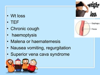
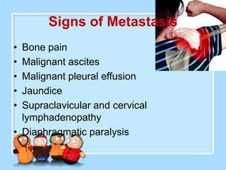
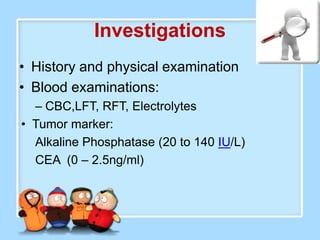
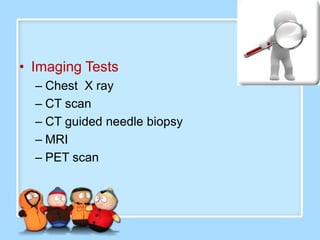
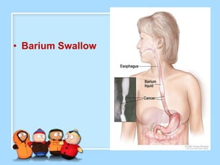
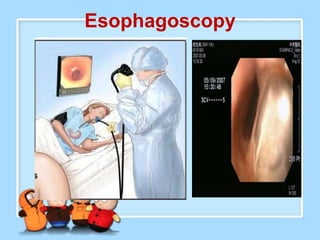
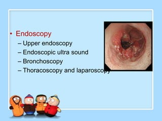
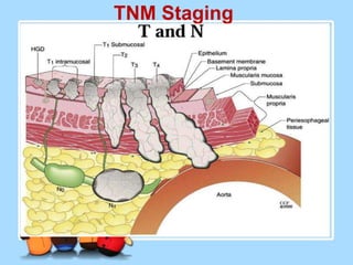
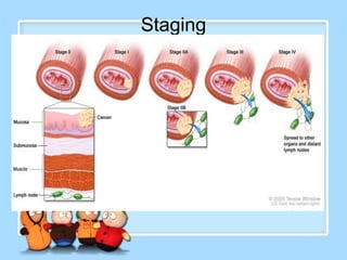
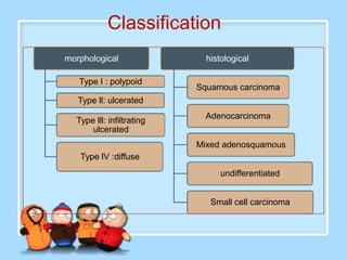
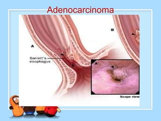
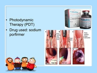
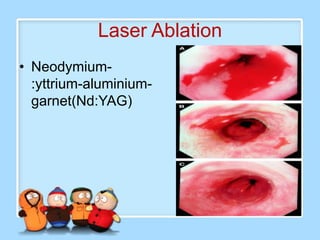
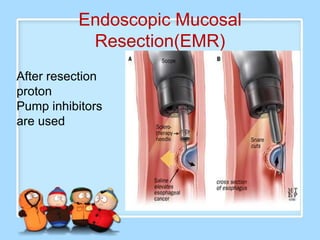
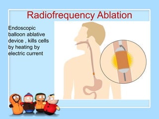
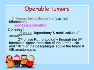
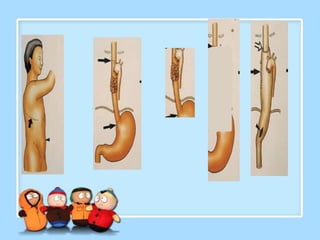
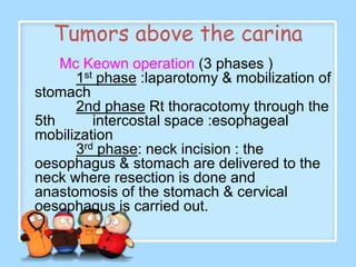
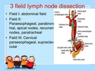
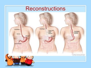
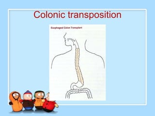
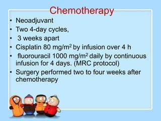
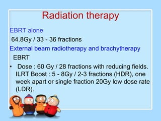
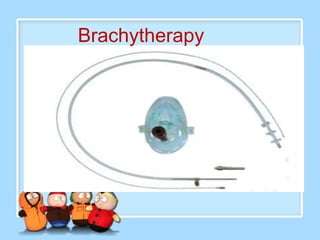
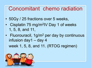
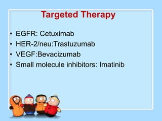
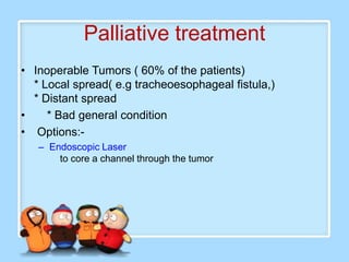
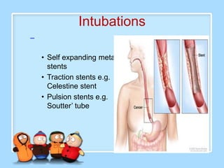
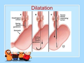
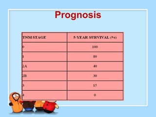
Ms Sujata Desai
Ms Sarita Kumari
Ms Shiney Sam
•   Ms Sujata Desai              •   Chemotherapy
•   Anatomy Physiology           •   Radiation therapy
•   Definition                   •   Palliative management
•   Epidemiology and incidence   •   Complications
•   Etiology                     •   Ms Shiney Sam
•   Prevention and Screening     •   Pre operative management
•   Pathophysiology              •   Post operative management
•   Clinical Manifestations      •   Rehabilitation
•   investigations               •   Prognosis
•   TNM staging                  •   Discharge planning
•   Ms Sarita Kumari             •   Follow up
•   Classification
•   Spread
•   Treatment :-
•   Surgery
Structure
• 4 layers               Mucosa

                         Submucosa

                         Muscularis
                         propria

                         Adventitia
Functions
Blood supply
Venous Drainage
Lymphatic drainage
Epidemiology
Country                          Incidence                    Link
US                               6 cases/100,000 men/year     (Black>white).

China (HenanProvince)            0.9% in the population       Nitrosamine in the soil     and
                                 older than 30 years of age   contamination of foods by fungi
                                                              (Geotrichum candidum) and yeast,
                                                              which produce mutagens

India, Pakistan, and Sri Lanka   9000 cases/year in 6         Chewing tobacco , smoking
                                 cancer registry
Singapore                                                     Hot beverages, Chinese tobacco and
                                                              wine

South African Bantus and Zulus                                Nitrosamine in the soil and
                                                              contamination       of     food      by
                                                              molds, especially the Fusarium species
Normandy, Brittany                                            Alcohol and smoking
Incidence
                         Squamous                  Adeno

New cases per year           16980                 12450
Male-to-female ratio         3:1                     7:1
Black-to-white ratio         6:1                     1:4
Most common locations middle distal
Major risk factors           smoking   Barrett’s
                             alcohol               esophagus
In TMH 1200 pts /year
2nd most common in men
4th most common in female
M0re than 180 Sx /yr
Etiology
• Unknown
• Hereditary & Genetics
• Smoking and alcohol
• Dietary factors
• *N-nitroso compounds (animal carcinogens)
  *Pickled vegetables and other food-products
  *Toxin-producing fungi
  *Betel nut chewing
  *Ingestion of very hot foods and beverages
  (such as tea)
• Obesity
• Work place exposure
ACHALASIA
Barrett’s Esophagus
Hiatus Hernia
Plummer Vinson Syndrome


Esophagitis
Anemia
dysphagia
Tylosis
Esophageal Web
Others
• Helicobacter pylori     •   h/o cancer
• Injury : ingestion of   •   HPV
  acids or alkalines      •   Aspirin
• Colic Disease           •   NSAIDS
• Chronic peptic sore
• Oral sepsis
• Syphilis
• Radiation Therapy
Prevention and screening
•   Counseling : Avoid alcohol and tobacco
•   Endemic mass screening programmes
•   Screening high risk factor
•   Surveillance Programme
    Barrett’s esophagus without dysplasia
     endoscopy 3 yearly
    Low grade Dysplasia: every year
    High Grade Dysplasia every 6months
       4 quadrant biopsy 2cm apart
Pathophysiology
Clinical manifestation
•   Dysphagia
•   Odynophagia
•   Hoarseness of voice
•   Dysphonia
•   Central chest pain
•   Wt loss
•   TEF
•   Chronic cough
•   haemoptysis
•   Malena or haematemesis
•   Nausea vomiting, regurgitation
•   Superior vena cava syndrome
Signs of Metastasis
• Bone pain
• Malignant ascites
• Malignant pleural effusion
• Jaundice
• Supraclavicular and cervical
  lymphadenopathy
• Diaphragmatic paralysis
Investigations
• History and physical examination
• Blood examinations:
  – CBC,LFT, RFT, Electrolytes
• Tumor marker:
  Alkaline Phosphatase (20 to 140 IU/L)
  CEA (0 – 2.5ng/ml)
• Imaging Tests
  – Chest X ray
  – CT scan
  – CT guided needle biopsy
  – MRI
  – PET scan
• Barium Swallow
Esophagoscopy
• Endoscopy
  – Upper endoscopy
  – Endoscopic ultra sound
  – Bronchoscopy
  – Thoracoscopy and laparoscopy
• OTHERS
  • Biopsy
  • HER2 Testing
TNM Staging
Staging
Classification
morphological                histological

   Type I : polypoid
                           Squamous carcinoma
   Type ll: ulcerated
                             Adenocarcinoma
  Type lll: infiltrating
      ulcerated
                           Mixed adenosquamous
   Type lV :diffuse
                                undifferentiated


                              Small cell carcinoma
Squamous cell carcinoma
a) Upper thirds of esophagus-20%
b) Middle thirds of esophagus-50%
c) Lower thirds of esophagus-30%
Adenocarcinoma
Spread
•   Commonly spread by Lymphatic system

    (1) Local spread
          Trachea                 tracheoesophageal fistula
           Aorta                    Fatal hemorrhage
           Recurrent laryngeal nerve       hoarseness of voice

•         (2) Lymphatic spread
             *Extensive submucosal lymphatic spread ( proximal
         line of resection should be 10cm proximal to the
         tumour).
             *Cervical ,mediastinal and coeliac LNs.

•        (3) Blood spread
             Lung, liver & brain.
Treatment modality
•   Surgery
•   Chemotherapy
•   Radiation therapy
•   Combination therapy
•   Palliative therapy
Management protocol
Surgery
•   1877- Czerny first surgeon to successfully resect a cervical esophageal
    cancer

•   Initially the anastomosis was done by bringing out the ends subcutaneously
    with external plastic tubes, skin tubes and flaps

•   1933- Ohsawa first stomach reconstruction

•   1946- Ivor Lewis two staged approach (rt thoracotomy and separate
    laparotomy)

•   1976- Mc Keown 3 stage operation

•   1982 & 1994 vagus nerve preservation

•   1997 – laparoscopic total esophagectomy
Management of
 early cancers
• Photodynamic
  Therapy (PDT)
• Drug used: sodium
  porfirmer
Laser Ablation
• Neodymium-
  :yttrium-aluminium-
  garnet(Nd:YAG)
Endoscopic Mucosal
           Resection(EMR)
After resection
proton
Pump inhibitors
are used
Radiofrequency Ablation
Endoscopic
balloon ablative
device , kills cells
by heating by
electric current
Surgery
Operable tumors
    1) Tumors below the carina (tracheal
  bifurcation)
       Ivor Lewis operation
(2 phases )
         1st phase :laparotomy & mobilization of
  stomach.
         2nd phase Rt thoracotomy through the 5th
  intercostal space resection of the tumor .LNs
  and 10cm of the oesophagus above the tumor &
  GE anastomosis.
Tumors above the carina
    Mc Keown operation (3 phases )
      1st phase :laparotomy & mobilization of
stomach
      2nd phase Rt thoracotomy through the
5th      intercostal space :esophageal
mobilization
      3rd phase: neck incision : the
oesophagus & stomach are delivered to the
neck where resection is done and
anastomosis of the stomach & cervical
oesophagus is carried out.
Transthoracic Esophagectomy
VATS
3) Tumors below the diaphragm (1
                           phase)
•
      lt thracoabdominal
  incision: the stomach
  & lower oesophagus
  are removed with
• Roux-en-Y
  esophagojujenostomy

• .
• Other options
  Transhiatal
 esophagectomy
     Thoracotomy is
 avoided by mobilizing
 the oesophagus from
 the abdomen via the
 diaphragmatic hiatus
 and via the neck
 incision
3 field lymph node dissection
• Field I: abdominal field
• Field II:
  Paraesophageal, parabronc
  hial, apical nodes, recurrent
  nodes, paratracheal
• Field III: Cervical
  paraesophageal, supraclavi
  cular
• Endopscopic removal
     through laparoscopy & thoracoscopy
Reconstructions
Colonic transposition
Chemotherapy
• Neoadjuvant
• Two 4-day cycles,
•  3 weeks apart
• Cisplatin 80 mg/m2 by infusion over 4 h
•  fluorouracil 1000 mg/m2 daily by continuous
  infusion for 4 days. (MRC protocol)
• Surgery performed two to four weeks after
  chemotherapy
Radiation therapy
EBRT alone
 64.8Gy / 33 - 36 fractions
External beam radiotherapy and brachytherapy
  EBRT
• Dose : 60 Gy / 28 fractions with reducing fields.
   ILRT Boost : 5 - 8Gy / 2-3 fractions (HDR), one
   week apart or single fraction 20Gy low dose rate
   (LDR).
Brachytherapy
Concomitant chemo radiation
• 50Gy / 25 fractions over 5 weeks,
• Cisplatin 75 mg/m2IV Day 1 of weeks
  1, 5, 8, and 11,
• Fluorouracil, 1g/m2 per day by continuous
  infusion day1 – day 4
  week 1, 5, 8, and 11. (RTOG regimen)
Targeted Therapy

•   EGFR: Cetuximab
•   HER-2/neu:Trastuzumab
•   VEGF:Bevacizumab
•   Small molecule inhibitors: Imatinib
Palliative treatment
• Inoperable Tumors ( 60% of the patients)
  * Local spread( e.g tracheoesophageal fistula,)
  * Distant spread
•    * Bad general condition
• Options:-
   – Endoscopic Laser
       to core a channel through the tumor
Intubations
–

    • Self expanding metal
      stents
    • Traction stents e.g.
      Celestine stent
    • Pulsion stents e.g.
      Soutter’ tube
Dilatation
– Radiotherapy for squamous cell ca
– Dose : 3000cGy /10 fractions /2 weeks
– Reduced field / boost : 2000cGy/10# / 2 weeks
– ILRT alone or in combination with EBRT.
– 5 - 8Gy/# in 2- 3 fractions, one week apart
– Chemotherapy :5 FU + Cisplatin
– 5Fu 1000mg/m2/day continuous IV infusion on day1-
  5Cisplatin 100mg/m2 iv on day 1
– Repeat cycles on 1,5, 8, 11 wks
PEG
Complications
•   Anastomotic leak
•   Respiratory insufficiency
•   Wound infection
•   Gastric outlet obstruction
•   Pulmonary embolism
•   Radiation pneumonitis
•   Stricture
•   Fistula
•   haemorrhage
Nursing Management
• Preoperative management
• Post operative management
Preoperative management
Psychological preparation
Assess level of anxiety
Answer the questions and concerns
regarding surgery
Allow time and privacy to prepare
psychologically
Provide support and assistance
Cultural aspect need to be considered
Discharge planning
Legal preparation

Informed consent by surgeon
No sedation should be administered
Documentation
Nutritional support
Aims : promote wt gain
Interventions
–   Assess wt , nutritional assessment
–   Sr Albumin , protein
–   Assessment of swallowing capacity
–   High calorie high protein diet in liquid and soft form
–   Enteral nutrition: NG feeds
–   Parentral nutrition
–   Hydration
–   Adjust diet according to existing problems-
    constipation/diarrhea
Prevent pulmonary complications
Patients are not able to clear secretions
Head elevation
Stent placement and dilatation
Physical and physiological
              preparation
•    Cleaning of surgical site
•    Shaving
•    Personal hygiene
•    Oral care
•    Nutrition: liquid diet x 3 days
•    Monitor vital signs
•    Intake /output chart
•    Antibiotics and regular medications
    NPO night before
    No enema and laxatives can be allowed
Pain management
Explain to notify pain
Pain medications will be prescribed
Non invasive pain relieve techniques
Preoperative exercises
Stop smoking
Chest physiotherapy
Incentive spirometry
Football bladder exercises
Coughing exercises
Deep breathing
Splinting
Getting out of bed
Pre anesthetic work up
All investigations & corrections to
Co morbidities
ECG
PFT
Arterial blood gas
2d echo
Mouth opening
Check list
Send all equipments to OT
Post operative management
• Immediate
• Intermediate
• Extended
Immediate
• Intensive care - 24 to 48 hrs
• Care of ventilated pt : patent airway
  Suctioning
• Care of drains
• Cardiopulmonary monitoring
Intermediate
• Neurological Status
• Assess neurological status every shift.
• Any neurological change should be
  carefully watched and
• Promptly reported
Pain Management
 Adequate pain control reduces the mortality and
  morbidity
 Asess the pain
 Initial pain management consist of morphine or
  bupivacaine given epidurally
 Patient-controlled analgesia with morphine, or a
  combination of both
 .Nothing by mouth for 5 to 7 days, intravenous or
  epidural pain medications are used.
Pain Management contd..
Oral pain medications are started on the
 fifth or seventh postoperative day
 The main classes : opoids, nonsteroidal
 anti-inflammatory drugs, and local
 anesthetics.
Non-pharmacological
         interventions
Distraction
 Relaxation
Positioning
Pulmonary Care
 Aggressive pulmonary toilet
 Pain control is paramount
 Patients are usually intubated after surgery monitor
  oxygenation closely (spo2)
 Suctioning
 Chest physiotherapy ,Nebulizers
 Coughing, deep breathing exercises, Incentive
  spirometer.
 Teach patients to splint their incision with a pillow.
 Early mobilization
 Monitor patients closely for fever

Chest tube care
 Assess the drainage every shift.
Serosanguinous within a few hours.
Not more than 100 to 200 ml/h on the first day.
A sudden change in the color of chest tube : milky (chyle
leak )
 Check the chest tube site for drainage,
 Keep the chest tube dressing clean, dry, and intact.
Keep the chest tube free of any kinks or dependent
loops
Subcutaneous emphysema
 Palpate the surrounding area
 Due to an air leak from a pleural injury
 Additional suction or placement of a new chest tube
 New-onset may indicate a leak of the esophageal
  anastomosis.
 . Fever, tachycardia, and hypoxemia
 Esophageal leak can be confirmed by barium swallow
 Postoperative chest radiographs for pneumothorax and
  for placement of any chest tube.
 Monitor abrupt changes in oxygenation
Hemodynamics
Intravenous maintenance fluid at a rate of 100 to 200 ml/h for the first
12 to 16 hours.
 Patients may require fluid boluses in the immediate postoperative
period.
Crystalloids or blood products may be used
Interstitial pulmonary edema.
Malnutrition and low protein levels can complicate the situation.
A delicate balance between adequate fluid replacement and fluid
overload.
 30 ml/h of urine output
Determination of body weight
Meticulous skin care is necessary.
Nasogastric Tubes
. Do not move, manipulate, or irrigate the
nasogastric tube.
Do not attempt to replace it.
 Monitor the tube for patency
 Assess the drainage for color and
amount.
Gastrointestinal Care

Restricted by mouth for 5 to 7 days
Oral medications, are crushed and put down the nasogastric tube on
the second day ; they are never swallowed.
Diligent mouth care
A jejunostomy feeding tube is often placed during surgery and is
used from the first post op day for feeding
Early enteral feeding helps in early healing
Jejunostomy site care
   At 5 to 7 days check the anastomosis for leaks
   Eat 6 to 8 small frequent meals each day,
   Avoid very hot or cold beverages and spicy foods.
   Protein supplements, high-energy foods, or a soft dysphagia diet
   Sit upright, chew slowly, and eat more than 3 hours before bedtime
    assists in reducing reflux.
   Drink fluids between meals rather than with meals
   Dumping syndrome, may arise in patients who have had their vagus
    nerves divided. After vagotomy is related to unregulated gastric
    emptying
   Minimizing liquids with meals
   Consumption of frequent, small, low-carbohydrate meals
   Discharged with plans for supplemental tube feeding.
Incision Care
Keep dressings clean, dry, and intact.
Change dressing 2 to 3 times a day
Saliva leak out through the cervical incision. Can
be managed by simple dressing
Large volumes (>250 ml every 8 hours),
application of a wound drainage bag
The leak is allowed to seal on its own,
Sealing could take several weeks.
Infection Risk
Compromised nutritional status,
They have invasive catheters
Risk of infection at the surgical sites.
Meticulous wound and skin care,
Hand washing,
Avoidance of cross-contamination
Changing of invasive catheters
Antibiotics
Adequate nutrition.
Prophylaxis of deep vein thrombosis

Heparin s/c BD
TED stockings
Early ambulation
Leg and ankle exercises
Discharge planning
• Do’s
   Check surgical incision
   Maintain personal hygiene
   Incision site care
   Resume daily activities, work and sexual
   activities
   Drink fluid b/w meals
   Eat 3 hrs before bedtime
   Check wt
Contd…
 Take stool softeners
 Crush all medications
 Observe complications: tarry stool,
 progressive wt loss, diarrhea
 Keep follow up appointments
• Don’ts
   Avoid smoking (join stop smoking group)
   Avoid strenuous activity for 12 wks
   Avoid driving for 3 wks
   Avoid hot & cold beverages , spicy food
   Drink fluid in between meals
Rehabilitation
Patient must sleep in a head high position
Get adapted to small frequent meals
Keep a difference of 2-3 hrs between
meals and bed time
Continue spirometer for 3 months
Donot carry weight more than 5 kgs
Resume daily activities
Prognosis
Follow up
Every 6 months
Plain X Ray, CBC
, Biochemistry on
visit
If symptomatic
CT, PET CT
Conclusion
Thank u

cancer esophagus

  • 1.
    Ms Sujata Desai MsSarita Kumari Ms Shiney Sam
  • 2.
    Ms Sujata Desai • Chemotherapy • Anatomy Physiology • Radiation therapy • Definition • Palliative management • Epidemiology and incidence • Complications • Etiology • Ms Shiney Sam • Prevention and Screening • Pre operative management • Pathophysiology • Post operative management • Clinical Manifestations • Rehabilitation • investigations • Prognosis • TNM staging • Discharge planning • Ms Sarita Kumari • Follow up • Classification • Spread • Treatment :- • Surgery
  • 5.
    Structure • 4 layers Mucosa Submucosa Muscularis propria Adventitia
  • 6.
  • 8.
  • 9.
  • 11.
  • 14.
    Epidemiology Country Incidence Link US 6 cases/100,000 men/year (Black>white). China (HenanProvince) 0.9% in the population Nitrosamine in the soil and older than 30 years of age contamination of foods by fungi (Geotrichum candidum) and yeast, which produce mutagens India, Pakistan, and Sri Lanka 9000 cases/year in 6 Chewing tobacco , smoking cancer registry Singapore Hot beverages, Chinese tobacco and wine South African Bantus and Zulus Nitrosamine in the soil and contamination of food by molds, especially the Fusarium species Normandy, Brittany Alcohol and smoking
  • 15.
    Incidence Squamous Adeno New cases per year 16980 12450 Male-to-female ratio 3:1 7:1 Black-to-white ratio 6:1 1:4 Most common locations middle distal Major risk factors smoking Barrett’s alcohol esophagus In TMH 1200 pts /year 2nd most common in men 4th most common in female M0re than 180 Sx /yr
  • 16.
    Etiology • Unknown • Hereditary& Genetics • Smoking and alcohol
  • 17.
    • Dietary factors •*N-nitroso compounds (animal carcinogens) *Pickled vegetables and other food-products *Toxin-producing fungi *Betel nut chewing *Ingestion of very hot foods and beverages (such as tea) • Obesity • Work place exposure
  • 18.
  • 19.
  • 20.
  • 22.
  • 23.
  • 24.
  • 25.
    Others • Helicobacter pylori • h/o cancer • Injury : ingestion of • HPV acids or alkalines • Aspirin • Colic Disease • NSAIDS • Chronic peptic sore • Oral sepsis • Syphilis • Radiation Therapy
  • 26.
    Prevention and screening • Counseling : Avoid alcohol and tobacco • Endemic mass screening programmes • Screening high risk factor • Surveillance Programme Barrett’s esophagus without dysplasia endoscopy 3 yearly Low grade Dysplasia: every year High Grade Dysplasia every 6months 4 quadrant biopsy 2cm apart
  • 27.
  • 28.
    Clinical manifestation • Dysphagia • Odynophagia • Hoarseness of voice • Dysphonia • Central chest pain
  • 29.
    Wt loss • TEF • Chronic cough • haemoptysis • Malena or haematemesis • Nausea vomiting, regurgitation • Superior vena cava syndrome
  • 30.
    Signs of Metastasis •Bone pain • Malignant ascites • Malignant pleural effusion • Jaundice • Supraclavicular and cervical lymphadenopathy • Diaphragmatic paralysis
  • 31.
    Investigations • History andphysical examination • Blood examinations: – CBC,LFT, RFT, Electrolytes • Tumor marker: Alkaline Phosphatase (20 to 140 IU/L) CEA (0 – 2.5ng/ml)
  • 32.
    • Imaging Tests – Chest X ray – CT scan – CT guided needle biopsy – MRI – PET scan
  • 33.
  • 34.
  • 35.
    • Endoscopy – Upper endoscopy – Endoscopic ultra sound – Bronchoscopy – Thoracoscopy and laparoscopy
  • 36.
    • OTHERS • Biopsy • HER2 Testing
  • 37.
  • 38.
  • 40.
    Classification morphological histological Type I : polypoid Squamous carcinoma Type ll: ulcerated Adenocarcinoma Type lll: infiltrating ulcerated Mixed adenosquamous Type lV :diffuse undifferentiated Small cell carcinoma
  • 41.
    Squamous cell carcinoma a)Upper thirds of esophagus-20% b) Middle thirds of esophagus-50% c) Lower thirds of esophagus-30%
  • 42.
  • 43.
    Spread • Commonly spread by Lymphatic system (1) Local spread Trachea tracheoesophageal fistula Aorta Fatal hemorrhage Recurrent laryngeal nerve hoarseness of voice • (2) Lymphatic spread *Extensive submucosal lymphatic spread ( proximal line of resection should be 10cm proximal to the tumour). *Cervical ,mediastinal and coeliac LNs. • (3) Blood spread Lung, liver & brain.
  • 44.
    Treatment modality • Surgery • Chemotherapy • Radiation therapy • Combination therapy • Palliative therapy
  • 45.
  • 46.
    Surgery • 1877- Czerny first surgeon to successfully resect a cervical esophageal cancer • Initially the anastomosis was done by bringing out the ends subcutaneously with external plastic tubes, skin tubes and flaps • 1933- Ohsawa first stomach reconstruction • 1946- Ivor Lewis two staged approach (rt thoracotomy and separate laparotomy) • 1976- Mc Keown 3 stage operation • 1982 & 1994 vagus nerve preservation • 1997 – laparoscopic total esophagectomy
  • 47.
  • 48.
    • Photodynamic Therapy (PDT) • Drug used: sodium porfirmer
  • 49.
    Laser Ablation • Neodymium- :yttrium-aluminium- garnet(Nd:YAG)
  • 50.
    Endoscopic Mucosal Resection(EMR) After resection proton Pump inhibitors are used
  • 51.
    Radiofrequency Ablation Endoscopic balloon ablative device, kills cells by heating by electric current
  • 52.
  • 53.
    Operable tumors 1) Tumors below the carina (tracheal bifurcation) Ivor Lewis operation (2 phases ) 1st phase :laparotomy & mobilization of stomach. 2nd phase Rt thoracotomy through the 5th intercostal space resection of the tumor .LNs and 10cm of the oesophagus above the tumor & GE anastomosis.
  • 55.
    Tumors above thecarina Mc Keown operation (3 phases ) 1st phase :laparotomy & mobilization of stomach 2nd phase Rt thoracotomy through the 5th intercostal space :esophageal mobilization 3rd phase: neck incision : the oesophagus & stomach are delivered to the neck where resection is done and anastomosis of the stomach & cervical oesophagus is carried out.
  • 56.
  • 57.
  • 58.
    3) Tumors belowthe diaphragm (1 phase) • lt thracoabdominal incision: the stomach & lower oesophagus are removed with • Roux-en-Y esophagojujenostomy • .
  • 59.
    • Other options Transhiatal esophagectomy Thoracotomy is avoided by mobilizing the oesophagus from the abdomen via the diaphragmatic hiatus and via the neck incision
  • 60.
    3 field lymphnode dissection • Field I: abdominal field • Field II: Paraesophageal, parabronc hial, apical nodes, recurrent nodes, paratracheal • Field III: Cervical paraesophageal, supraclavi cular
  • 61.
    • Endopscopic removal through laparoscopy & thoracoscopy
  • 62.
  • 63.
  • 64.
    Chemotherapy • Neoadjuvant • Two4-day cycles, • 3 weeks apart • Cisplatin 80 mg/m2 by infusion over 4 h • fluorouracil 1000 mg/m2 daily by continuous infusion for 4 days. (MRC protocol) • Surgery performed two to four weeks after chemotherapy
  • 65.
    Radiation therapy EBRT alone 64.8Gy / 33 - 36 fractions External beam radiotherapy and brachytherapy EBRT • Dose : 60 Gy / 28 fractions with reducing fields. ILRT Boost : 5 - 8Gy / 2-3 fractions (HDR), one week apart or single fraction 20Gy low dose rate (LDR).
  • 66.
  • 67.
    Concomitant chemo radiation •50Gy / 25 fractions over 5 weeks, • Cisplatin 75 mg/m2IV Day 1 of weeks 1, 5, 8, and 11, • Fluorouracil, 1g/m2 per day by continuous infusion day1 – day 4 week 1, 5, 8, and 11. (RTOG regimen)
  • 68.
    Targeted Therapy • EGFR: Cetuximab • HER-2/neu:Trastuzumab • VEGF:Bevacizumab • Small molecule inhibitors: Imatinib
  • 69.
    Palliative treatment • InoperableTumors ( 60% of the patients) * Local spread( e.g tracheoesophageal fistula,) * Distant spread • * Bad general condition • Options:- – Endoscopic Laser to core a channel through the tumor
  • 70.
    Intubations – • Self expanding metal stents • Traction stents e.g. Celestine stent • Pulsion stents e.g. Soutter’ tube
  • 71.
  • 72.
    – Radiotherapy forsquamous cell ca – Dose : 3000cGy /10 fractions /2 weeks – Reduced field / boost : 2000cGy/10# / 2 weeks – ILRT alone or in combination with EBRT. – 5 - 8Gy/# in 2- 3 fractions, one week apart – Chemotherapy :5 FU + Cisplatin – 5Fu 1000mg/m2/day continuous IV infusion on day1- 5Cisplatin 100mg/m2 iv on day 1 – Repeat cycles on 1,5, 8, 11 wks
  • 73.
  • 74.
    Complications • Anastomotic leak • Respiratory insufficiency • Wound infection • Gastric outlet obstruction • Pulmonary embolism • Radiation pneumonitis • Stricture • Fistula • haemorrhage
  • 76.
    Nursing Management • Preoperativemanagement • Post operative management
  • 77.
  • 78.
    Psychological preparation Assess levelof anxiety Answer the questions and concerns regarding surgery Allow time and privacy to prepare psychologically Provide support and assistance Cultural aspect need to be considered Discharge planning
  • 79.
    Legal preparation Informed consentby surgeon No sedation should be administered Documentation
  • 80.
    Nutritional support Aims :promote wt gain Interventions – Assess wt , nutritional assessment – Sr Albumin , protein – Assessment of swallowing capacity – High calorie high protein diet in liquid and soft form – Enteral nutrition: NG feeds – Parentral nutrition – Hydration – Adjust diet according to existing problems- constipation/diarrhea
  • 81.
    Prevent pulmonary complications Patientsare not able to clear secretions Head elevation Stent placement and dilatation
  • 82.
    Physical and physiological preparation • Cleaning of surgical site • Shaving • Personal hygiene • Oral care • Nutrition: liquid diet x 3 days • Monitor vital signs • Intake /output chart • Antibiotics and regular medications NPO night before No enema and laxatives can be allowed
  • 83.
    Pain management Explain tonotify pain Pain medications will be prescribed Non invasive pain relieve techniques
  • 84.
    Preoperative exercises Stop smoking Chestphysiotherapy Incentive spirometry Football bladder exercises Coughing exercises Deep breathing Splinting Getting out of bed
  • 85.
    Pre anesthetic workup All investigations & corrections to Co morbidities ECG PFT Arterial blood gas 2d echo Mouth opening Check list Send all equipments to OT
  • 86.
  • 87.
  • 88.
    Immediate • Intensive care- 24 to 48 hrs • Care of ventilated pt : patent airway Suctioning • Care of drains • Cardiopulmonary monitoring
  • 89.
  • 90.
    • Neurological Status •Assess neurological status every shift. • Any neurological change should be carefully watched and • Promptly reported
  • 91.
    Pain Management  Adequatepain control reduces the mortality and morbidity  Asess the pain  Initial pain management consist of morphine or bupivacaine given epidurally  Patient-controlled analgesia with morphine, or a combination of both  .Nothing by mouth for 5 to 7 days, intravenous or epidural pain medications are used.
  • 92.
    Pain Management contd.. Oralpain medications are started on the fifth or seventh postoperative day  The main classes : opoids, nonsteroidal anti-inflammatory drugs, and local anesthetics.
  • 93.
    Non-pharmacological interventions Distraction  Relaxation Positioning
  • 94.
    Pulmonary Care  Aggressivepulmonary toilet  Pain control is paramount  Patients are usually intubated after surgery monitor oxygenation closely (spo2)  Suctioning  Chest physiotherapy ,Nebulizers  Coughing, deep breathing exercises, Incentive spirometer.  Teach patients to splint their incision with a pillow.  Early mobilization  Monitor patients closely for fever 
  • 95.
    Chest tube care Assess the drainage every shift. Serosanguinous within a few hours. Not more than 100 to 200 ml/h on the first day. A sudden change in the color of chest tube : milky (chyle leak ) Check the chest tube site for drainage, Keep the chest tube dressing clean, dry, and intact. Keep the chest tube free of any kinks or dependent loops
  • 96.
    Subcutaneous emphysema  Palpatethe surrounding area  Due to an air leak from a pleural injury  Additional suction or placement of a new chest tube  New-onset may indicate a leak of the esophageal anastomosis.  . Fever, tachycardia, and hypoxemia  Esophageal leak can be confirmed by barium swallow  Postoperative chest radiographs for pneumothorax and for placement of any chest tube.  Monitor abrupt changes in oxygenation
  • 97.
    Hemodynamics Intravenous maintenance fluidat a rate of 100 to 200 ml/h for the first 12 to 16 hours. Patients may require fluid boluses in the immediate postoperative period. Crystalloids or blood products may be used Interstitial pulmonary edema. Malnutrition and low protein levels can complicate the situation. A delicate balance between adequate fluid replacement and fluid overload. 30 ml/h of urine output Determination of body weight Meticulous skin care is necessary.
  • 98.
    Nasogastric Tubes . Donot move, manipulate, or irrigate the nasogastric tube. Do not attempt to replace it. Monitor the tube for patency Assess the drainage for color and amount.
  • 99.
    Gastrointestinal Care Restricted bymouth for 5 to 7 days Oral medications, are crushed and put down the nasogastric tube on the second day ; they are never swallowed. Diligent mouth care A jejunostomy feeding tube is often placed during surgery and is used from the first post op day for feeding Early enteral feeding helps in early healing Jejunostomy site care
  • 100.
    At 5 to 7 days check the anastomosis for leaks  Eat 6 to 8 small frequent meals each day,  Avoid very hot or cold beverages and spicy foods.  Protein supplements, high-energy foods, or a soft dysphagia diet  Sit upright, chew slowly, and eat more than 3 hours before bedtime assists in reducing reflux.  Drink fluids between meals rather than with meals  Dumping syndrome, may arise in patients who have had their vagus nerves divided. After vagotomy is related to unregulated gastric emptying  Minimizing liquids with meals  Consumption of frequent, small, low-carbohydrate meals  Discharged with plans for supplemental tube feeding.
  • 101.
    Incision Care Keep dressingsclean, dry, and intact. Change dressing 2 to 3 times a day Saliva leak out through the cervical incision. Can be managed by simple dressing Large volumes (>250 ml every 8 hours), application of a wound drainage bag The leak is allowed to seal on its own, Sealing could take several weeks.
  • 102.
    Infection Risk Compromised nutritionalstatus, They have invasive catheters Risk of infection at the surgical sites. Meticulous wound and skin care, Hand washing, Avoidance of cross-contamination Changing of invasive catheters Antibiotics Adequate nutrition.
  • 103.
    Prophylaxis of deepvein thrombosis Heparin s/c BD TED stockings Early ambulation Leg and ankle exercises
  • 104.
  • 105.
    • Do’s Check surgical incision Maintain personal hygiene Incision site care Resume daily activities, work and sexual activities Drink fluid b/w meals Eat 3 hrs before bedtime Check wt
  • 106.
    Contd… Take stoolsofteners Crush all medications Observe complications: tarry stool, progressive wt loss, diarrhea Keep follow up appointments
  • 107.
    • Don’ts Avoid smoking (join stop smoking group) Avoid strenuous activity for 12 wks Avoid driving for 3 wks Avoid hot & cold beverages , spicy food Drink fluid in between meals
  • 108.
    Rehabilitation Patient must sleepin a head high position Get adapted to small frequent meals Keep a difference of 2-3 hrs between meals and bed time Continue spirometer for 3 months Donot carry weight more than 5 kgs Resume daily activities
  • 109.
  • 110.
    Follow up Every 6months Plain X Ray, CBC , Biochemistry on visit If symptomatic CT, PET CT
  • 111.
  • 113.