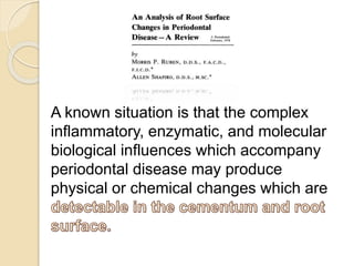
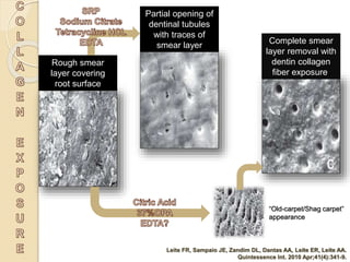
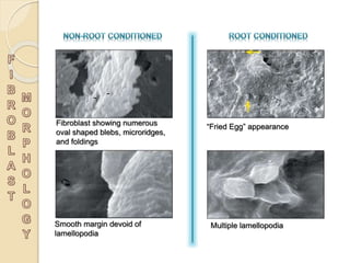
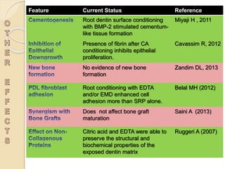
kənˈdɪʃ(ə)n/
Verb
1. have a significant influence on or
determine the manner or outcome of
something.
2. Bring something into the desired
state for use.
“Presumable Clinical Reattachment”
with Aromatic H2SO4
"loss of Cemental Vitality”
“Cemental Cuticle”
“Cemental Pins”
“Fibrin Linkage”
A known situation is that the complex
inflammatory, enzymatic, and molecular
biological influences which accompany
periodontal disease may produce
physical or chemical changes which are
Goes P, Melo IM, Dutra CS, Lima AP, Lima V.
Effect of alendronate on bone-specific alkaline phosphatase on periodontal bone
loss in rats. Arch Oral Biol. 2012 Nov;57(11):1537-44.
A zone of partial destruction of the
connective tissue fibers
A zone of intact connective tissue
fiber attachment to cementum
A zone of complete destruction
A space of 0.1 to 0.2 µ containing
some granular material but no
recognizable fibrils, can be seen.
This is the “Cemental Cuticle”
Loss of collagen cross banding,
Breakdown of dentogingival fibers
Dissolution of mineral components
Atypical development and
orientation of large apatite crystals
characterize the surface structure
of hypermineralized cementum in
periodontitis
Cervical root resorption or
idiopathic root resorption, and
its nature has not been adequately
elucidated
The root surface is covered with a
“cementum cuticle” formed
because of fiber destruction and
hypermineralization.
Cementum which has undergone
longer exposure shows a complete
covering of the normal projections
with what appears to be flat sheet-
like plaque and calculus
formations.
Plaque bacterial products have been
detected in the deep
surface layer
Bacterial lipopolysaccharide can be
identified at a distance of from
the surface of the periodontally
diseased roots
Invasion of bacteria, enzymes and
noxious products into cemental
matrix has been demonstrated in
periodontitis
The composition of the smear layer has
not yet been well defined*, but it
presumably reflects the composition of
the underlying cementum from which it is
produced.
The Ca/P ratio is 1.69 in normal cementum
vs 1.13** in smear layer over cementum.
*Ruggeri A Jr, Prati C, Mazzoni A, Nucci C, Di Lenarda R, Mazzotti G, Breschi L. Arch Oral Biol.
2007 Jan;52(1):1-8.
**Eliades G, Palaghias G,Vougiouklakis G. J Dent Res 1991; 70:383,Abstract 940.
Poor clot
stabilization on
root surface
Impaired
attachment of
periodontal fibers
Poor migration
and proliferation
of fibroblasts
Almost zero
expression of root
surface collagen
*Endodontics, Volume 1
edited by John Ide Ingle, Leif K. Bakland
Prevents
hypersensitivity**
Prevents
further bacterial
ingress**
**Rusin RP, Agee K, Suchko M, Pashley DH.
Dent Mater. 2010 Jun;26(6):600-7.
Although meticulous root
Instrumentation has been advocated,
root surface will inevitably
for
connective tissue attachment to the root
surface.
Thus, for regeneration to occur,
in order to
restore its biocompatibility becomes the
necessity.
Kassab M, Cohen RE. Compend Contin Educ Dent. 2003 Jan;24(1):31-4
Rough smear
layer covering
root surface
“Old-carpet/Shag carpet”
appearance
Partial opening of
dentinal tubules
with traces of
smear layer
Complete smear
layer removal with
dentin collagen
fiber exposure.
Leite FR, Sampaio JE, Zandim DL, Dantas AA, Leite ER, Leite AA.
Quintessence Int. 2010 Apr;41(4):341-9.
Absence of Fibrin
Network
Dense Cellular and Fibrin
Network
Scarce Fibrin Network
Moderate Cellular and
Fibrin Network
Dantas AA et al. J Contemp Dent Pract. 2012 May 1;13(3):332-8.
Fibroblast showing numerous
oval shaped blebs, microridges,
and foldings
“Fried Egg” appearance
Smooth margin devoid of
lamellopodia
Multiple lamellopodia
Feature Current Status Reference
Root dentin surface conditioning
with BMP-2 stimulated cementum-
like tissue formation
Miyaji H , 2011
Presence of fibrin after CA
conditioning inhibits epithelial
proliferation.
Cavassim R, 2012
No evidence of new bone
formation
Zandim DL, 2013
Root conditioning with EDTA
and/or EMD enhanced cell
adhesion more than SRP alone.
Belal MH (2012)
Does not affect bone graft
maturation
Saini A (2013)
Citric acid and EDTA were able to
preserve the structural and
biochemical properties of the
exposed dentin matrix
Ruggeri A (2007)
In vivo animal and human histological
studies have shown
when decalcifying
agents are used to condition the root
surface.
Nevertheless, the
employed by clinicians and
researchers has prevented consistent
comparisons among them*.
*do Amaral. J Appl Oral Sci vol.19 no.5 Bauru Sept./Oct. 2011
There still is a
concerning to the
which justifies the search for
parameters that can support the
option for this procedure in periodontal
treatments.
Still, and perhaps because clinical and
histological data ,
many clinicians include root conditioning
as part of their combination procedures for
periodontal regeneration*.
There is stronger evidence for smoking,
plaque control and maintenance
compliance
.
*Saito A, Saito E. Oral Health Dent Manag. 2014 Jun;13(2):543-7.
**de Sanctis M, Clementini M. J Clin Periodontol. 2014 Apr;41 Suppl 15:S108-22
Root conditioning in periodontics

Root conditioning in periodontics

  • 2.
    kənˈdɪʃ(ə)n/ Verb 1. have asignificant influence on or determine the manner or outcome of something. 2. Bring something into the desired state for use.
  • 3.
    “Presumable Clinical Reattachment” withAromatic H2SO4 "loss of Cemental Vitality” “Cemental Cuticle” “Cemental Pins” “Fibrin Linkage”
  • 4.
    A known situationis that the complex inflammatory, enzymatic, and molecular biological influences which accompany periodontal disease may produce physical or chemical changes which are
  • 5.
    Goes P, MeloIM, Dutra CS, Lima AP, Lima V. Effect of alendronate on bone-specific alkaline phosphatase on periodontal bone loss in rats. Arch Oral Biol. 2012 Nov;57(11):1537-44.
  • 6.
    A zone ofpartial destruction of the connective tissue fibers A zone of intact connective tissue fiber attachment to cementum A zone of complete destruction A space of 0.1 to 0.2 µ containing some granular material but no recognizable fibrils, can be seen. This is the “Cemental Cuticle”
  • 7.
    Loss of collagencross banding, Breakdown of dentogingival fibers Dissolution of mineral components Atypical development and orientation of large apatite crystals characterize the surface structure of hypermineralized cementum in periodontitis Cervical root resorption or idiopathic root resorption, and its nature has not been adequately elucidated
  • 8.
    The root surfaceis covered with a “cementum cuticle” formed because of fiber destruction and hypermineralization. Cementum which has undergone longer exposure shows a complete covering of the normal projections with what appears to be flat sheet- like plaque and calculus formations.
  • 9.
    Plaque bacterial productshave been detected in the deep surface layer Bacterial lipopolysaccharide can be identified at a distance of from the surface of the periodontally diseased roots Invasion of bacteria, enzymes and noxious products into cemental matrix has been demonstrated in periodontitis
  • 13.
    The composition ofthe smear layer has not yet been well defined*, but it presumably reflects the composition of the underlying cementum from which it is produced. The Ca/P ratio is 1.69 in normal cementum vs 1.13** in smear layer over cementum. *Ruggeri A Jr, Prati C, Mazzoni A, Nucci C, Di Lenarda R, Mazzotti G, Breschi L. Arch Oral Biol. 2007 Jan;52(1):1-8. **Eliades G, Palaghias G,Vougiouklakis G. J Dent Res 1991; 70:383,Abstract 940.
  • 14.
    Poor clot stabilization on rootsurface Impaired attachment of periodontal fibers Poor migration and proliferation of fibroblasts Almost zero expression of root surface collagen *Endodontics, Volume 1 edited by John Ide Ingle, Leif K. Bakland Prevents hypersensitivity** Prevents further bacterial ingress** **Rusin RP, Agee K, Suchko M, Pashley DH. Dent Mater. 2010 Jun;26(6):600-7.
  • 15.
    Although meticulous root Instrumentationhas been advocated, root surface will inevitably for connective tissue attachment to the root surface. Thus, for regeneration to occur, in order to restore its biocompatibility becomes the necessity.
  • 17.
    Kassab M, CohenRE. Compend Contin Educ Dent. 2003 Jan;24(1):31-4
  • 20.
    Rough smear layer covering rootsurface “Old-carpet/Shag carpet” appearance Partial opening of dentinal tubules with traces of smear layer Complete smear layer removal with dentin collagen fiber exposure. Leite FR, Sampaio JE, Zandim DL, Dantas AA, Leite ER, Leite AA. Quintessence Int. 2010 Apr;41(4):341-9.
  • 21.
    Absence of Fibrin Network DenseCellular and Fibrin Network Scarce Fibrin Network Moderate Cellular and Fibrin Network Dantas AA et al. J Contemp Dent Pract. 2012 May 1;13(3):332-8.
  • 23.
    Fibroblast showing numerous ovalshaped blebs, microridges, and foldings “Fried Egg” appearance Smooth margin devoid of lamellopodia Multiple lamellopodia
  • 24.
    Feature Current StatusReference Root dentin surface conditioning with BMP-2 stimulated cementum- like tissue formation Miyaji H , 2011 Presence of fibrin after CA conditioning inhibits epithelial proliferation. Cavassim R, 2012 No evidence of new bone formation Zandim DL, 2013 Root conditioning with EDTA and/or EMD enhanced cell adhesion more than SRP alone. Belal MH (2012) Does not affect bone graft maturation Saini A (2013) Citric acid and EDTA were able to preserve the structural and biochemical properties of the exposed dentin matrix Ruggeri A (2007)
  • 26.
    In vivo animaland human histological studies have shown when decalcifying agents are used to condition the root surface. Nevertheless, the employed by clinicians and researchers has prevented consistent comparisons among them*. *do Amaral. J Appl Oral Sci vol.19 no.5 Bauru Sept./Oct. 2011
  • 31.
    There still isa concerning to the which justifies the search for parameters that can support the option for this procedure in periodontal treatments.
  • 32.
    Still, and perhapsbecause clinical and histological data , many clinicians include root conditioning as part of their combination procedures for periodontal regeneration*. There is stronger evidence for smoking, plaque control and maintenance compliance . *Saito A, Saito E. Oral Health Dent Manag. 2014 Jun;13(2):543-7. **de Sanctis M, Clementini M. J Clin Periodontol. 2014 Apr;41 Suppl 15:S108-22