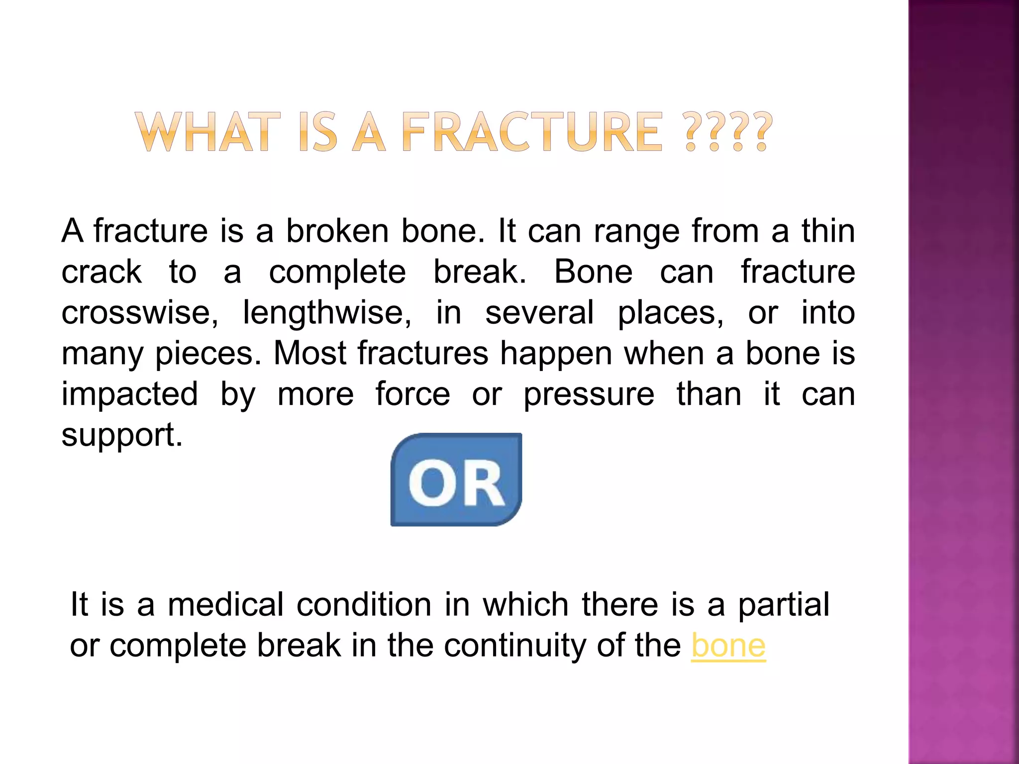
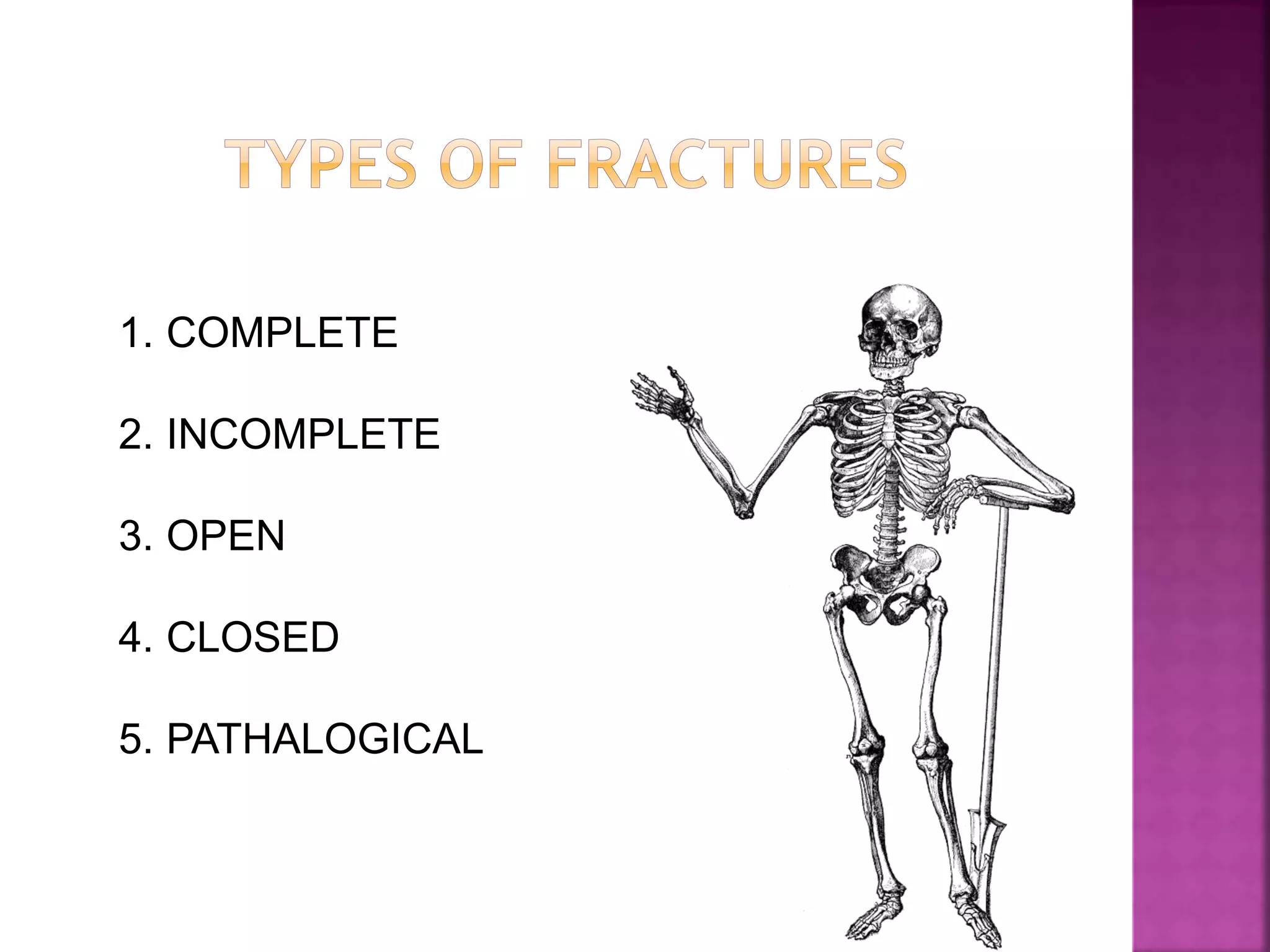
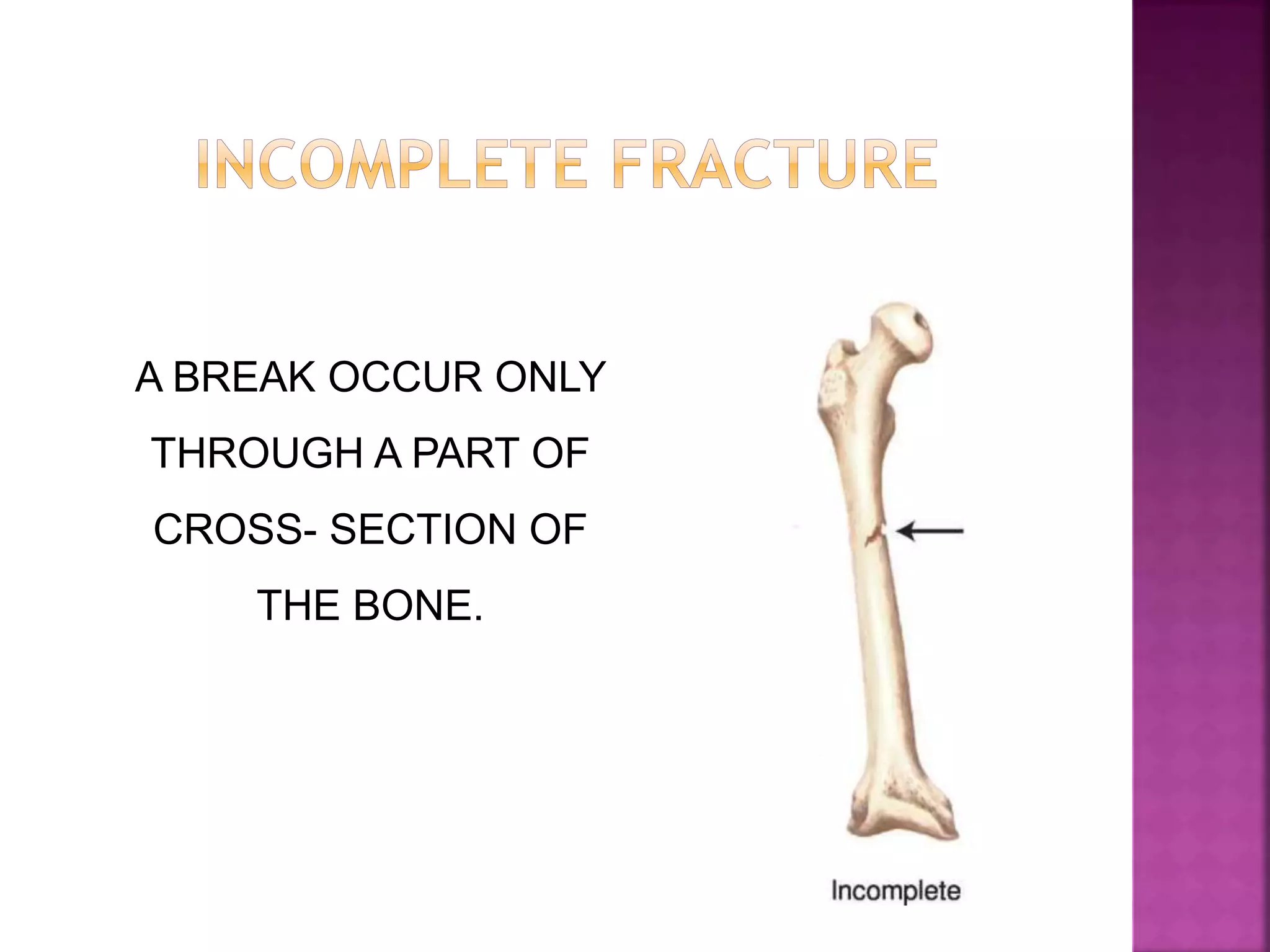
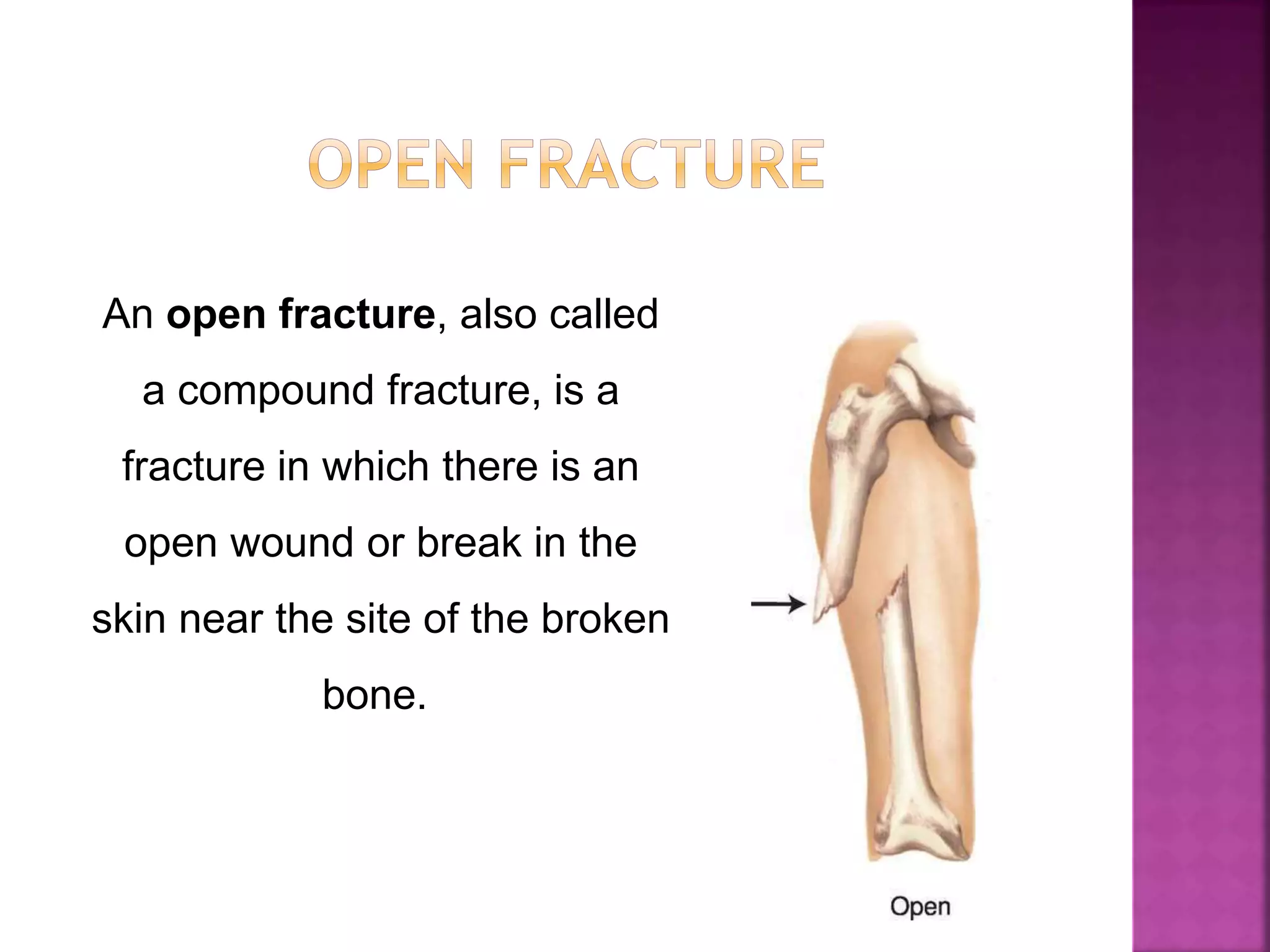
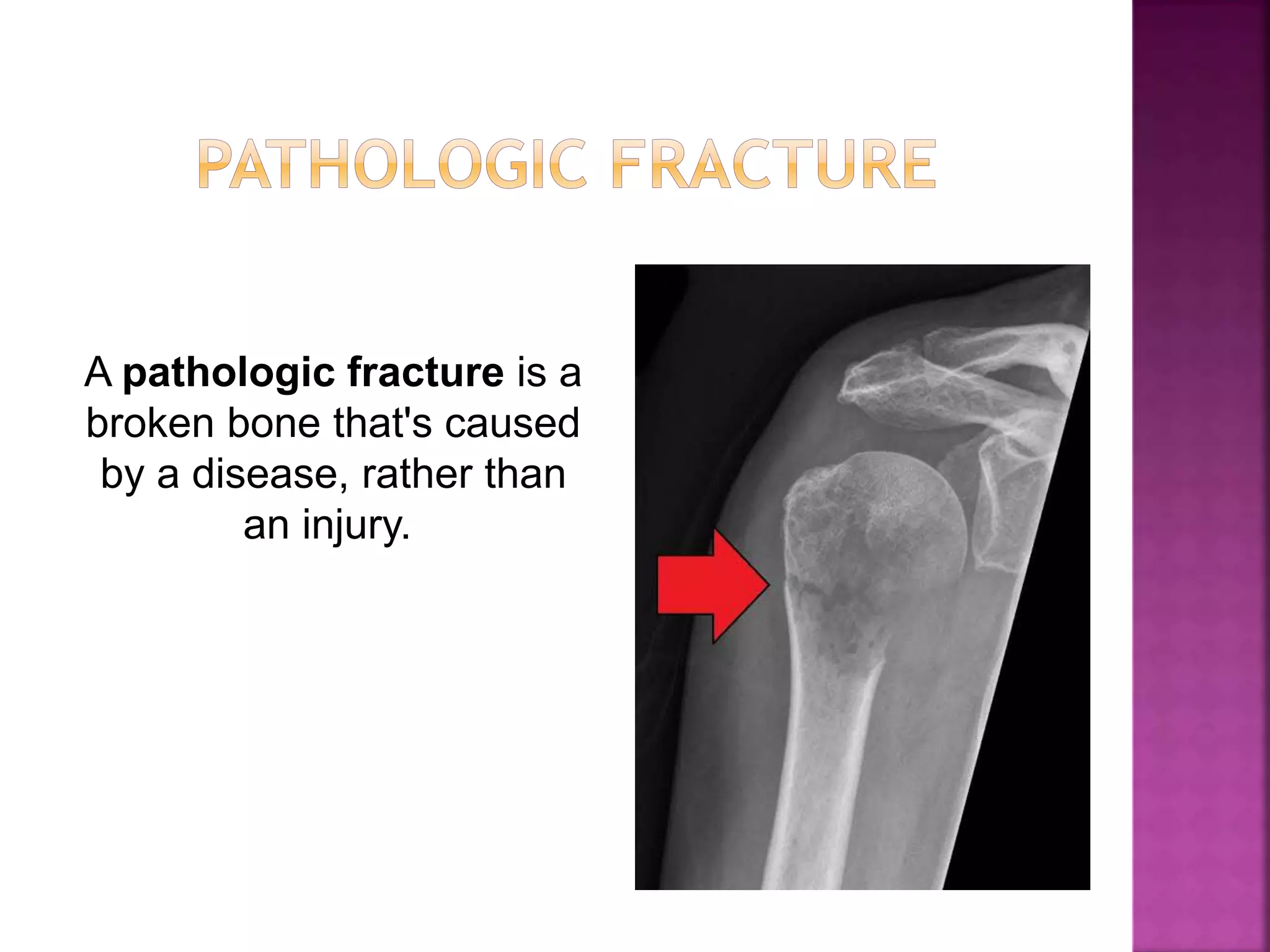
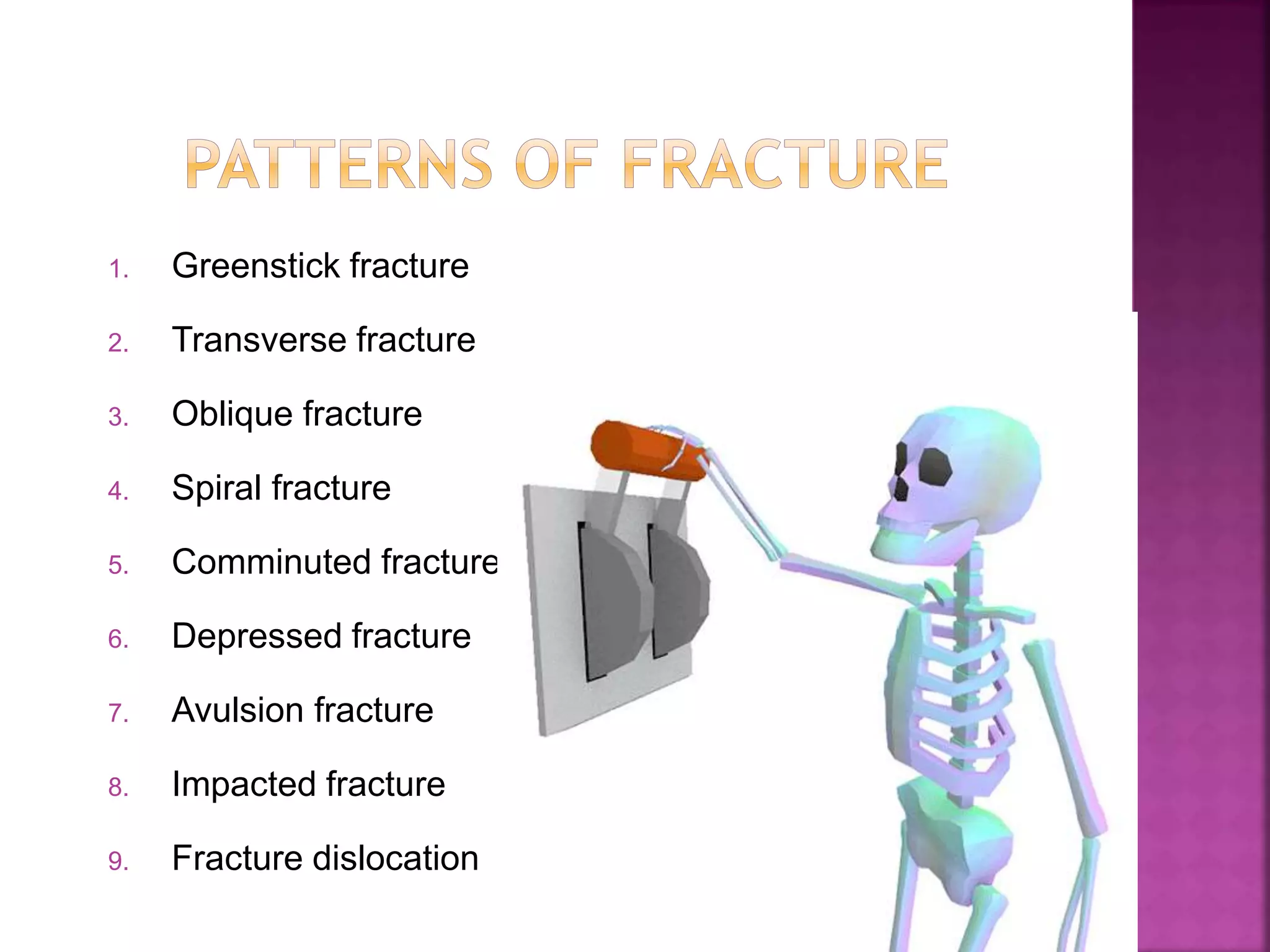
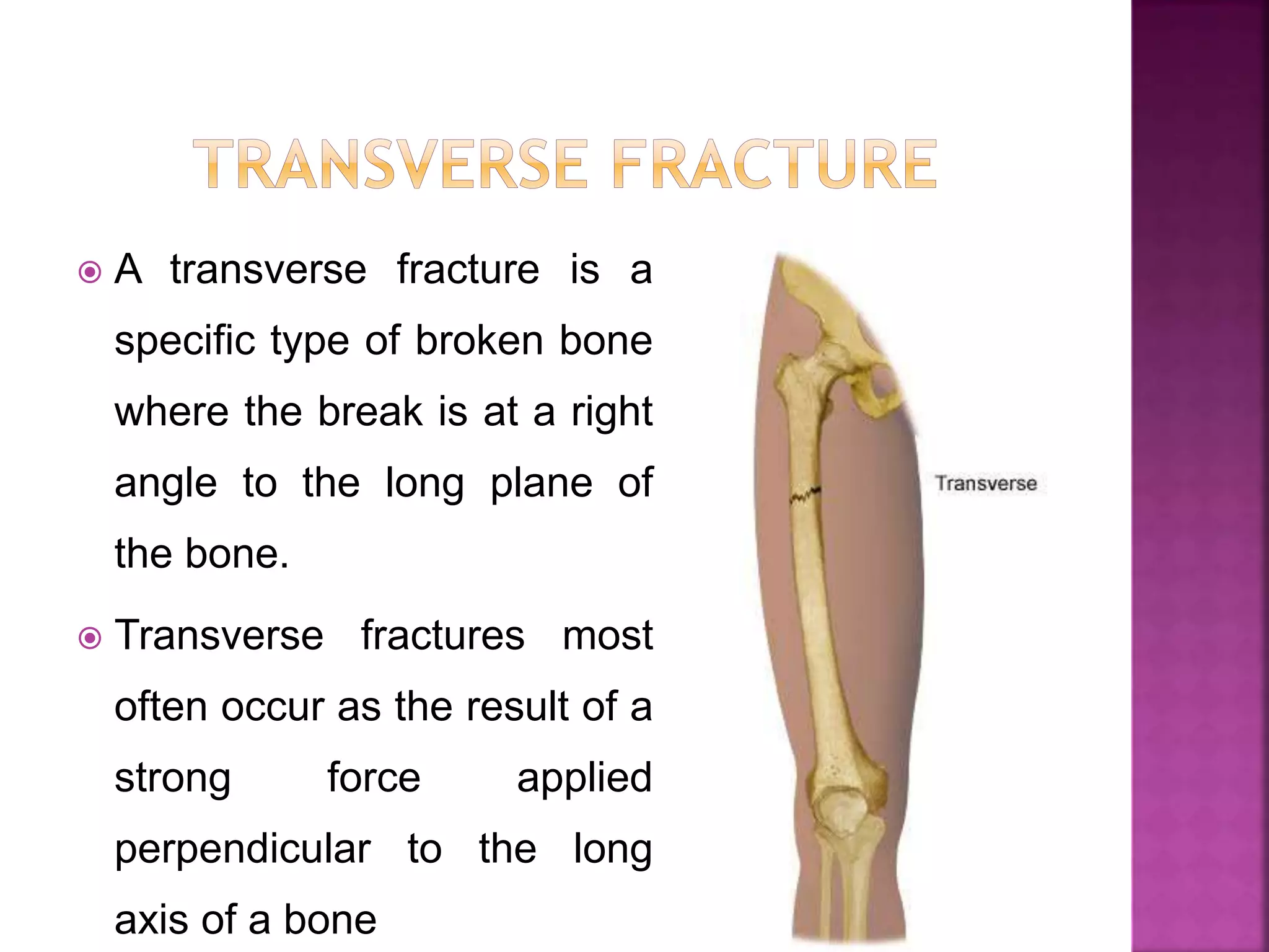
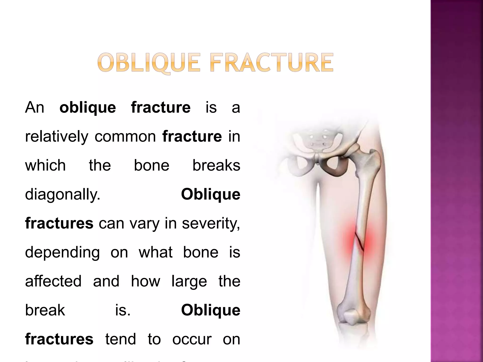
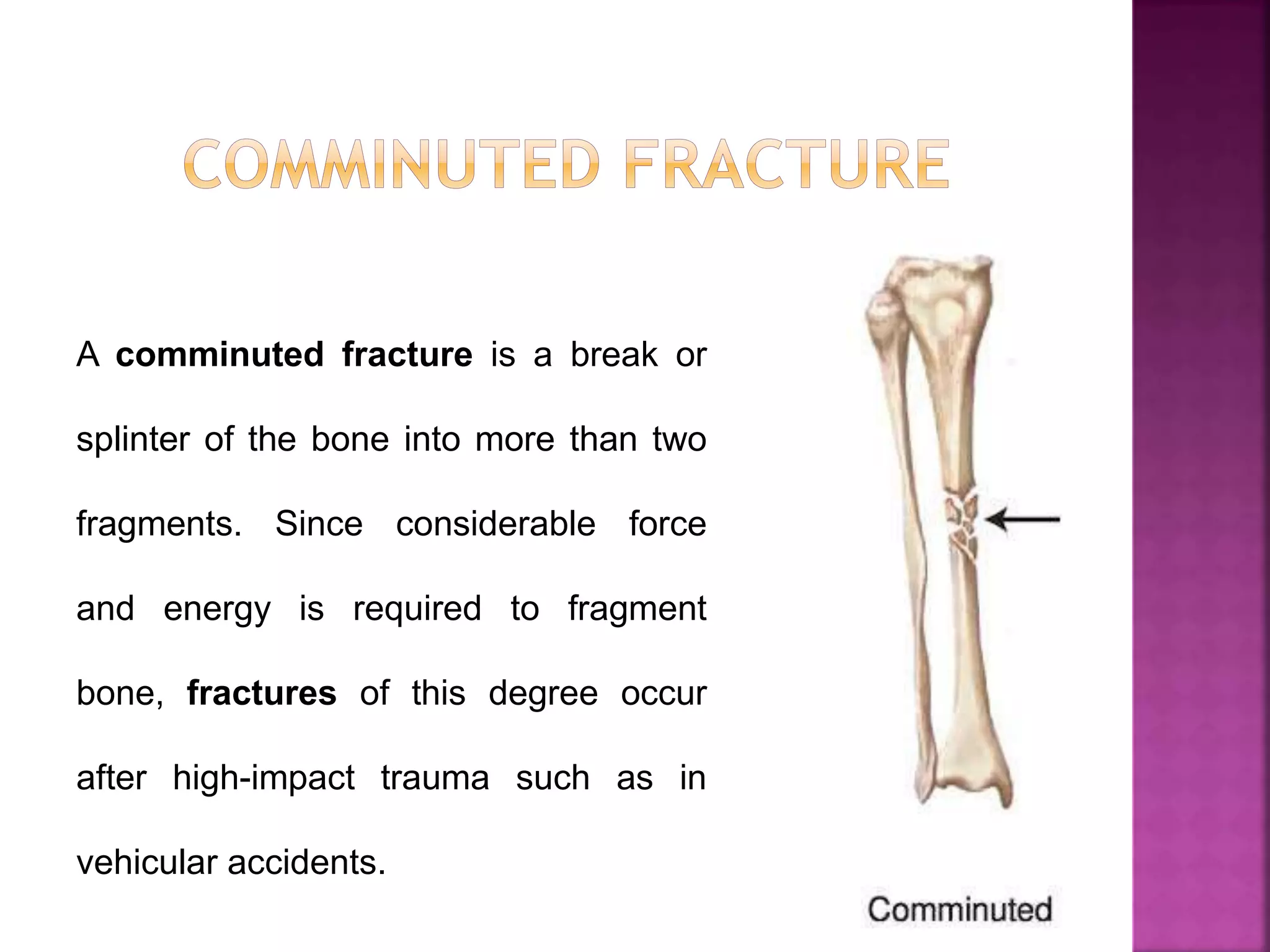
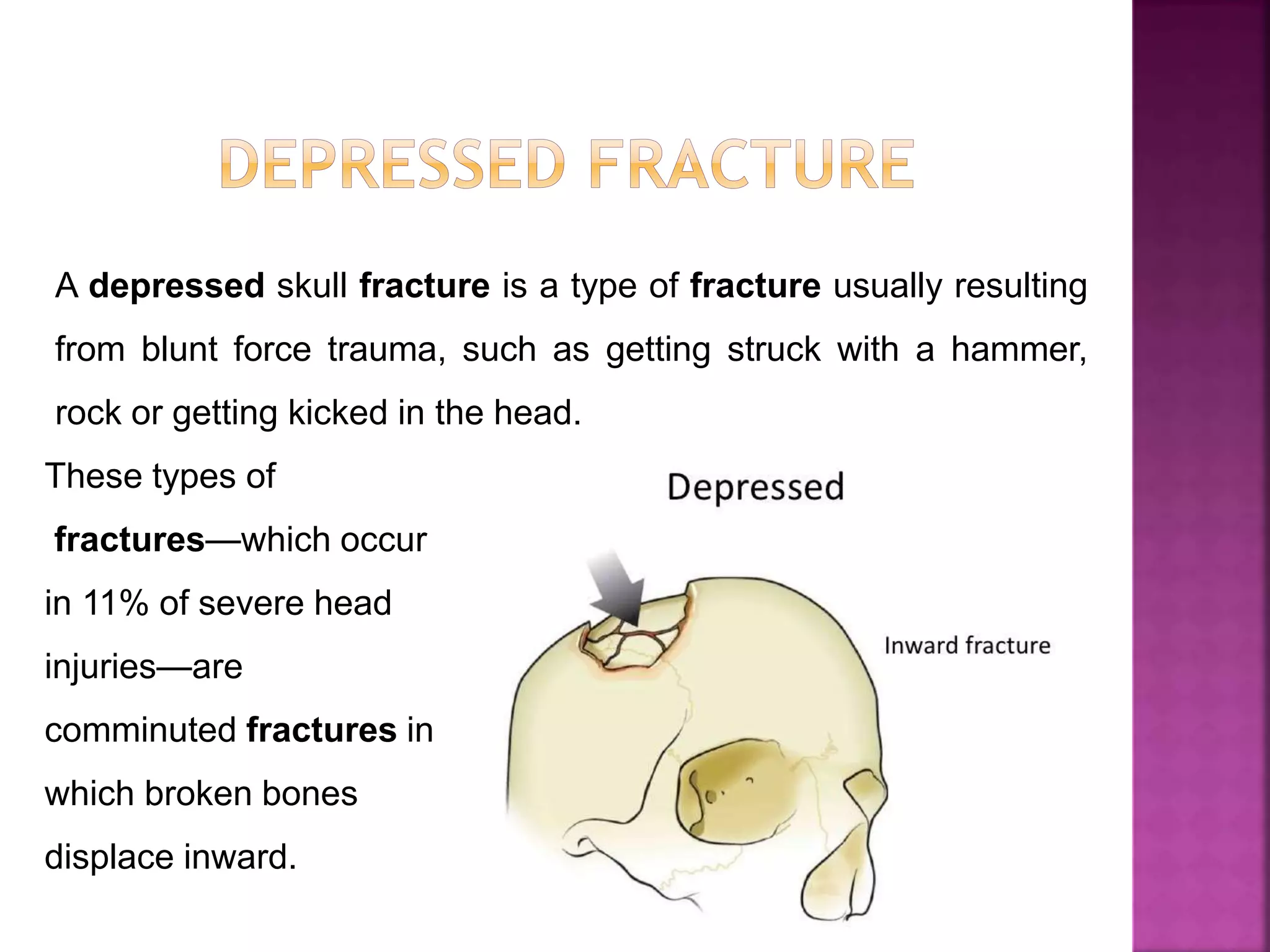
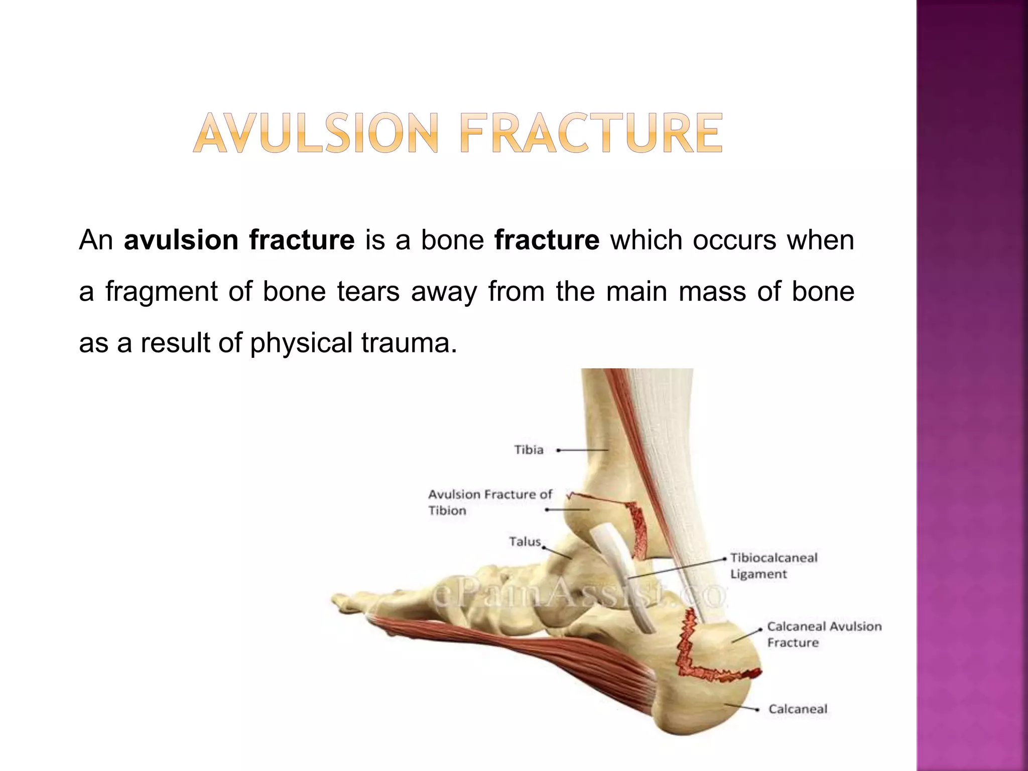
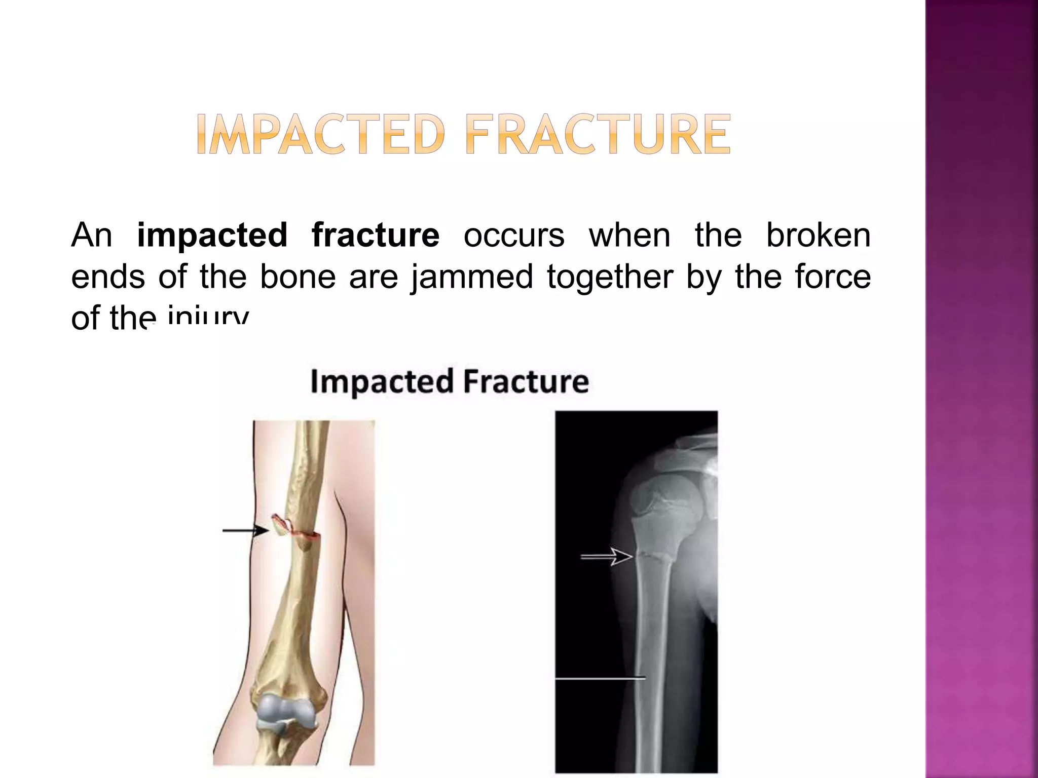
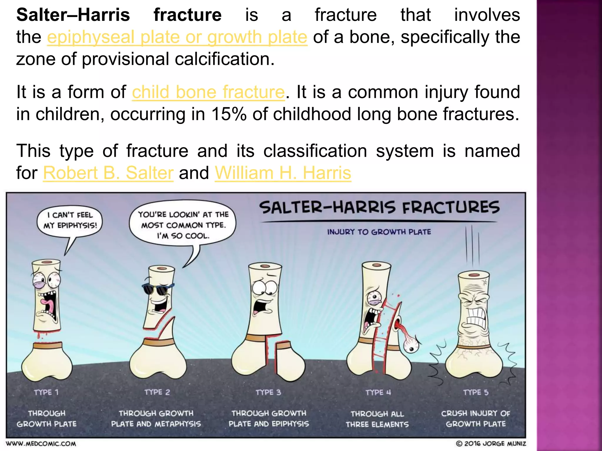
Fractures & dislocations general principlesDr KAMBLE
This document provides an overview of fractures and dislocations, including:
- A fracture is a break in the continuity of a bone, while a dislocation is the complete displacement of articular surfaces from one another.
- Fractures can be classified based on etiology (traumatic, pathological, stress), displacement, relationship to external environment (closed, open), complexity of treatment (simple, complex), and pattern (transverse, oblique, etc.).
- Pathological fractures occur through weakened bone from underlying disease. Treatment involves addressing the underlying cause and stabilizing the fracture.
- Dislocations can cause immediate complications like neurovascular injury or long-term issues like recurrence, stiffness and arthritis.
The document provides an overview of general principles and techniques for amputation. It discusses:
1. The definition of amputation and its historical development from ancient times to modern antiseptic techniques.
2. The common indications for amputation including peripheral vascular disease, trauma, infections, and tumors.
3. Surgical principles such as ideal stump shape and length, techniques for blood vessels, nerves, muscles and bone, and postoperative care including rigid dressings.
4. Potential complications like hematoma, infection, wound necrosis, contractures and phantom limb pain.
1. Open fractures are classified using the Gustilo-Anderson Classification based on the wound size and degree of soft tissue damage.
2. Essential treatment of open fractures includes administration of antibiotics, prompt wound debridement and irrigation, stabilization of the fracture, and early definitive wound cover.
3. Potential complications of open fractures include infection, osteomyelitis, compartment syndrome, and vascular injury which may require further intervention.
1. Open fractures require urgent evaluation, debridement within 6 hours, and antibiotic therapy.
2. Initial debridement removes contaminated tissue, copious irrigation is used, and repeat debridement occurs within 24-72 hours.
3. Fracture stabilization is determined initially while leaving the wound open. Early bone grafting and aggressive rehabilitation of the extremity are also recommended.
This document discusses the management of open fractures. It begins by defining an open fracture and classifying open fractures using the Gustilo and Tscherne systems. It then outlines the treatment principles of open fractures which include antibiotic prophylaxis, wound debridement, and fracture stabilization. The initial management, primary surgery including further debridement, irrigation, and skeletal stabilization are described. Factors determining limb salvage versus amputation are provided. The document concludes with discussions on external fixation, internal fixation, and wound closure approaches.
Crush injury results from prolonged physical compression of parts of the body, which can lead to crush syndrome - systemic manifestations including acute kidney injury or multisystem organ failure. Common causes are being trapped under vehicles or heavy machinery in industrial, construction, or farming accidents, building collapses during earthquakes, or crowd stampedes, where victims' bodies are compressed for extended periods.
The document discusses the principles of fracture management. It covers fracture classification, emergency care including splinting and analgesia, and definitive treatment including closed and open reduction, immobilization methods like casting, traction, and fixation. The goals of treatment are to heal the bone in proper position and alignment to restore function with minimal time, expense and complications.
Open pelvic trauma and associated injury managementUday Bangalore
This document discusses the management of open pelvic trauma from pelvic fractures. It notes that open pelvic fractures have a high mortality rate of 30-50% and require a multidisciplinary team approach and early, aggressive treatment. A key part of treatment is controlling hemorrhage through measures like external fixation, pelvic packing, angiography, and embolization to address bleeding from various pelvic arteries and veins. Other important aspects of treatment include antibiotics to prevent infection, debridement of soft tissue wounds, and repair of any associated injuries to organs like the bladder, urethra, bowel or vagina. Definitive treatment to stabilize the pelvis is done later, once the patient is stabilized.
This document discusses the anatomy, blood supply, classification, clinical features, treatment, and complications of fractures of the neck of the femur. It notes that these fractures most commonly occur in elderly women due to low-energy falls. The goals of treatment are to minimize discomfort, restore hip function, achieve early stable fixation or prosthetic replacement, and allow rapid mobilization. Surgical options include internal fixation with screws or plates for younger patients or total hip replacement or hemiarthroplasty for elderly patients. Complications can include non-union, osteonecrosis, fixation failure, and issues associated with prolonged immobilization.
The document discusses fracture healing through both primary (direct) and secondary (indirect) mechanisms. Primary healing occurs with absolute stability and involves regeneration of bone through cutting cones, while secondary healing involves callus formation and occurs with relative stability. The phases of secondary healing are hematoma and inflammation, soft callus formation through intramembranous ossification, hard callus formation through endochondral ossification, and remodeling. A variety of growth factors are involved in recruiting cells and stimulating new bone formation during the healing process.
This document provides an overview of fractures, including definitions, related terms, causes, clinical features, classifications, and the healing process. It defines a fracture as a break or disruption in bone continuity. Fractures are classified based on etiology, soft tissue involvement, displacement, fracture pattern, and number of bone fragments. The healing process involves hematoma formation, granulation tissue growth, callus formation, ossification, consolidation, and remodeling over several months.
Open fractures are unique, complex, and emergently presenting injuries that expose sterile bone to the contaminated environment.
Because a fracture disrupts the intramedullary blood supply, the additionally stripped soft tissue envelope further devitalizes the bone.
The more severe the soft tissue injury or open wound, the more severe the osseous injury.
Historically, open fractures were associated with infection, delayed union, nonunion, amputation, or death.
1) A fracture is a break in the bone that can range from a crack to a complete break with bone fragments displaced. Closed fractures involve an intact skin surface while open fractures breach the skin or a body cavity.
2) Treatment of fractures involves three phases - emergency care, definitive care, and rehabilitation. Emergency care focuses on splinting, RICE therapy, and stabilizing the patient. Definitive care includes reducing the fracture, using various methods to hold the reduction in place, and early mobilization.
3) Management of closed fractures generally prioritizes the patient's general condition first before addressing the fracture. Methods to hold a closed fracture in place include casting, traction, internal fixation, external
Dr. Suman Paul provides a historical overview of the treatment of open fractures. Early civilizations like Egyptians recognized the need to cover open fracture wounds to minimize morbidity. Through the 19th century, amputation was often used for treatment after open fractures. Advances in the 20th century included debridement and stabilization principles from World War I, and the introduction of local antibiotics in World War II which reduced wound sepsis rates. The Gustilo classification system, introduced in 1976, provides guidelines for prognosis and treatment of open fractures based on the degree of soft tissue injury. Later classifications like the Bowen system in 2005 incorporated host risk factors to better predict infection risk. Common bacteria associated with open fractures include staph, strep,
Principles and conservative treatment of fractures.2560Ukris Ortho
This document provides information about fracture classification and treatment. It discusses describing fractures based on anatomy seen on radiographs. Key details include location of the fracture line, bone condition, and deformities. Treatment may involve conservative methods like traction, casting, or bracing to immobilize the fracture for healing. The goal is healing the fracture in proper anatomical position while allowing function.
This document discusses outcomes of treating distal tibia fractures using minimally invasive plate osteosynthesis (MIPO) technique. It provides an overview of the MIPO surgical procedure and reviews several studies comparing MIPO to traditional open reduction and internal fixation. The studies found MIPO resulted in high union rates, shorter healing times, and fewer complications like infection compared to open reduction. MIPO preserves the fracture site's blood supply and limits soft tissue damage, allowing for better callus formation and healing.
ortho 01 management of open fracture-update by kk 31052010vora kun
The document discusses the management of open fractures, including their classification, principles of treatment, debridement and irrigation procedures, fracture stabilization methods, wound coverage techniques, and antibiotic protocols. Open fractures expose the bone and surrounding tissues to contamination and have higher risks of infection, soft tissue damage, and bone and functional loss compared to closed fractures. Gustilo-Anderson classification determines treatment choices and prognosticates complications based on factors like wound size, bone loss, and high-energy mechanisms. Management involves early debridement and irrigation to remove nonviable tissue and contamination, assessing injury extent, repairing structures, stabilizing fractures, and providing wound coverage.
1. The Advanced Trauma Life Support (ATLS) protocol focuses on simultaneously identifying and treating life-threatening injuries within the crucial "Golden Hour" period after trauma.
2. The ATLS protocol involves two surveys - the Primary Survey to address airway, breathing, circulation, disability, and exposure issues, and the Secondary Survey for a full history and physical exam after initial resuscitation is complete.
3. Key components of the Primary Survey include assessing the airway, identifying tension pneumothorax and hemorrhage, and providing spinal immobilization, followed by full exposure to identify all injuries.
The document discusses open fractures, defining them as fractures where a break in the skin leads directly into the fracture site. It covers the classification system for open fractures, management approaches including prevention of infection, soft tissue coverage, and fracture stabilization, and acute compartment syndrome caused by increased pressure within a confined anatomical space.
The document discusses the ATLS (Advanced Trauma Life Support) concept for managing trauma. It describes ATLS as an internationally recognized system that teaches a methodical approach to rapidly assess and treat life-threatening injuries in trauma patients. The steps include: (1) conducting a primary survey to evaluate the patient's airway, breathing, circulation, disability and exposure; (2) performing a secondary survey involving a full physical exam and history; and (3) re-evaluating the patient to ensure all injuries are identified and managed. The goal of ATLS and trauma management overall is to prevent death by treating life-threatening conditions as the top priority, especially within the "Golden Hour" period after injury.
An open fracture occurs when a broken bone pierces the skin, exposing the fracture site. Left untreated, open fractures can lead to high rates of infection, wound complications, and failure of the bone to heal properly. Gustilo-Anderson classification grades open fractures based on wound size, soft tissue damage, level of contamination, and fracture pattern, with higher grades indicating more severe injuries associated with worse outcomes. Proper management of open fractures involves emergency wound care, antibiotics, splinting or stabilization of the fracture, and often surgical debridement and irrigation to reduce the risk of infection.
The document discusses fractures of the forearm and their treatment. It summarizes that the forearm functions as a joint with six articulations. Forearm fractures can result in deformities like shortening, angulation, and loss of alignment if not treated properly. Treatment goals are anatomical reduction, restoration of length and rotation, and early return of function. Plate fixation is the gold standard and provides stable fixation, allowing early motion to restore function with high union rates over 95%.
This document provides guidelines for the treatment of open fractures. It defines open fractures and classifies them based on wound contamination and soft tissue injury. It emphasizes the importance of early intravenous antibiotics within 3 hours, serial examinations, vascular repair within 6 hours, and treatment by a multidisciplinary team for complex fractures. The guidelines recommend early wound debridement and stabilization, along with wound coverage within 7 days to prevent infection.
Open fractures involve a break in the bone that communicates with the external environment through a break in the skin and soft tissue. They are often caused by high-energy trauma like traffic accidents or falls. The initial management involves thorough debridement to remove all non-viable tissue, irrigation to clean the wound, fracture stabilization, and antibiotic treatment. Further debridement may be needed over subsequent days to fully clean the wound. The goal is to prevent infection while stabilizing the fracture and achieving soft tissue coverage. Outcomes depend on adequate initial management and reconstruction as needed.
Open fractures occur when a bone fracture communicates with the external environment through a break in the skin. They were classified and early management was discussed, including irrigation, debridement, antibiotics, tetanus prophylaxis, and splinting. Options for fracture stabilization include external fixation, plating, and intramedullary nails. Wound closure considerations involve primary versus delayed closure, skin grafting, and flaps. The goal is thorough debridement and stable fixation to prevent infection while obtaining soft tissue coverage.
Hip dislocations are classified by the direction of femoral head displacement as posterior, anterior, or central. Posterior dislocations are the most common, often resulting from high-energy trauma like motor vehicle accidents. They require closed reduction under anesthesia which may be difficult due to bone fractures. Anterior dislocations are rare but can occur from abduction and external rotation of the hip. Central dislocations actually involve an acetabular fracture displacing the femoral head medially. All hip dislocations require prompt reduction to prevent long-term complications like avascular necrosis or osteoarthritis.
This document provides an overview of the management of open fractures. It defines an open fracture as a soft tissue injury complicated by a broken bone with communication to the external environment. The history of open fracture treatment is discussed, from ancient practices like debridement to modern advances with antibiotics and fixation methods. Classification systems for open fractures are presented, including the Gustilo-Anderson classification which correlates the degree of soft tissue injury with infection risk. Key steps in managing open fractures are described, including thorough debridement and irrigation, antibiotic administration, fracture stabilization options like external or internal fixation depending on the injury, and wound management. Overall infection rates and healing times are correlated with the classification of the soft tissue injury.
The document outlines the principles of management of open fractures. It begins with definitions of open fractures and epidemiological data. It then discusses the classification system developed by Gustilo and Anderson which categorizes open fractures based on the degree of soft tissue damage and level of contamination. The key principles of management are also summarized which include emergency care, debridement, stabilization of the fracture, wound care, and rehabilitation. Complications and prognosis are also briefly covered.
This document defines and describes different types of bone fractures. It begins by defining a fracture as a complete or partial break in the bone from excessive force. Fractures are usually caused by traumatic injury and can be classified as simple, compound, incomplete or complete. It then describes three specific types of fractures: comminuted fractures involve the bone breaking into three or more pieces; greenstick fractures occur when the bone bends and cracks instead of breaking completely; and impacted fractures happen when broken bone fragments are driven into other fragments.
Bone fractures are a common injury that can result from a variety of causes, such as accidents, sports injuries, or osteoporosis. Here we will discuss 10 different types of bone fractures. For more info, you can contact us at (+44) 0161 4597 034 or visit us at:- https://www.ctclinic.co.uk/
A fracture is a broken bone that occurs when physical force exceeds the strength of the bone. Fractures are common and their risk depends on age. Greenstick fractures are partial breaks in young, soft bones caused by bending forces from falls or blows. They are treated by reducing the fracture and immobilizing the bone in a cast. Transverse fractures completely break the bone straight across from direct impacts, and require closed or open reduction and casting to heal properly. Simple or closed fractures involve a single break where the skin is intact, and are treated by aligning the bone and casting.
The document discusses different types of fractures including greenstick fractures, compound fractures, and compression fractures. It provides details on the characteristics of each type of fracture such as an incomplete break on one side for a greenstick fracture and exposure of the bone for a compound fracture. Treatment options are also summarized which include stopping bleeding, immobilizing the injured area, applying ice, and treating for shock.
Power Point 2 - Bone Fractures by Sandra Landinguinsandrute3
A greenstick fracture occurs in young bones that are still soft. It involves the bone bending and partially breaking on the outside. As bones age they become harder and more brittle. A greenstick fracture has three basic forms: a transverse fracture that extends into the midportion of the bone, a torus or buckling fracture caused by impaction, and a bow fracture where the bone becomes curved.
A fracture is a break in the bone. There are several factors that can influence fractures, such as age, disease, and location of the break. There are different types of fractures including simple (closed break in the bone), compound (open break where the bone pierces the skin), complete (bone broken into two or more pieces), and incomplete (bone is cracked but not completely broken). Specific types of fractures include comminuted (bone shattered into fragments), impacted (broken bones jammed together), and pathologic (caused by weakened bones from disease like osteoporosis).
A fracture is a broken bone. There are different types of fractures depending on factors like the energy of the event and the strength of the bone. Key types include greenstick fractures, where one side of the bone is bent instead of completely broken; comminuted fractures, where the bone is shattered into many fragments; and compound fractures, where the broken bone pierces the skin. Compound fractures require immediate surgery to clean the wound and realign the bone to prevent infection, and they generally take longer to heal than other types of fractures.
This document discusses different types of bone fractures including open or compound fractures where the bone is exposed, increasing risk of infection. Compression fractures occur when bones are forced against each other from falls or osteoporosis. Impacted fractures are similar but occur within the same bone from pressure on both ends causing fragments to jam together, common after car accidents or falls. Comminuted fractures result in several bone fragments.
Fractures occur when a bone cannot withstand physical force exerted on it. There are three main types of fractures: stress fractures which are hairline breaks often invisible on x-rays; compound fractures where the bone protrudes through the skin; and greenstick fractures where the bone cracks but does not break all the way through. Stress fractures are caused by overuse while compound fractures often result from high-energy injuries. Treatment depends on the type of fracture but generally involves rest, braces, immobilization and sometimes surgery to repair the bone and prevent infection in compound fractures.
Fracture types - Plaster Of Paris tecniques and ComplicationsVenkatesh Ghantasala
This document discusses fracture types, classifications, and POP techniques and complications. It begins by explaining the purposes of fracture classification systems which include characterizing fractures, guiding treatment, predicting outcomes, and having a common language. It then discusses the history of classifications and different classification systems including fracture-specific, generic, and those based on soft tissue injury. The rest of the document details different fracture types such as based on mechanism, soft tissue involvement, displacement, pattern, fragments, and pediatric fractures. It also provides examples of fracture classifications and eponyms.
fracture introduction, aetiology, complete and incomplete fractures, traumatic and pathologic fractures, simple and compound fractures, patterns of fractures and types of displacement
A fracture is a break in a bone caused by physical force exceeding the bone's strength. Fractures are common, with the average person experiencing two in their lifetime. They can result from high impact trauma or trivial injury if a medical condition weakens the bones. There are three main types: simple fractures where the skin is intact; compound fractures where the skin is broken, risking infection; and transverse fractures which occur at a right angle to the bone, often from sharp blows or stress. Symptoms include pain, swelling, bruising, deformity, limited movement, and numbness near the injury.
A fracture is a break in a bone caused by physical force exceeding the bone's strength. Fractures are common, with the average person experiencing two in their lifetime. They can result from high impact trauma or trivial injury if a medical condition weakens the bones. There are three main types: simple fractures where the skin is intact; compound fractures where the skin is broken, risking infection; and transverse fractures which occur at right angles to the bone's axis, often from sharp blows or stress. Symptoms include pain, swelling, bruising, impaired movement and sensation in the injured area.
A fracture is a break in a bone caused by physical force exceeding the bone's strength. Fractures are common, with the average person experiencing two in their lifetime. They can result from high impact trauma or trivial injury if a medical condition weakens the bones. There are three main types: simple fractures where the skin is intact; compound fractures where the skin is broken, risking infection; and transverse fractures which occur at a right angle to the bone's axis, often from sharp blows or stress. Symptoms include pain, swelling, bruising, impaired movement or weight-bearing, numbness, and paleness near the injury.
A fracture is a break in a bone caused by physical force exceeding the bone's strength. Fractures are common, with the average person experiencing two in their lifetime. They can result from high impact trauma or trivial injury if a medical condition weakens the bones. There are three main types: simple fractures where the skin is intact; compound fractures where the skin is broken, risking infection; and transverse fractures which occur at right angles to the bone's axis, often from sharp blows or stress. Symptoms include pain, swelling, bruising, limited movement, and numbness near the injury.
A fracture is a break in a bone caused by physical force exceeding the bone's strength. Fractures are common, with the average person experiencing two in their lifetime. They can result from high impact trauma or trivial injury if a medical condition weakens the bones. There are three main types: simple fractures where the skin is intact; compound fractures where the skin is broken, risking infection; and transverse fractures which occur at right angles to the bone's axis, often from sharp blows or stress. Symptoms include pain, swelling, bruising, impaired movement and sensation in the injured area.
Fractures occur when external forces cause the bone to break. Common causes include falls, impacts from objects, or twisting of the bone. There are different types of fractures such as greenstick, spiral, and compound fractures. Greenstick fractures involve a partial break of soft bone like a young branch. Spiral fractures occur when the bone twists, leaving a corkscrew shape. Compound fractures are the most serious as the broken bone pierces the skin, increasing risk of infection. Immediate treatment is needed to clean the wound and realign bones for compound fractures.
A fracture is a broken bone that occurs when physical force exceeds the strength of the bone. There are many types of fractures including open or closed, comminuted, greenstick, stress, spiral, and more. Treatment depends on the severity and location, with common options being casts, splints, braces, or surgery to internally fix the bone using plates, screws, rods or wires. Fractures are common injuries that increase in risk with age due to bone density loss.
A bone fracture occurs when a force exceeds a bone's strength, causing it to break. The most common fracture sites are the wrist, ankle, and hip. Children, the elderly, and women with osteoporosis are at highest risk. There are several types of fractures, including greenstick (incomplete break on one side), oblique (at an angle), spiral (twisted break), and impacted (many small fragments driven into each other). Bone fractures heal through stages including hematoma formation, fibrocartilaginous callus growth, bony callus formation, and remodeling over several months.
Transverse fractures occur when a bone is broken straight across at a right angle to its long axis, often from a direct blow. Oblique fractures occur at an angle to the bone, usually from a twisting motion or angled impact, and are more common in the elderly with osteoporosis. Compound or open fractures break the skin, requiring immediate treatment to prevent infection from entering the bone. Pathologic fractures are caused by weakened bone from conditions like tumors or infection and can occur during normal activities.
An electrocardiogram — abbreviated as EKG or ECG — is a test that measures the electrical activity of the heartbeat. With each beat, an electrical impulse (or “wave”) travels through the heart.
In human anatomy, the thigh is the area between the hip (pelvis) and the knee. Anatomically, it is part of the lower limb. The single bone in the thigh is called the femur.
Magnesium deficiency can cause a wide variety of features including hypocalcaemia, hypokalaemia and cardiac and neurological manifestations. Chronic low magnesium state has been associated with a number of chronic diseases including diabetes, hypertension, coronary heart disease, and osteoporosis.
Neonatal resuscitation also known as newborn resuscitation is an emergency procedure focused on supporting the approximately 10% of newborn children who do not readily begin breathing, putting them at risk of irreversible organ injury and death.
The medical history may include an underlying disorder that predisposes the child to a condition that results in altered mental status. A child with diabetes may have ketoacidosis or hypoglycemia.
A mechanical ventilator is a machine that helps a patient breathe (ventilate) when they are having surgery or cannot breathe on their own due to a critical illness. The patient is connected to the ventilator with a hollow tube (artificial airway) that goes in their mouth and down into their main airway or trachea
Basic life support is a level of medical care which is used for victims of life-threatening illnesses or injuries until they can be given full medical care at a hospital.
This document provides information about arterial blood gas (ABG) analysis, including what it is, its purpose, interpretation, and conditions it can assess like diabetic ketoacidosis (DKA). ABG measures oxygen, carbon dioxide, pH in blood and helps evaluate lung and kidney function in acid-base balance. It determines pH levels and the partial pressures of carbon dioxide and oxygen. The 6 steps to interpret ABGs are analyzing pH, pCO2, HCO3, matching acid-base disturbances, checking for compensation, and analyzing pO2 and oxygen saturation. DKA is a life-threatening complication of diabetes where lack of insulin causes ketone production from fat breakdown.
The document discusses acute pancreatitis. It begins by describing the anatomy of the pancreas and pancreatic duct. It then defines acute and chronic pancreatitis, with acute pancreatitis presenting as an acute abdominal condition. Common causes of acute pancreatitis include gallstones and excessive alcohol use. Clinical features include abdominal pain, nausea, vomiting, and laboratory findings such as elevated pancreatic enzymes. Diagnosis involves symptoms, laboratory tests, and imaging like CT scans or ultrasound. The document discusses Ranson's criteria for predicting prognosis and describes Cullen's and Grey Turner's signs. Treatment aims to support the patient and treat any underlying causes or complications.
Appendicitis is an inflammation of the vermiform appendix caused by obstruction of its lumen by a fecalith or other blockage. Common symptoms include pain that begins around the navel and shifts to the lower right abdomen, nausea, vomiting, and loss of appetite. Diagnosis is suggested by physical exam findings like Murphy's sign and confirmed through investigations like blood tests, urine analysis, ultrasound, and CT scan. Treatment involves antibiotics for mild cases but surgery to remove the appendix (appendicectomy) is usually required to prevent rupture or complications.
RSI is the process of simultaneous administration of an induction and a neuromuscular blocking agent to Facilitate Tracheal Intubation And Is Preferred For Emergency intubation
Acute respiratory distress syndrome (ARDS) is a sudden and progressive form of acute respiratory failure in which the alveolar capillary membrane becomes damaged and more permeable to intravascular fluid resulting in severe dyspnoea, hypoxemia and diffuse pulmonary infiltrates.
share - Lions, tigers, AI and health misinformation, oh my!.pptxTina Purnat
• Pitfalls and pivots needed to use AI effectively in public health
• Evidence-based strategies to address health misinformation effectively
• Building trust with communities online and offline
• Equipping health professionals to address questions, concerns and health misinformation
• Assessing risk and mitigating harm from adverse health narratives in communities, health workforce and health system
- Video recording of this lecture in English language: https://youtu.be/kqbnxVAZs-0
- Video recording of this lecture in Arabic language: https://youtu.be/SINlygW1Mpc
- Link to download the book free: https://nephrotube.blogspot.com/p/nephrotube-nephrology-books.html
- Link to NephroTube website: www.NephroTube.com
- Link to NephroTube social media accounts: https://nephrotube.blogspot.com/p/join-nephrotube-on-social-media.html
Integrating Ayurveda into Parkinson’s Management: A Holistic ApproachAyurveda ForAll
Explore the benefits of combining Ayurveda with conventional Parkinson's treatments. Learn how a holistic approach can manage symptoms, enhance well-being, and balance body energies. Discover the steps to safely integrate Ayurvedic practices into your Parkinson’s care plan, including expert guidance on diet, herbal remedies, and lifestyle modifications.
Basavarajeeyam is a Sreshta Sangraha grantha (Compiled book ), written by Neelkanta kotturu Basavaraja Virachita. It contains 25 Prakaranas, First 24 Chapters related to Rogas& 25th to Rasadravyas.
Here is the updated list of Top Best Ayurvedic medicine for Gas and Indigestion and those are Gas-O-Go Syp for Dyspepsia | Lavizyme Syrup for Acidity | Yumzyme Hepatoprotective Capsules etc
TEST BANK For Basic and Clinical Pharmacology, 14th Edition by Bertram G. Kat...rightmanforbloodline
TEST BANK For Basic and Clinical Pharmacology, 14th Edition by Bertram G. Katzung, Verified Chapters 1 - 66, Complete Newest Version.
2. A fracture is a broken bone. It can range from a thin
crack to a complete break. Bone can fracture
crosswise, lengthwise, in several places, or into
many pieces. Most fractures happen when a bone is
impacted by more force or pressure than it can
support.
It is a medical condition in which there is a partial
or complete break in the continuity of the bone
5. A BREAK OCCUR ONLY
THROUGH A PART OF
CROSS- SECTION OF
THE BONE.
6. An open fracture, also called
a compound fracture, is a
fracture in which there is an
open wound or break in the
skin near the site of the broken
bone.
7. IS A SIMPLE FRACTURE THAT DOES NOT
CAUSE BREAK IN THE CONTINUITY OF THE
SKIN.
8. A pathologic fracture is a
broken bone that's caused
by a disease, rather than
an injury.
10. A greenstick fracture is
a fracture in a young, soft
bone in which the bone
bends and breaks.
Greenstick fractures
usually occur most often
during infancy and
childhood when bones
are soft. The name is by
analogy with green (i.e.,
fresh) wood which
similarly breaks on the
outside when bent.
11. A transverse fracture is a
specific type of broken bone
where the break is at a right
angle to the long plane of
the bone.
Transverse fractures most
often occur as the result of a
strong force applied
perpendicular to the long
axis of a bone
12. An oblique fracture is a
relatively common fracture in
which the bone breaks
diagonally. Oblique
fractures can vary in severity,
depending on what bone is
affected and how large the
break is. Oblique
fractures tend to occur on
13. A spiral fracture is a bone fracture that occurs when a long bone is
broken by a twisting force.
It usually takes a combination of surgery, rest, and physical therapy to
recover
14. A comminuted fracture is a break or
splinter of the bone into more than two
fragments. Since considerable force
and energy is required to fragment
bone, fractures of this degree occur
after high-impact trauma such as in
vehicular accidents.
15. A depressed skull fracture is a type of fracture usually resulting
from blunt force trauma, such as getting struck with a hammer,
rock or getting kicked in the head.
These types of
fractures—which occur
in 11% of severe head
injuries—are
comminuted fractures in
which broken bones
displace inward.
16. An avulsion fracture is a bone fracture which occurs when
a fragment of bone tears away from the main mass of bone
as a result of physical trauma.
17. An impacted fracture occurs when the broken
ends of the bone are jammed together by the force
of the injury.
18. Fracture–dislocation, a severe injury in which
both fracture and dislocation take place
simultaneously.
Frequently, a
loose piece of
bone remains
jammed between
the ends of
the dislocated b
ones and may
have to be
removed
surgically before
the dislocation c
19.
20. Salter–Harris fracture is a fracture that involves
the epiphyseal plate or growth plate of a bone, specifically the
zone of provisional calcification.
It is a form of child bone fracture. It is a common injury found
in children, occurring in 15% of childhood long bone fractures.
This type of fracture and its classification system is named
for Robert B. Salter and William H. Harris