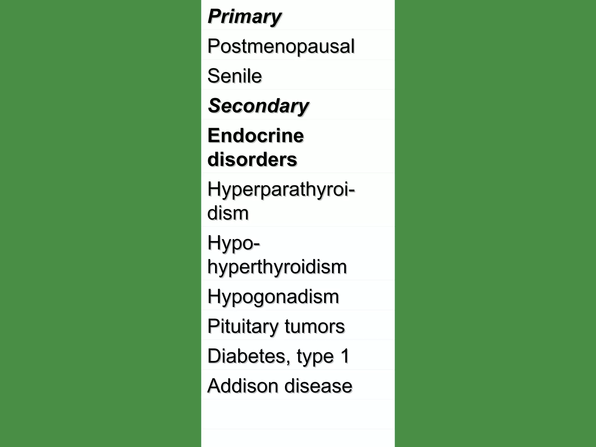
BONE & DEGENERATIVE DISORDERS DEPARTMENT OF PATHOLOGY www.freelivedoctor.com
O STEOPOROSIS Decreased  b one mass (  o steopenia.)  r esulting in thin,fragile bones that are susceptible to fracture. Most common bone disorder in  U.S.A.(about 15 million individuals have primary type)   Most commonly occurs in postmenopausal Caucasian women and the elderly. www.freelivedoctor.com
www.freelivedoctor.com Primary Postmenopausal Senile Secondary Endocrine disorders Hyperparathyroi-dism Hypo-hyperthyroidism Hypogonadism Pituitary tumors Diabetes, type 1 Addison disease
www.freelivedoctor.com Neoplasia Multiple myeloma Carcinomatosis Gastrointestinal Malnutrition Malabsorption Hepatic insufficiency Vitamin C, D deficiencies Idiopathic
www.freelivedoctor.com Rheumatologic disease Drugs Anticoagulants Chemotherapy Corticosteroids Anticonvulsants Alcohol Miscellaneous Osteogenesis imperfecta Immobilization Pulmonary disease Homocystinuria Anemia
www.freelivedoctor.com
O STEOPOROSIS(cont.) Patients may experience bone pain and fractures. Weight bearing bones are predisposed to fractures Vertebrae ( compression fractures) Femoral neck (  h ip fracture) Distal radius ( Colles fracture) Pulmonary embolization www.freelivedoctor.com
www.freelivedoctor.com
O STEOPOROSIS(cont.) Loss of height ,  kyphos coliosis, lordosis X-rays: generalized radiolucency of bone (  osteopenia ) Dual Energy X Ray Absorptiometry  ( DEXA.) NORMAL SERUM CALCIUM,PHOSPHORUS,and ALKALINE PHOSPHATASE. Micro:  t hinned cortical and trabecular bone  Treatment :  e strogen replacement therapy ,  w eight bearing exercise,Calcium and Vitamin D ,  Biphosphonate ( Alendro  nate)  ,  Calcitonin.  www.freelivedoctor.com
www.freelivedoctor.com
O STEOMALACIA  and R ICKETS Both diseases are characterized by decreased mineralization of newly formed bone, usually caused by deficiency or abnormal metabolism of vitamin D.  www.freelivedoctor.com
www.freelivedoctor.com
O STEOMALACIA  and R ICKETS .  Etiology:  d ietary deficiency of Vitamin D Intestinal malabsorption (   bilis,pancreatic insuff.,celiac sprue,regional enteritis) Lack of sunlight Renal and liver disease Chronic use of antacids(Al OH binds to P) Drugs(incr.rate of degradation of sterols:phe  nytoin,phenobarbital,rifampin) www.freelivedoctor.com
R ICKETS  ( Children) Prior to closure of the epiphyses Both remodeled bone and bone formed at the epypheseal growth plate are undermineralized Enchondral bone fromation is affected leading to skeletal deformities. www.freelivedoctor.com
R ICKETS  (children)  Craniotabes and frontal bossing: skull deformities. Rachitic rosary: deformity of the chest wall as a result of an overgrowth of cartilage at the costochondral junction Pectus carinatum ( pigeon breast deformiy); outward protrusion of the sternum. Lumbar lordosis:  s pinal curvature Bowing of the legs:  c urvature of femur, tibia due to weight bearing Fractures may also occur. www.freelivedoctor.com
O STEOMALACIA . ( Adults)  Impaired mineralization of the osteoid matrix results in thin, fragile bones that are susceptible to fracture. www.freelivedoctor.com
O STEOMALACIA  ( Adults)  Clinical  p resentation: -- Bone pain -- Fractures of the vertebrae, hips and wrist. -- X-rays  :  diffuse radiolucency of bone (osteo penia ) --Lab: l ow serum calcium and phosphorus  and/or raised  alkaline phosphatase www.freelivedoctor.com
B ONE  I NFECTIONS . O STEOMYELITIS ,TB, Syphilis. P YOGENIC  O STEOMYELITIS . Routes of infection:  Hematogenous spread : Most  common     seeding of bone after bacteremia, commonly affects the metaphysis. www.freelivedoctor.com
www.freelivedoctor.com
O STEOMYELITIS (cont . ) Routes of Infection: Hematogenous: from skin pustule,infected gums/teeth,IV puncture, UTIs, urologic procedures  ends of long bones Direct inoculation Spread from an adjacent site of infection Microbiology:Syaphylococcus aureus, ( m ost common), Escherichia coli, Streptococci,Gonococci, Haemophilus influenzae, Salmonella (common in sickle cell disease) Pseudomonas ( common in intravenous drug abusers and diabetics ) www.freelivedoctor.com
O STEOMYELITIS (cont.) Clinical features:  f ever , localized pain, erythema and swelling, leukocytosis   X-ray :  m ay be normal for up to 2 weeks , but it  m ay initially show periosteal elevation Later: l ytic focus with surrounding sclerosis. www.freelivedoctor.com
O STEOMYELITIS (cont . ) Pathology:  s uppurative  i nflammation Vascular insufficiency Ischemic necrosis of bone Sequestrum:  necrotic bone. Involucrum:  n ew bone formation that surrounds the sequestrum www.freelivedoctor.com
O STEOMYELITIS(cont.) Diagnosis:  Blood culture. Bone biopsy and culture Treatment: Antibiotics:+  s urgical drainage. www.freelivedoctor.com
www.freelivedoctor.com
O STEOMYELITIS(cont.) Complications: Fracture Intraosseous ( Brodie) abscess. Amyloidosis Sinus tract formation Squamous cell Ca.  o f the skin at the site of persistent draining sinus tract Osteogenic sarcoma ( rare. )  www.freelivedoctor.com
T B  O STEOMYELITIS . Affected individuals : adolescents or young adults, inmigrants. U.S.A :   V ictims tend to be older, excepting  I mmunosupressed.  Occurs in 1% of cases of TB. www.freelivedoctor.com
www.freelivedoctor.com
T B  O STEOMYELITIS(cont.) Pain or tenderness, fever, night sweats, weight loss. Caseating granulomas with extensive destruction of the bones. Common site : Thora c ic and lumbar vertebrae ( Pott ´s  Disease)  www.freelivedoctor.com
T B  O STEOMYELITIS(cont.) Complications. Vertebral compression fracture Psoas abscesses Amyloidosis www.freelivedoctor.com
S KELETAL  S YPHILIS Infrequent. Congenital : 5th month of gestation  Fully developed at birth. Osteochondritis Periostitis Acquired syphilis: Involvement of the bone in the tertiary stage. www.freelivedoctor.com
S KELETAL  S YPHILIS(cont.) 2-5 years after the infection. Bones involved: Nose, palate, skull and extremities.( Saber shin) Histology: Edematous granulation tissue , plasma cells and necrotic bone Gummata Silver stain for spirochetes. www.freelivedoctor.com
Paget´s disease(Osteitis deform.) Begins after 40´s and is commonly seen in caucasians(3%), with some hereditary predisposition(autosomal dominant trait) Paramyxovirus may have a role as cause of the disease showing 2 forms of presentation  a. Localized in tibia, femur, iliac, humerus, vertebrae & skull(monostotic) in 15% of cases.  b. Localized in several bones(polyostotic) in 85% of cases. www.freelivedoctor.com
Paget´s disease(Osteitis deform.) Pathology.  There are 3 stages:  --Osteolytic  --Osteolytic-osteoblastic  --Osteosclerotic(“cold” stage) Micro:  --Thickened trabecular & cortical bone  --Abnormal arrangement of lamellar bone  (mosaic pattern)  --Woven(reactive) bone  --Giant multinucleated osteoclasts(w/viral incl.)  --Fibrotic marrow  www.freelivedoctor.com
www.freelivedoctor.com
Paget´s disease(Osteitis deform.) Complications.  --Myelophthisic anemia  --Optic & auditory nerve compression  --Fx.  --Cardiac failure(high output due to large A/V shunts from blood vessels in fibrotic marrow  --Malignancies(about 1% of cases)  osteo  genic sarcoma, fibrosarcoma(jaw,femur, pelvis) www.freelivedoctor.com
www.freelivedoctor.com
BONE & DEGENERATIVE  DISORDERS There are 206 bones diverse in size & shape that have a role in mineral homeostasis, contains hematopoietic elements, determine the body size/shape and provide support for movement and protection. Long bones have - Epiphysis  - Epiphysial plate - Metaphysis  - Diaphysis(shaft) www.freelivedoctor.com
BONE & DEGENERATIVE  DISORDERS Also, a bone shows 2 gross anatomical forms in proportion to a particular function: 1. Compact bone:dense outer shell(cortex) with basic units(osteons) arranged in vertical columns and lamellar structure that contain osteocytes & capillaries forming a haversyan system. 2. Cancellous bone(spongious) arranged in trabecules(osteocytes+osteoblasts+capillaries) along with marrow www.freelivedoctor.com
BONE & DEGENERATIVE  DISORDERS 3. Woven bone(non-lamellar), a primitive form laid down in fetal development, with irregular trabecula in a primitive matrix. In adult life this type is seen in bone regeneration(later replaced by lamellar bone) and tumors www.freelivedoctor.com
www.freelivedoctor.com
BONE & DEGENERATIVE  DISORDERS Bone cells constitute about 2% of bone weight: Osteoprogenitor(STEM) cells in vacinity of surfaces  Osteoblasts in surface of bones(synthesis&  arrange of proteins) w/receptors for hormo  nes, GFs Osteocytes(control of Ca++, P) Multinucleated osteoclasts(from granulocyte-mo  nocyte precursor in marrow) with resorption pits  (Howship´s lacunae) www.freelivedoctor.com
BONE & DEGENERATIVE  DISORDERS Bone proteins include type I collagen(90% of organic component) and non-collagenous proteins derived from osteoblasts  matrix Bone is a blend of organic matrix(35%)and inorganic(65%) Inorganic components are:Ca++hydroxyapa  tite(stores 99% of body Ca++, 80% of P and 65% of Na & Mg) www.freelivedoctor.com
BONE & DEGENERATIVE  DISORDERS BONE DEPOSITION.  - From rows of osteoblasts laying down lamellar collagen matrix  incorporated into the bone and housed in small lacuna as osteocyte  non-calcified osteoid in the centre of the osteon/surface of trabecula. Normally calcification follows very quickly  -During osteoblastic activity ALKALINE PHOSPATASE is liberated into blood flow  www.freelivedoctor.com
BONE & DEGENERATIVE  DISORDERS Bone & calcium homeostasis:  -  parathyroids blood Ca++  PTH  blood Ca++    bone (osteocyte rel/Ca++  www.freelivedoctor.com
BONE & DEGENERATIVE  DISORDERS MINERAL HOMEOSTASIS.  1. PTH in parathyroid glands  Ca++    PO++++  2. 1.25(OH2)D3 in proximal conv.& straight tubules  Ca+++    PO++++  3. Calcitonin in “C” cells  Ca++    PO++++ www.freelivedoctor.com

Bone degenerative disorders

  • 1.
    BONE & DEGENERATIVEDISORDERS DEPARTMENT OF PATHOLOGY www.freelivedoctor.com
  • 2.
    O STEOPOROSIS Decreased b one mass ( o steopenia.) r esulting in thin,fragile bones that are susceptible to fracture. Most common bone disorder in U.S.A.(about 15 million individuals have primary type) Most commonly occurs in postmenopausal Caucasian women and the elderly. www.freelivedoctor.com
  • 3.
    www.freelivedoctor.com Primary PostmenopausalSenile Secondary Endocrine disorders Hyperparathyroi-dism Hypo-hyperthyroidism Hypogonadism Pituitary tumors Diabetes, type 1 Addison disease
  • 4.
    www.freelivedoctor.com Neoplasia Multiplemyeloma Carcinomatosis Gastrointestinal Malnutrition Malabsorption Hepatic insufficiency Vitamin C, D deficiencies Idiopathic
  • 5.
    www.freelivedoctor.com Rheumatologic diseaseDrugs Anticoagulants Chemotherapy Corticosteroids Anticonvulsants Alcohol Miscellaneous Osteogenesis imperfecta Immobilization Pulmonary disease Homocystinuria Anemia
  • 6.
  • 7.
    O STEOPOROSIS(cont.) Patientsmay experience bone pain and fractures. Weight bearing bones are predisposed to fractures Vertebrae ( compression fractures) Femoral neck ( h ip fracture) Distal radius ( Colles fracture) Pulmonary embolization www.freelivedoctor.com
  • 8.
  • 9.
    O STEOPOROSIS(cont.) Lossof height , kyphos coliosis, lordosis X-rays: generalized radiolucency of bone ( osteopenia ) Dual Energy X Ray Absorptiometry ( DEXA.) NORMAL SERUM CALCIUM,PHOSPHORUS,and ALKALINE PHOSPHATASE. Micro: t hinned cortical and trabecular bone Treatment : e strogen replacement therapy , w eight bearing exercise,Calcium and Vitamin D , Biphosphonate ( Alendro nate) , Calcitonin. www.freelivedoctor.com
  • 10.
  • 11.
  • 12.
  • 13.
  • 14.
    O STEOMALACIA and R ICKETS Both diseases are characterized by decreased mineralization of newly formed bone, usually caused by deficiency or abnormal metabolism of vitamin D. www.freelivedoctor.com
  • 15.
  • 16.
    O STEOMALACIA and R ICKETS . Etiology: d ietary deficiency of Vitamin D Intestinal malabsorption ( bilis,pancreatic insuff.,celiac sprue,regional enteritis) Lack of sunlight Renal and liver disease Chronic use of antacids(Al OH binds to P) Drugs(incr.rate of degradation of sterols:phe nytoin,phenobarbital,rifampin) www.freelivedoctor.com
  • 17.
    R ICKETS ( Children) Prior to closure of the epiphyses Both remodeled bone and bone formed at the epypheseal growth plate are undermineralized Enchondral bone fromation is affected leading to skeletal deformities. www.freelivedoctor.com
  • 18.
    R ICKETS (children) Craniotabes and frontal bossing: skull deformities. Rachitic rosary: deformity of the chest wall as a result of an overgrowth of cartilage at the costochondral junction Pectus carinatum ( pigeon breast deformiy); outward protrusion of the sternum. Lumbar lordosis: s pinal curvature Bowing of the legs: c urvature of femur, tibia due to weight bearing Fractures may also occur. www.freelivedoctor.com
  • 19.
    O STEOMALACIA .( Adults) Impaired mineralization of the osteoid matrix results in thin, fragile bones that are susceptible to fracture. www.freelivedoctor.com
  • 20.
    O STEOMALACIA ( Adults) Clinical p resentation: -- Bone pain -- Fractures of the vertebrae, hips and wrist. -- X-rays : diffuse radiolucency of bone (osteo penia ) --Lab: l ow serum calcium and phosphorus and/or raised alkaline phosphatase www.freelivedoctor.com
  • 21.
    B ONE I NFECTIONS . O STEOMYELITIS ,TB, Syphilis. P YOGENIC O STEOMYELITIS . Routes of infection: Hematogenous spread : Most common  seeding of bone after bacteremia, commonly affects the metaphysis. www.freelivedoctor.com
  • 22.
  • 23.
  • 24.
    O STEOMYELITIS (cont. ) Routes of Infection: Hematogenous: from skin pustule,infected gums/teeth,IV puncture, UTIs, urologic procedures  ends of long bones Direct inoculation Spread from an adjacent site of infection Microbiology:Syaphylococcus aureus, ( m ost common), Escherichia coli, Streptococci,Gonococci, Haemophilus influenzae, Salmonella (common in sickle cell disease) Pseudomonas ( common in intravenous drug abusers and diabetics ) www.freelivedoctor.com
  • 25.
    O STEOMYELITIS (cont.)Clinical features: f ever , localized pain, erythema and swelling, leukocytosis X-ray : m ay be normal for up to 2 weeks , but it m ay initially show periosteal elevation Later: l ytic focus with surrounding sclerosis. www.freelivedoctor.com
  • 26.
    O STEOMYELITIS (cont. ) Pathology: s uppurative i nflammation Vascular insufficiency Ischemic necrosis of bone Sequestrum: necrotic bone. Involucrum: n ew bone formation that surrounds the sequestrum www.freelivedoctor.com
  • 27.
    O STEOMYELITIS(cont.) Diagnosis: Blood culture. Bone biopsy and culture Treatment: Antibiotics:+ s urgical drainage. www.freelivedoctor.com
  • 28.
  • 29.
  • 30.
    O STEOMYELITIS(cont.) Complications:Fracture Intraosseous ( Brodie) abscess. Amyloidosis Sinus tract formation Squamous cell Ca. o f the skin at the site of persistent draining sinus tract Osteogenic sarcoma ( rare. ) www.freelivedoctor.com
  • 31.
    T B O STEOMYELITIS . Affected individuals : adolescents or young adults, inmigrants. U.S.A : V ictims tend to be older, excepting I mmunosupressed. Occurs in 1% of cases of TB. www.freelivedoctor.com
  • 32.
  • 33.
  • 34.
  • 35.
  • 36.
  • 37.
  • 38.
  • 39.
    T B O STEOMYELITIS(cont.) Pain or tenderness, fever, night sweats, weight loss. Caseating granulomas with extensive destruction of the bones. Common site : Thora c ic and lumbar vertebrae ( Pott ´s Disease) www.freelivedoctor.com
  • 40.
    T B O STEOMYELITIS(cont.) Complications. Vertebral compression fracture Psoas abscesses Amyloidosis www.freelivedoctor.com
  • 41.
    S KELETAL S YPHILIS Infrequent. Congenital : 5th month of gestation Fully developed at birth. Osteochondritis Periostitis Acquired syphilis: Involvement of the bone in the tertiary stage. www.freelivedoctor.com
  • 42.
    S KELETAL S YPHILIS(cont.) 2-5 years after the infection. Bones involved: Nose, palate, skull and extremities.( Saber shin) Histology: Edematous granulation tissue , plasma cells and necrotic bone Gummata Silver stain for spirochetes. www.freelivedoctor.com
  • 43.
    Paget´s disease(Osteitis deform.)Begins after 40´s and is commonly seen in caucasians(3%), with some hereditary predisposition(autosomal dominant trait) Paramyxovirus may have a role as cause of the disease showing 2 forms of presentation a. Localized in tibia, femur, iliac, humerus, vertebrae & skull(monostotic) in 15% of cases. b. Localized in several bones(polyostotic) in 85% of cases. www.freelivedoctor.com
  • 44.
    Paget´s disease(Osteitis deform.)Pathology. There are 3 stages: --Osteolytic --Osteolytic-osteoblastic --Osteosclerotic(“cold” stage) Micro: --Thickened trabecular & cortical bone --Abnormal arrangement of lamellar bone (mosaic pattern) --Woven(reactive) bone --Giant multinucleated osteoclasts(w/viral incl.) --Fibrotic marrow www.freelivedoctor.com
  • 45.
  • 46.
    Paget´s disease(Osteitis deform.)Complications. --Myelophthisic anemia --Optic & auditory nerve compression --Fx. --Cardiac failure(high output due to large A/V shunts from blood vessels in fibrotic marrow --Malignancies(about 1% of cases)  osteo genic sarcoma, fibrosarcoma(jaw,femur, pelvis) www.freelivedoctor.com
  • 47.
  • 48.
  • 49.
  • 50.
  • 51.
  • 52.
  • 53.
    BONE & DEGENERATIVE DISORDERS There are 206 bones diverse in size & shape that have a role in mineral homeostasis, contains hematopoietic elements, determine the body size/shape and provide support for movement and protection. Long bones have - Epiphysis - Epiphysial plate - Metaphysis - Diaphysis(shaft) www.freelivedoctor.com
  • 54.
    BONE & DEGENERATIVE DISORDERS Also, a bone shows 2 gross anatomical forms in proportion to a particular function: 1. Compact bone:dense outer shell(cortex) with basic units(osteons) arranged in vertical columns and lamellar structure that contain osteocytes & capillaries forming a haversyan system. 2. Cancellous bone(spongious) arranged in trabecules(osteocytes+osteoblasts+capillaries) along with marrow www.freelivedoctor.com
  • 55.
    BONE & DEGENERATIVE DISORDERS 3. Woven bone(non-lamellar), a primitive form laid down in fetal development, with irregular trabecula in a primitive matrix. In adult life this type is seen in bone regeneration(later replaced by lamellar bone) and tumors www.freelivedoctor.com
  • 56.
  • 57.
  • 58.
    BONE & DEGENERATIVE DISORDERS Bone cells constitute about 2% of bone weight: Osteoprogenitor(STEM) cells in vacinity of surfaces Osteoblasts in surface of bones(synthesis& arrange of proteins) w/receptors for hormo nes, GFs Osteocytes(control of Ca++, P) Multinucleated osteoclasts(from granulocyte-mo nocyte precursor in marrow) with resorption pits (Howship´s lacunae) www.freelivedoctor.com
  • 59.
    BONE & DEGENERATIVE DISORDERS Bone proteins include type I collagen(90% of organic component) and non-collagenous proteins derived from osteoblasts  matrix Bone is a blend of organic matrix(35%)and inorganic(65%) Inorganic components are:Ca++hydroxyapa tite(stores 99% of body Ca++, 80% of P and 65% of Na & Mg) www.freelivedoctor.com
  • 60.
    BONE & DEGENERATIVE DISORDERS BONE DEPOSITION. - From rows of osteoblasts laying down lamellar collagen matrix  incorporated into the bone and housed in small lacuna as osteocyte  non-calcified osteoid in the centre of the osteon/surface of trabecula. Normally calcification follows very quickly -During osteoblastic activity ALKALINE PHOSPATASE is liberated into blood flow www.freelivedoctor.com
  • 61.
    BONE & DEGENERATIVE DISORDERS Bone & calcium homeostasis: - parathyroids blood Ca++ PTH blood Ca++  bone (osteocyte rel/Ca++ www.freelivedoctor.com
  • 62.
    BONE & DEGENERATIVE DISORDERS MINERAL HOMEOSTASIS. 1. PTH in parathyroid glands Ca++ PO++++ 2. 1.25(OH2)D3 in proximal conv.& straight tubules Ca+++ PO++++ 3. Calcitonin in “C” cells Ca++ PO++++ www.freelivedoctor.com