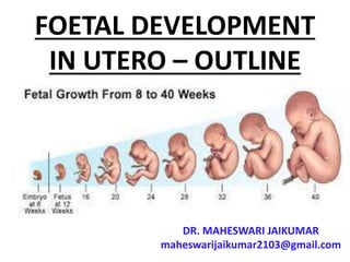
FOETAL DEVELOPMENT
IN UTERO – OUTLINE
DR. MAHESWARI JAIKUMAR
maheswarijaikumar2103@gmail.com
PRIMER ON CONCEPTION
• Each month a group of eggs
(called oocytes) is recruited from
the ovary for ovulation (release
of the egg)
• The eggs develop in small fluid-
filled cysts called follicles
• Normally, one follicle in the
group is selected to complete
maturation
• This dominant follicle suppresses
all the other follicles in the
group, which stop growing and
degenerate
• The mature follicle opens and
releases the egg from the ovary
(ovulation)
• Ovulation generally occurs about
two weeks before a woman's
next menstrual period begins
After ovulation, the ruptured
follicle develops into a structure
called the corpus luteum, which
secretes progesterone and
estrogen
• The progesterone helps prepare
the endometrium (lining of the
uterus) for the embryo to
implant
• On average, fertilization occurs
about two weeks after your last
menstrual period
• When the sperm penetrates the
egg, changes occur in the protein
coating around it to prevent
other sperm from entering
FERTILIZATION
• At the moment of fertilization,
your baby's genetic make-up is
complete, including its gender
• If a Y sperm fertilizes the egg,
your baby will be a boy; if an X
sperm fertilizes the egg, your
baby will be a girl
• Human chorionic gonadotrophin
(hCG) is a hormone present in
your blood from the time of
conception
• It is produced by cells that form
the placenta and is the hormone
detected in a pregnancy test
• However, it usually takes three
to four weeks from the first day
of your last period for the hCG to
increase enough to be detected
by pregnancy tests
• Within 24 hours after
fertilization, the egg begins
dividing rapidly into many cells
• It remains in the fallopian tube
for about three days
• The fertilized egg (called a
blastocyte) continues to divide
as it passes slowly through the
fallopian tube to the uterus
where its next job is to attach to
the endometrium (a process
called implantation)
• Before this happens, the
blastocyte breaks out of its
protective covering
• When the blastocyte establishes
contact with the endometrium,
an exchange of hormones helps
the blastocyte attach
• Some women notice spotting (or
slight bleeding) for one or two
days around the time of
implantation
• The endometrium becomes
thicker and the cervix is sealed
by a plug of mucus
• Within three weeks, the
blastocyte cells ultimately form a
little ball, or an embryo, and the
baby's first nerve cells have
already formed
• The developing baby is called an
embryo from the moment of
conception to the eighth week of
pregnancy
• After the eighth week and until
the moment of birth, the
developing baby is called a fetus
• The development stages of
pregnancy are called trimesters,
or three-month periods, because
of the distinct changes that occur
in each stage
8 WEEK FOETUS
• Foetal growth and maturation
from the 9th week foetal growth
is rapid
• Tissues grow by cell
proliferation, cell enlargement
and accretion of extracellular
material
• An adequate supply of nutrients
and oxygen from the placenta to
the foetus is crucial for growth
• As foetal is an indicator of foetal
health and wellbeing,
monitoring of growth is crucial
• This is done by visual
observation of the uterus for
size, fundal height measurement
and ultrasonograpy
0-4 WEEKS
• Primitive streak appears
• Somebody systems laid down in
primitive form
• Primitive central nervous system
forms
• Heart develops and begins to
beat
• Covered with a layer of skin
• Limb buds form gender
determined
4-8 WEEKS
•Rapid cell division
•More body systems laid
down in primitive form
•Blood is pumped around the
vessels
•Lower respiratory system begins
•Head and facial features develop
•Early movements
•Visible on ultrasound from
6weeks.
8 WEEK FOETUS
8-12 WEEKS
•Rapid weight gain
•Eyelids fuse
•External genitalia present but
gender not distinguishable
9 WEEKS
•Finger nails develop
•Lanugo appears
•Some primitive reflexes
present
12-16 WEEKS
•Rapid skeletal development –
visible on x-ray
•Meconium present in gut
•Nasal septum and palate
fuse
•Gender distinguishable
16-20 WEEKS
•Constant weight gain
•Quickening mother feels
foetal movements
•Foetal heart sound heard
on auscultation
•Skin covered with lanugo
vermin caseosa is present
20-24 WEEKS
• Legally viable and survival
may be expected is born
• Eyelids open
17-20 WEEKS
•Most organs functioning
well
•Eyes complete
•Periods of sleep and
activity
•Ear apparatus developing
•Responds to sound
•Skin red and wrinkled
24-28 WEEKS
• Legally visible and survival may
be expected if born
• Eyelids open
• Respiratory movements
24 WEEKS
28-32 WEEKS
• Begins to store fat and iron
• Tests descend into scrotum
• Lanugo disappears from face skin
and is less wrinkled
32-36 WEEKS
•Testicle usually descend into
the scrotum
•One Lanugo tends to
disappear
• Head hair lengthens
32 WEEKS
36-40 WEEKS
•Both the testicle descends into
the scrotum
•Shape rounded
•Skull formed but soft and
palpable
40 WEEKS
GROWTH OF THE FOETUS
• Normal foetal growth is
characterised by cellular
hyperplasia followed by
hyperplasia and hypertrophy and
lastly by hypertrophy alone
• The foetal growth increases
linearly until 37th week
• It is controlled by genetic factor
in the first half and by
environmental factors in the
second half of pregnancy
IMPORTANT
PHYSIOLOGICAL FACTORS
•RACE : European babies are
heavier than Indians
GENDER
• Male baby weighs greater
Female baby
PARENTAL HEIGHT AND
WEIGHT
• Tall and Heavier mother have
heavier babies
BIRTH ORDER
• Weight rises from first to
second pregnancy
SOCIOECONOMIC FACTORS
• Heavier babies in higher socio
economic class
THANK YOU

FOETAL DEVELOPMENT - OUTLINE

  • 1.
    FOETAL DEVELOPMENT IN UTERO– OUTLINE DR. MAHESWARI JAIKUMAR maheswarijaikumar2103@gmail.com
  • 2.
    PRIMER ON CONCEPTION •Each month a group of eggs (called oocytes) is recruited from the ovary for ovulation (release of the egg) • The eggs develop in small fluid- filled cysts called follicles
  • 3.
    • Normally, onefollicle in the group is selected to complete maturation • This dominant follicle suppresses all the other follicles in the group, which stop growing and degenerate
  • 4.
    • The maturefollicle opens and releases the egg from the ovary (ovulation) • Ovulation generally occurs about two weeks before a woman's next menstrual period begins
  • 5.
    After ovulation, theruptured follicle develops into a structure called the corpus luteum, which secretes progesterone and estrogen
  • 6.
    • The progesteronehelps prepare the endometrium (lining of the uterus) for the embryo to implant
  • 7.
    • On average,fertilization occurs about two weeks after your last menstrual period • When the sperm penetrates the egg, changes occur in the protein coating around it to prevent other sperm from entering
  • 8.
  • 9.
    • At themoment of fertilization, your baby's genetic make-up is complete, including its gender • If a Y sperm fertilizes the egg, your baby will be a boy; if an X sperm fertilizes the egg, your baby will be a girl
  • 10.
    • Human chorionicgonadotrophin (hCG) is a hormone present in your blood from the time of conception • It is produced by cells that form the placenta and is the hormone detected in a pregnancy test
  • 11.
    • However, itusually takes three to four weeks from the first day of your last period for the hCG to increase enough to be detected by pregnancy tests
  • 12.
    • Within 24hours after fertilization, the egg begins dividing rapidly into many cells • It remains in the fallopian tube for about three days
  • 13.
    • The fertilizedegg (called a blastocyte) continues to divide as it passes slowly through the fallopian tube to the uterus where its next job is to attach to the endometrium (a process called implantation)
  • 14.
    • Before thishappens, the blastocyte breaks out of its protective covering • When the blastocyte establishes contact with the endometrium, an exchange of hormones helps the blastocyte attach
  • 15.
    • Some womennotice spotting (or slight bleeding) for one or two days around the time of implantation • The endometrium becomes thicker and the cervix is sealed by a plug of mucus
  • 16.
    • Within threeweeks, the blastocyte cells ultimately form a little ball, or an embryo, and the baby's first nerve cells have already formed
  • 17.
    • The developingbaby is called an embryo from the moment of conception to the eighth week of pregnancy
  • 18.
    • After theeighth week and until the moment of birth, the developing baby is called a fetus • The development stages of pregnancy are called trimesters, or three-month periods, because of the distinct changes that occur in each stage
  • 19.
  • 20.
    • Foetal growthand maturation from the 9th week foetal growth is rapid • Tissues grow by cell proliferation, cell enlargement and accretion of extracellular material
  • 21.
    • An adequatesupply of nutrients and oxygen from the placenta to the foetus is crucial for growth • As foetal is an indicator of foetal health and wellbeing, monitoring of growth is crucial
  • 22.
    • This isdone by visual observation of the uterus for size, fundal height measurement and ultrasonograpy
  • 23.
    0-4 WEEKS • Primitivestreak appears • Somebody systems laid down in primitive form • Primitive central nervous system forms
  • 25.
    • Heart developsand begins to beat • Covered with a layer of skin • Limb buds form gender determined
  • 26.
    4-8 WEEKS •Rapid celldivision •More body systems laid down in primitive form •Blood is pumped around the vessels
  • 28.
    •Lower respiratory systembegins •Head and facial features develop •Early movements •Visible on ultrasound from 6weeks.
  • 29.
  • 30.
    8-12 WEEKS •Rapid weightgain •Eyelids fuse •External genitalia present but gender not distinguishable
  • 31.
  • 32.
    •Finger nails develop •Lanugoappears •Some primitive reflexes present
  • 33.
    12-16 WEEKS •Rapid skeletaldevelopment – visible on x-ray •Meconium present in gut
  • 34.
    •Nasal septum andpalate fuse •Gender distinguishable
  • 35.
    16-20 WEEKS •Constant weightgain •Quickening mother feels foetal movements
  • 36.
    •Foetal heart soundheard on auscultation •Skin covered with lanugo vermin caseosa is present
  • 37.
    20-24 WEEKS • Legallyviable and survival may be expected is born • Eyelids open
  • 38.
  • 39.
    •Most organs functioning well •Eyescomplete •Periods of sleep and activity
  • 40.
    •Ear apparatus developing •Respondsto sound •Skin red and wrinkled
  • 41.
    24-28 WEEKS • Legallyvisible and survival may be expected if born • Eyelids open • Respiratory movements
  • 42.
  • 43.
    28-32 WEEKS • Beginsto store fat and iron • Tests descend into scrotum • Lanugo disappears from face skin and is less wrinkled
  • 44.
    32-36 WEEKS •Testicle usuallydescend into the scrotum •One Lanugo tends to disappear • Head hair lengthens
  • 45.
  • 46.
    36-40 WEEKS •Both thetesticle descends into the scrotum •Shape rounded •Skull formed but soft and palpable
  • 47.
  • 48.
    GROWTH OF THEFOETUS • Normal foetal growth is characterised by cellular hyperplasia followed by hyperplasia and hypertrophy and lastly by hypertrophy alone
  • 49.
    • The foetalgrowth increases linearly until 37th week • It is controlled by genetic factor in the first half and by environmental factors in the second half of pregnancy
  • 50.
    IMPORTANT PHYSIOLOGICAL FACTORS •RACE :European babies are heavier than Indians
  • 51.
    GENDER • Male babyweighs greater Female baby
  • 52.
    PARENTAL HEIGHT AND WEIGHT •Tall and Heavier mother have heavier babies
  • 53.
    BIRTH ORDER • Weightrises from first to second pregnancy
  • 54.
    SOCIOECONOMIC FACTORS • Heavierbabies in higher socio economic class
  • 55.