Download to read offline
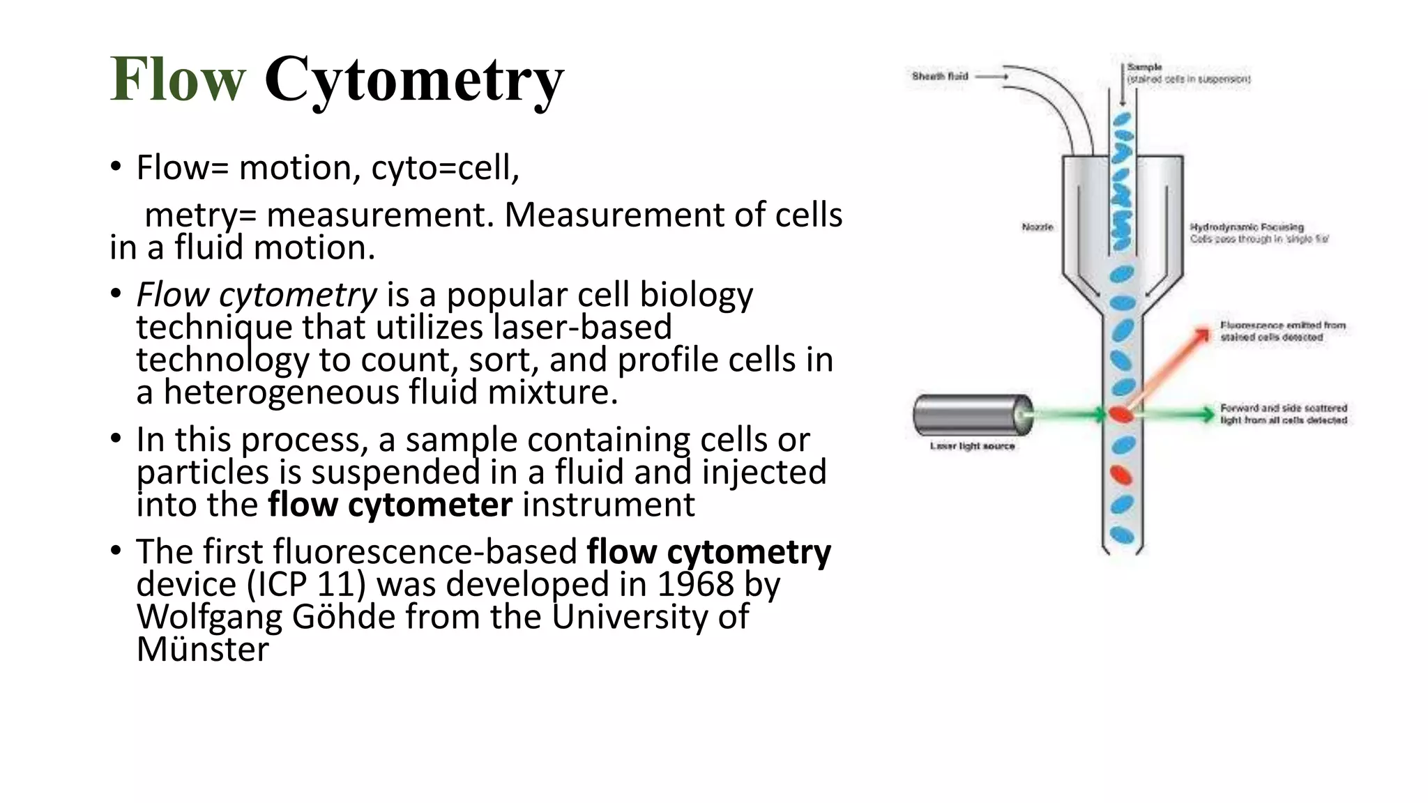
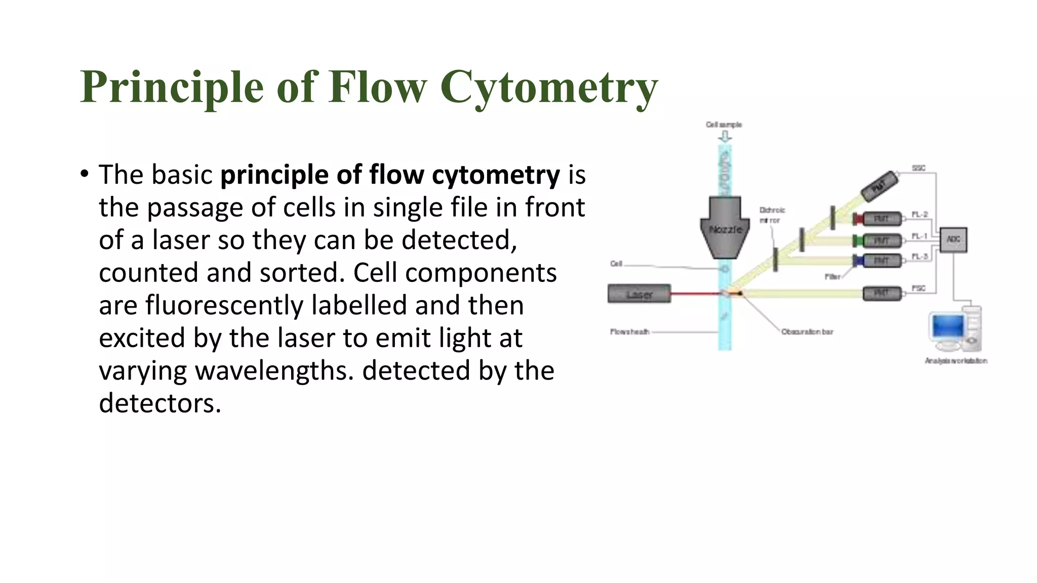
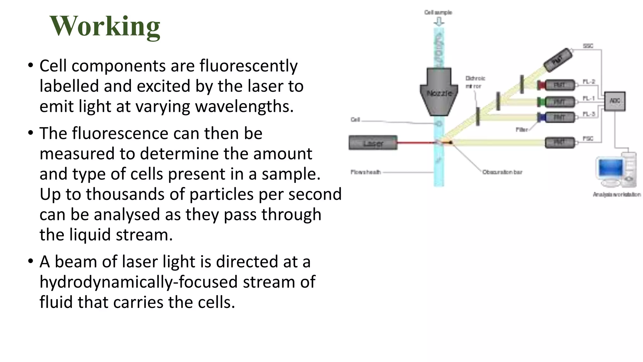


Flow cytometry is a laser-based technique used to measure and analyze cells in a fluid mixture, developed initially in 1968. It works by exciting fluorescently labeled cell components with a laser to measure scattered and emitted light, providing information on cell types and structures. The technique is vital for applications such as immunophenotyping, malignancy detection, and cell cycle analysis, though it has limitations, including high costs and dependence on skilled technicians.


![FlowBasics2[1]](https://cdn.slidesharecdn.com/ss_thumbnails/7f56678c-0f61-43d6-bbfe-d51ebe159eed-160219222349-thumbnail.jpg?width=640&height=640&fit=bounds)


