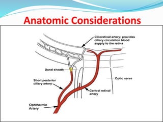
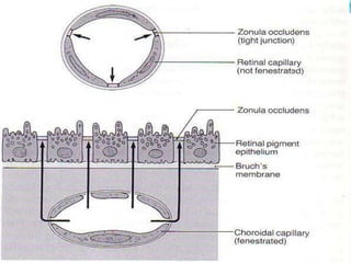
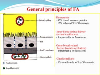
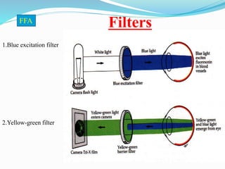
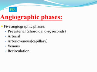
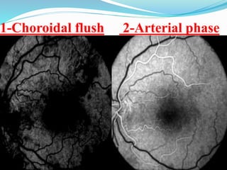
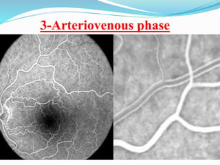
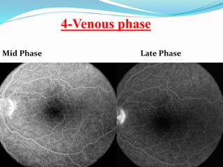
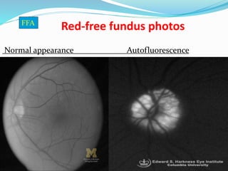
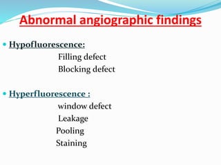
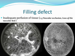
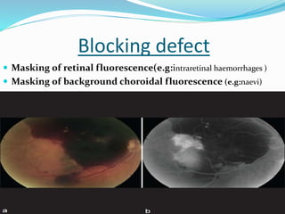
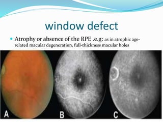
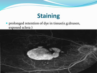
Mohamed Abdel-Aziz (MSc,ICO,Egyption Board)
Mataryia Teaching Hospital
 Fluorescence:
is the property of certain
molecules to emit light of
a longer wavelength when
stimulated by light of a
shorter wavelength.
FFA
Characteristics of Fluorescein
 Alkaline solution
 Highly fluorescent
 Absorbs blue light (490 nm)
 Emits yellow-green (530nm)
 Removal from blood by kidneys and liver within
24-48 hrs.
 injection of 5 cc of 10% Na Fl, or 3 cc of 25%
solution
FFA
Hazards
 Minimal relatively safe drug
 Temporary tan skin color
 Fl. Urine discoloration
 Transient nausea and vomiting
 Laryngeal edema
 Rarely – Syncope, resp. or Cardiac arrest
 Rx – I.V. Cortisone
 A physician in the 1st few minutes
FFA
Anatomic Considerations
FFA
General principles of FA
Fluorescein
• 85% bound to serum proteins
• 15% unbound ‘free’ fluorescein
• Impermeable to fluorescein
Outer blood-retinal
barrier (zonula occludens)
• Impermeable to fluorescein
Choriocapillaris
• Permeable only to ‘free’ fluorescein
Inner blood-retinal barrier
(retinal capillaries)
Filters
1.Blue excitation filter
2.Yellow-green filter
FFA
Angiographic phases
Angiographic phases:
 Five angiographic phases:
• Pre arterial (choroidal 9-15 seconds)
• Arterial
• Arteriovenous(capillary)
• Venous
• Recirculation
FFA
1-Choroidal flush 2-Arterial phase
3-Arteriovenous phase
4-Venous phase
Mid Phase Late Phase
Interpretation of FA
Red-free fundus photos
Normal appearance Autofluorescence
FFA
Abnormal angiographic findings
 Hypofluorescence:
Filling defect
Blocking defect
 Hyperfluorescence :
window defect
Leakage
Pooling
Staining
Hypofluorescence
Filling defect
 Inadequate perfusion of tissue (e.g:Vascular occlusion, Loss of the
vascular bed )
Blocking defect
 Masking of retinal fluorescence(e.g:intraretinal haemorrhages )
 Masking of background choroidal fluorescence (e.g:naevi)
Hyperfluorescence
window defect
 Atrophy or absence of the RPE .e.g: as in atrophic age-
related macular degeneration, full-thickness macular holes
Pooling
 breakdown of the outer blood–retinal barrier (RPE tight
junctions)
 A) In the sub-retinal space: increases in intensity and
area, the maximum extent remaining relatively well-
defined.(e.g:CSR)
Pooling
 B ) In the sub-RPE space: early hyper-fluorescence which
increases in intensity but not in size .(e.g:PED)
Leakage
 early hyper-fluorescence which increases in both area and
intensity
 A) Dysfunction or loss of existing vascular endothelial
tight junctions(e.g:CME)
Leakage
 B) Primary absence of vascular endothelial tight
junctions (e.g:CNV,PDR)
Staining
 prolonged retention of dye in tissue(e.g:drusen,
exposed sclera )
Systematic approach to reporting
angiograms
a- Patient's age and gender.
b-Right or left eyes.
c-Comment on any color and red free images.
d -Timing of filling, especially arm-to-
eye transit time.
e-Briefly scan through the sequence of images
Eye Flourecein Angiography

Eye Flourecein Angiography