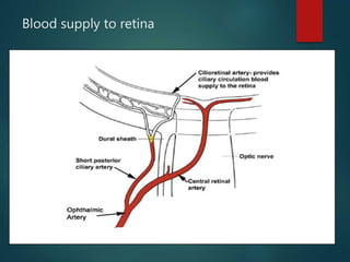
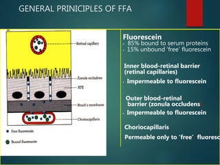
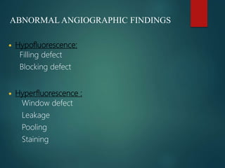
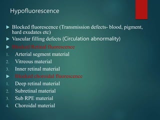
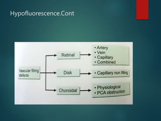
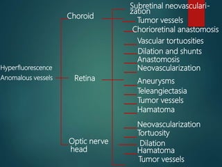
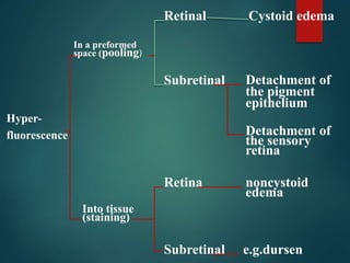
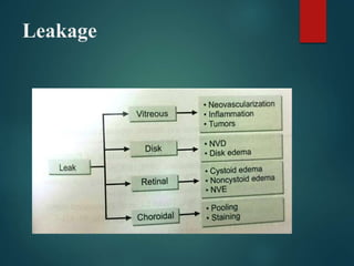
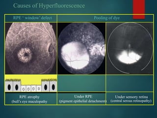
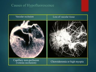
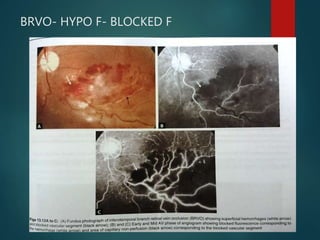
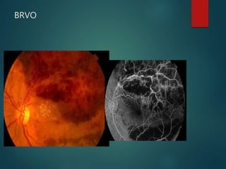
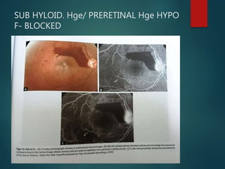
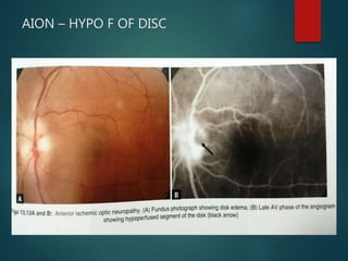
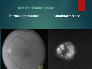
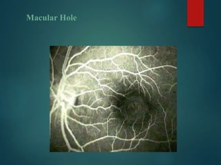
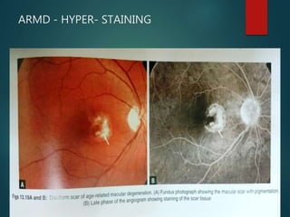
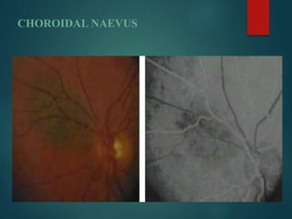
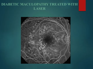
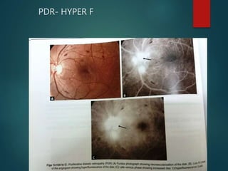
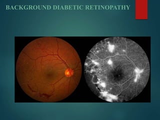
Fundus Fluorescein
Angiography
Dr Md Afzal Mahfuzullah
MCPS,FCPS,Felow Vitreo-Retina
Retina Specialist & Surgeon
Bangabandhu Sheikh Mujib Medical University
WHY FFA
 To confirms the elements already revealed by clinical
examination.
 Flow characteristics in the blood vessels as the dye
reaches and circulates through the retina and choroid.
 Gives a clear picture of the retinal vessels and assessment
of their functional integrity.
 To monitor the disease intensity and impact of therapy.
 Provides guidance for application of focal laser in
photocoagulation therapy.
 To detect the leakages without clinical manifestation of
edema
What are the informations we get from FFA
FFA reveals:
 Inflammatory status of retinal and choroidal blood
vessels.
 Localization of intra retinal lesions e.g. depth of
pathological involvement in DR.
 CNV
 Neovascularisation in disc or retina
FFA reveals: Cont
 Subretinal fluid status
 Cystoid macular edema
 Intraretinal/ preretinal haemorrhages
 Optic nerve disorders
 RPE integrity and disorders
 Choroidal neovessels and chorioretinal atrophy
BASIC PRINCIPLES:
BASIC PRINCIPLES:
 Based on luminescence and fluorescence.
 Luminescence – is the emission of light from any source
other than high temperature.
 Fluorescence is luminescence that is maintained only by
continuous excitation. Excitation occurs at one wave length
and immediate emission occurs through a longer wave
length.
FLUORESCENCE
 Refers to fluorescein sodium (C20H10Na2O5)
 A brown or orange red crystalline substance, alkaline in
nature.
 MW- 376 dalton
 Readily diffuses through body fluids and choriocapillaries
but does not diffuse through vascular endothelial cells
and RPE (Blood retina barriers)
OPTICAL PRINCIPLE
 Absorbs blue light (465-
490nm ) and Emits
yellow-green light (520-
530nm)
 Metabolized by liver and
exerted by kidney
FILTERS
1.Blue excitation filter
2.Yellow-green filter
Blood supply to retina
Layers & blood supply
GENERAL PRINICIPLES OF FFA
Fluorescein
• 85% bound to serum proteins
• 15% unbound ‘free’ fluorescein
• Impermeable to fluorescein
Outer blood-retinal
barrier (zonula occludens)
• Impermeable to fluorescein
Choriocapillaris
Permeable only to ‘free’ fluoresce
Inner blood-retinal barrier
(retinal capillaries)
Circulation of NaF
Dye injected from peripheral vein
Venous circulation
Heart
Arterial system
INTERNAL CAROTID ARTERY
Ophthalmic artery
Short posterior ciliary artery) Central retinal artery
(choroidal circulation.) ( retinal circulation)
N.B. The choroidal filling is 1 second prior to the retinal filling.
ANGIOGRAPHIC PHASES
 Five angiographic phases:
• Pre arterial (Choroidal 9-15 seconds)
• Arterial
• Arteriovenous(capillary)
• Venous
• Recirculation
Basic anatomy :
 The inner retina contains the retinal blood vessels.
 The larger vessels in the Nerve fiber layer.
 The retinal capillaries in the Inner nuclear layer.
 Both are impermeable to dye.
 The outer retina is primarily interstitial space of the retina, where
hemorrhages, edematous fluid and hard exudates accumulate.
 In normal conditions this layer does contain NaF as because of RPE
tight junctions(Outer BRB)
 Large choroidal vessels do not leake NaF but choriocapillaris does
leak.
BASIC ANATOMY
PRE-ARTERIAL/ CHOROIDAL PHASE
 Choroidal flush
 Patchy Choroidal filling because of
lobular arrangements of
choriocapillaris
 10-12 sec in young
 12-15 sec in old
 Cilioretinal artery fills at the same
time with choroid circulation
 Macula remains dark due to tall
RPE and more pigments.
ARTERIAL PHASE
ARTERIAL PHASE Cont…
ARTERIOVENOUS PHASE
VENOUS PHASE
Recirculation phase
ABNORMAL ANGIOGRAPHIC FINDINGS
 Hypofluorescence:
Filling defect
Blocking defect
 Hyperfluorescence :
Window defect
Leakage
Pooling
Staining
Hypofluorescence
 Blocked fluorescence (Transmission defects- blood, pigment,
hard exudates etc)
 Vascular filling defects (Circulation abnormality)
 Blocked Retinal fluorescence
1. Arterial segment material
2. Vitreous material
3. Inner retinal material
 Blocked choroidal fluorescence
1. Deep retinal material
2. Subretinal material
3. Sub RPE material
4. Choroidal material
Hypofluorescence.Cont
Hyperfluorescence
Anomalous vessels
Choroid
Retina
Optic nerve
head
Subretinal neovasculari-
zation
Tumor vessels
Chorioretinal anastomosis
Vascular tortuosities
Dilation and shunts
Anastomosis
Neovascularization
Aneurysms
Teleangiectasia
Tumor vessels
Hamatoma
Neovascularization
Tortuosity
Dilation
Hamatoma
Tumor vessels
Hyper-
fluorescence
In a preformed
space (pooling)
Into tissue
(staining)
Retinal
Subretinal
Retina
Subretinal
Cystoid edema
Detachment of
the pigment
epithelium
Detachment of
the sensory
retina
noncystoid
edema
e.g.dursen
Leakage
Fluorescence
without the
administration
of fluorescein
Autofluorescence
Pseudofluorescence
Drusen of the optic
nerve head
Hamatoma
Scleral exudate
Myelinate nerve
Fibers, optic nerve drusen
Scar tissue
Foreign body
Causes of dark appearance of fovea
-Avascularity
• Increased density of
xanthophyll
• Large RPE cells with more
melanin
-Blockage of background
choroidal fluorescence
by:
Causes of Hyperfluorescence
RPE ‘ window’ defect
RPE atrophy
(bull’s eye maculopathy
Pooling of dye
Under RPE
(pigment epithelial detachment)
Under sensory retina
(central serous retinopathy)
Causes of Hyperfluorescence
Leakage of dye Prolonged dye retention
( staining )
Into sensory retina
(cystoid macular oedema)
From new vessels
(choroidal neovascularization
Associated with drusen
Vascular occlusion
Capillary non-perfusion
(venous occlusion)
Loss of vascular tissue
Choroideremia or high myopia
Causes of Hypofluorescence
BRVO- HYPO F- BLOCKED F
BRVO
SUB HYLOID. Hge/ PRERETINAL Hge HYPO
F- BLOCKED
STARGARDT'S DISEASE
 Fundusflavimaculatus: (Stargardt disease (STGD) is the most
common childhood recessively inherited macular dystrophy.
 Blocked the choroidal fluorescence, so fundus background looks
black.
AION – HYPO F OF DISC
Red-free Fundus photos
Normal appearance Autofluorescence
Macular Hole
ARMD - HYPER- STAINING
CHOROIDAL NAEVUS
DIABETIC RETINOPATHY
Diabetic retinopathy gives a combination of both
hyper/ hypofluorescence. Several pathologies are seen
in this frame:
DIABETIC RETINOPATHY
 Hypofluorescence:
Retinal haemorrhage (1)
Ischaemia (2).
 Hyperfluorescence:
microaneurysms (3) and
neovascularization (4)
In addition, there are IRMA
(5) between the retinal
artery and vein and venous
beading (6)
DIABETIC MACULOPATHY TREATED WITH
LASER
PDR- HYPER F
BACKGROUND DIABETIC RETINOPATHY
CENTRAL SEROUS RETINOPATHY
POOLING/ HYPER F
Late phase of FFA shows a spot of pigment
epithelium leakage has enlarged & fuzzy,in this
case there is pooling of fluorescein under the
detched retina
HYPER F- WINDOW/ POOLING EFFECT
 Fundus photography shows PED & late phase of
angiogram showing the corresponding well defined
hyperfluorescent lesion
HYPERTENSIVE RETINOPATHY
Limitations of FFA
1) Does not permit study of choroidal circulation details due to
a) melanin in RPE
b) low mol wt of fluorescein
2) More adverse reaction
3) Inability to obtain angiogram in patient with excess
hemoglobin or serum protein.
Thank you

FFA Dr Md Afzal Mahfuzullah

  • 1.
    Fundus Fluorescein Angiography Dr MdAfzal Mahfuzullah MCPS,FCPS,Felow Vitreo-Retina Retina Specialist & Surgeon Bangabandhu Sheikh Mujib Medical University
  • 2.
    WHY FFA  Toconfirms the elements already revealed by clinical examination.  Flow characteristics in the blood vessels as the dye reaches and circulates through the retina and choroid.  Gives a clear picture of the retinal vessels and assessment of their functional integrity.  To monitor the disease intensity and impact of therapy.  Provides guidance for application of focal laser in photocoagulation therapy.  To detect the leakages without clinical manifestation of edema
  • 3.
    What are theinformations we get from FFA FFA reveals:  Inflammatory status of retinal and choroidal blood vessels.  Localization of intra retinal lesions e.g. depth of pathological involvement in DR.  CNV  Neovascularisation in disc or retina
  • 4.
    FFA reveals: Cont Subretinal fluid status  Cystoid macular edema  Intraretinal/ preretinal haemorrhages  Optic nerve disorders  RPE integrity and disorders  Choroidal neovessels and chorioretinal atrophy
  • 5.
  • 6.
    BASIC PRINCIPLES:  Basedon luminescence and fluorescence.  Luminescence – is the emission of light from any source other than high temperature.  Fluorescence is luminescence that is maintained only by continuous excitation. Excitation occurs at one wave length and immediate emission occurs through a longer wave length.
  • 7.
    FLUORESCENCE  Refers tofluorescein sodium (C20H10Na2O5)  A brown or orange red crystalline substance, alkaline in nature.  MW- 376 dalton  Readily diffuses through body fluids and choriocapillaries but does not diffuse through vascular endothelial cells and RPE (Blood retina barriers)
  • 8.
    OPTICAL PRINCIPLE  Absorbsblue light (465- 490nm ) and Emits yellow-green light (520- 530nm)  Metabolized by liver and exerted by kidney
  • 9.
  • 10.
  • 11.
  • 12.
    GENERAL PRINICIPLES OFFFA Fluorescein • 85% bound to serum proteins • 15% unbound ‘free’ fluorescein • Impermeable to fluorescein Outer blood-retinal barrier (zonula occludens) • Impermeable to fluorescein Choriocapillaris Permeable only to ‘free’ fluoresce Inner blood-retinal barrier (retinal capillaries)
  • 13.
    Circulation of NaF Dyeinjected from peripheral vein Venous circulation Heart Arterial system INTERNAL CAROTID ARTERY Ophthalmic artery Short posterior ciliary artery) Central retinal artery (choroidal circulation.) ( retinal circulation) N.B. The choroidal filling is 1 second prior to the retinal filling.
  • 14.
    ANGIOGRAPHIC PHASES  Fiveangiographic phases: • Pre arterial (Choroidal 9-15 seconds) • Arterial • Arteriovenous(capillary) • Venous • Recirculation
  • 15.
    Basic anatomy : The inner retina contains the retinal blood vessels.  The larger vessels in the Nerve fiber layer.  The retinal capillaries in the Inner nuclear layer.  Both are impermeable to dye.  The outer retina is primarily interstitial space of the retina, where hemorrhages, edematous fluid and hard exudates accumulate.  In normal conditions this layer does contain NaF as because of RPE tight junctions(Outer BRB)  Large choroidal vessels do not leake NaF but choriocapillaris does leak.
  • 16.
  • 17.
    PRE-ARTERIAL/ CHOROIDAL PHASE Choroidal flush  Patchy Choroidal filling because of lobular arrangements of choriocapillaris  10-12 sec in young  12-15 sec in old  Cilioretinal artery fills at the same time with choroid circulation  Macula remains dark due to tall RPE and more pigments.
  • 18.
  • 19.
  • 20.
  • 21.
  • 22.
  • 23.
    ABNORMAL ANGIOGRAPHIC FINDINGS Hypofluorescence: Filling defect Blocking defect  Hyperfluorescence : Window defect Leakage Pooling Staining
  • 24.
    Hypofluorescence  Blocked fluorescence(Transmission defects- blood, pigment, hard exudates etc)  Vascular filling defects (Circulation abnormality)  Blocked Retinal fluorescence 1. Arterial segment material 2. Vitreous material 3. Inner retinal material  Blocked choroidal fluorescence 1. Deep retinal material 2. Subretinal material 3. Sub RPE material 4. Choroidal material
  • 25.
  • 26.
    Hyperfluorescence Anomalous vessels Choroid Retina Optic nerve head Subretinalneovasculari- zation Tumor vessels Chorioretinal anastomosis Vascular tortuosities Dilation and shunts Anastomosis Neovascularization Aneurysms Teleangiectasia Tumor vessels Hamatoma Neovascularization Tortuosity Dilation Hamatoma Tumor vessels
  • 27.
    Hyper- fluorescence In a preformed space(pooling) Into tissue (staining) Retinal Subretinal Retina Subretinal Cystoid edema Detachment of the pigment epithelium Detachment of the sensory retina noncystoid edema e.g.dursen
  • 28.
  • 29.
    Fluorescence without the administration of fluorescein Autofluorescence Pseudofluorescence Drusenof the optic nerve head Hamatoma Scleral exudate Myelinate nerve Fibers, optic nerve drusen Scar tissue Foreign body
  • 30.
    Causes of darkappearance of fovea -Avascularity • Increased density of xanthophyll • Large RPE cells with more melanin -Blockage of background choroidal fluorescence by:
  • 31.
    Causes of Hyperfluorescence RPE‘ window’ defect RPE atrophy (bull’s eye maculopathy Pooling of dye Under RPE (pigment epithelial detachment) Under sensory retina (central serous retinopathy)
  • 32.
    Causes of Hyperfluorescence Leakageof dye Prolonged dye retention ( staining ) Into sensory retina (cystoid macular oedema) From new vessels (choroidal neovascularization Associated with drusen
  • 33.
    Vascular occlusion Capillary non-perfusion (venousocclusion) Loss of vascular tissue Choroideremia or high myopia Causes of Hypofluorescence
  • 34.
    BRVO- HYPO F-BLOCKED F
  • 35.
  • 36.
    SUB HYLOID. Hge/PRERETINAL Hge HYPO F- BLOCKED
  • 37.
    STARGARDT'S DISEASE  Fundusflavimaculatus:(Stargardt disease (STGD) is the most common childhood recessively inherited macular dystrophy.  Blocked the choroidal fluorescence, so fundus background looks black.
  • 38.
    AION – HYPOF OF DISC
  • 39.
    Red-free Fundus photos Normalappearance Autofluorescence
  • 40.
  • 41.
    ARMD - HYPER-STAINING
  • 42.
  • 43.
    DIABETIC RETINOPATHY Diabetic retinopathygives a combination of both hyper/ hypofluorescence. Several pathologies are seen in this frame:
  • 44.
    DIABETIC RETINOPATHY  Hypofluorescence: Retinalhaemorrhage (1) Ischaemia (2).  Hyperfluorescence: microaneurysms (3) and neovascularization (4) In addition, there are IRMA (5) between the retinal artery and vein and venous beading (6)
  • 45.
  • 46.
  • 47.
  • 48.
    CENTRAL SEROUS RETINOPATHY POOLING/HYPER F Late phase of FFA shows a spot of pigment epithelium leakage has enlarged & fuzzy,in this case there is pooling of fluorescein under the detched retina
  • 49.
    HYPER F- WINDOW/POOLING EFFECT  Fundus photography shows PED & late phase of angiogram showing the corresponding well defined hyperfluorescent lesion
  • 50.
  • 51.
  • 52.
    1) Does notpermit study of choroidal circulation details due to a) melanin in RPE b) low mol wt of fluorescein 2) More adverse reaction 3) Inability to obtain angiogram in patient with excess hemoglobin or serum protein.
  • 53.