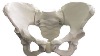
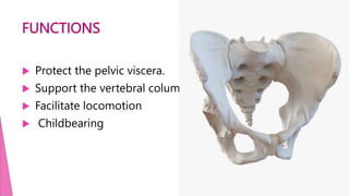
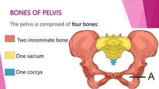
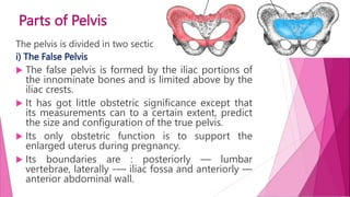
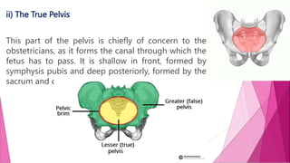
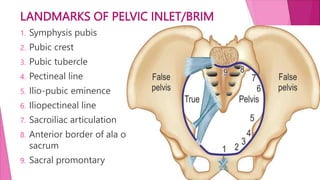
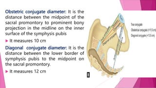
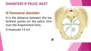
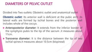
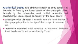
FEMALE PELVIS
Presenting by
V.Jhansi
M.Sc(N)
I Year
GCON
FEMALE PELVIS
Knowledge of the female pelvis is very essential for
the midwife , especially during conduction of the
labor.
SHAPE
The pelvis is a basin shaped structure that supports
the spinal column and protects the abdominal
organs.
FUNCTIONS
 Protect the pelvic viscera.
 Support the vertebral column
 Facilitate locomotion
 Childbearing
BONES OF PELVIS
The pelvis is comprised of four bones:
Two innominate bones
One sacrum
One coccyx
PELVIC JOINTS
The symphysis pubis-1
The sacroiliac joints-2
The sacrococcygeal joints-
1
PELVIC
LIGAMENTS
 Interpubic ligament
 Sacroiliac ligaments
 Sacrococcygeal ligament
 Sacrotuberous ligament
 Sacrospinous ligament
I.INNOMINATE BONE
Each innominate bone composed of three parts
INNOMINATE BONE-ILIUM
The ilium is the large flared out part covering 1/3rd part of
acetabulum.
 Inner upper wing like structure known wing/ala of ilium
 The concave anterior surface of the ilium is the iliac
fossa.
 Anterior lower border of ilium known iliopectineal line
which continues with pectineal line of pubis
 Upper crest and at the front of the Crest can be felt a
bony prominence known as the anterior superior iliac
spine.
 A short distance below it is the anterior inferior iliac
spine.
 There are two similar points at the other end of the
iliac crest, namely the posterior superior and the
posterior inferior iliac spines.
INNOMINATE BONE-ISCHIUM
The Ischium is the thick lowest part formed by
lower 2/3rd lower part of acetabulum and ½
portion of obturator foramen.
 It has a large prominence known as the
ischial tuberosity on which the body rests
when sitting.
 In relation to the ischial spines In labour the
station of the foetal head is estimated
INNOMINATE BONE-PUBIS
The Pubic bone forms the anterior part by 1/3rd of
acetabulum and ½ obturator foramen.
 It has a body and two rami—the superior and
inferior.
 The projection between ilium and pubis known
as ileo pubic eminence
 The rough portion just beside symphysis pubis is
pubic crest
 Just beside pubic crest a projection called
pubic tubercle
 The inferior borderline of the pubis is
pectineal line
 The two pubic bones meet at the symphysis
pubis and two inferior rami form the pubic
arch, merging into a similar ramus on the
ischium.
 On the lower border of the innominate bone
are found two curves the greater sciatic
notch and the lesser sciatic notch
II. THE SACRUM
 The Sacrum is a wedge-shaped bone
consisting of five fused vertebrae.
 The upper border of the first sacral
vertebra projects forward and is
known as the sacral promontory.
 The anterior surface of the sacrum is
concave and is referred as the hollow
of the sacrum.
 It has two wings or ala.
III.THE COCCYX
It consists of four fused
vertebrae, forming a small
triangular bone.
Parts of Pelvis
The pelvis is divided in two sections:
i) The False Pelvis
 The false pelvis is formed by the iliac portions of
the innominate bones and is limited above by the
iliac crests.
 It has got little obstetric significance except that
its measurements can to a certain extent, predict
the size and configuration of the true pelvis.
 Its only obstetric function is to support the
enlarged uterus during pregnancy.
 Its boundaries are : posteriorly — lumbar
vertebrae, laterally -— iliac fossa and anteriorly —
anterior abdominal wall.
ii) The True Pelvis
This part of the pelvis is chiefly of concern to the
obstetricians, as it forms the canal through which the
fetus has to pass. It is shallow in front, formed by
symphysis pubis and deep posteriorly, formed by the
sacrum and coccyx.
True pelvis is
divided into
Inlet
cavity
outlet.
LANDMARKS OF PELVIC INLET/BRIM
1. Symphysis pubis
2. Pubic crest
3. Pubic tubercle
4. Pectineal line
5. Ilio-pubic eminence
6. Iliopectineal line
7. Sacroiliac articulation
8. Anterior border of ala of
sacrum
9. Sacral promontary
DIAMETERS IF PELVIC INLET
1.Antero posterior diameter: it is
divided into 3 conjugates
 Anatomical or true conjugate
diameter: the : It is the distance
between the midpoint of the sacral
promontory to the inner margin of
the upper border of symphysis
 It measures 11 cm
Obstetric conjugate diameter: It is the
distance between the midpoint of the
sacral promontory to prominent bony
projection in the midline on the inner
surface of the symphysis pubis
 It measures 10 cm
Diagonal conjugate diameter: It is the
distance between the lower border of
symphysis pubis to the midpoint on
the sacral promontory.
 It measures 12 cm
DIAMETERS IF PELVIC INLET
II. Oblique diameter:
 There are two oblique diameters
— right and left.
 Each one extends from one
sacroiliac joint to the opposite
iliopubic eminence
 It measures 12 cm
DIAMETERS IF PELVIC INLET
III.Transverse diameter:
It is the distance between the two
farthest points on the pelvic brim
over the iliopectineal lines.
It measures 13 cm
DIAMETERS IF PELVIC CAVITY
Cavity is the segment of the pelvis bounded above by the
inlet and below by plane of least pelvic dimensions.
Antero-posterior diameter (12 cm) : It measures from the
mid-point on the posterior surface of the symphysis pubis
to the junction of 2nd and 3rd sacral vertebrae
Transverse diameter (12cm) : It cannot be precisely
measured as the points lie over the soft tissues covering
obturator foramen
DIAMETERS OF PELVIC OUTLET
Divided into Two outlets: Obstetric outlet and anatomical outlet
Obstetric outlet: Its anterior wall is deficient at the pubic arch; its
lateral walls are formed by ischial bones and the posterior wall
includes whole of the coccyx.
 Anteroposterior diameter: It extends from the inferior border of
the symphysis pubis to the tip of the sacrum .it measures about
11cm.
 Transverse diameter: It is the distance between the tip of two
ischial spines.it measures about 10.5cm (bispineal)
Anatomical outlet: It is otherwise known as bony outlet. It is
bounded in front by the lower border of the symphysis pubis;
laterally by the ischiopubic rami, ischial tuberosity and
sacrotuberous ligament and posteriorly by the tip of coccyx
 Anteroposterior diameter: It extends from the lower border of
the symphysis pubis to the tip of the coccyx. It measures 13
cm
 Transverse diameter: Inter tuberous .It measures between
inner borders of ischial tuberosities by 11cm.
FEMALE PELVIS.pptx
FEMALE PELVIS.pptx

FEMALE PELVIS.pptx

  • 2.
  • 3.
    FEMALE PELVIS Knowledge ofthe female pelvis is very essential for the midwife , especially during conduction of the labor. SHAPE The pelvis is a basin shaped structure that supports the spinal column and protects the abdominal organs.
  • 5.
    FUNCTIONS  Protect thepelvic viscera.  Support the vertebral column  Facilitate locomotion  Childbearing
  • 6.
    BONES OF PELVIS Thepelvis is comprised of four bones: Two innominate bones One sacrum One coccyx
  • 7.
    PELVIC JOINTS The symphysispubis-1 The sacroiliac joints-2 The sacrococcygeal joints- 1
  • 8.
    PELVIC LIGAMENTS  Interpubic ligament Sacroiliac ligaments  Sacrococcygeal ligament  Sacrotuberous ligament  Sacrospinous ligament
  • 9.
    I.INNOMINATE BONE Each innominatebone composed of three parts
  • 10.
    INNOMINATE BONE-ILIUM The iliumis the large flared out part covering 1/3rd part of acetabulum.  Inner upper wing like structure known wing/ala of ilium  The concave anterior surface of the ilium is the iliac fossa.  Anterior lower border of ilium known iliopectineal line which continues with pectineal line of pubis  Upper crest and at the front of the Crest can be felt a bony prominence known as the anterior superior iliac spine.  A short distance below it is the anterior inferior iliac spine.  There are two similar points at the other end of the iliac crest, namely the posterior superior and the posterior inferior iliac spines.
  • 11.
    INNOMINATE BONE-ISCHIUM The Ischiumis the thick lowest part formed by lower 2/3rd lower part of acetabulum and ½ portion of obturator foramen.  It has a large prominence known as the ischial tuberosity on which the body rests when sitting.  In relation to the ischial spines In labour the station of the foetal head is estimated
  • 12.
    INNOMINATE BONE-PUBIS The Pubicbone forms the anterior part by 1/3rd of acetabulum and ½ obturator foramen.  It has a body and two rami—the superior and inferior.  The projection between ilium and pubis known as ileo pubic eminence  The rough portion just beside symphysis pubis is pubic crest
  • 13.
     Just besidepubic crest a projection called pubic tubercle  The inferior borderline of the pubis is pectineal line  The two pubic bones meet at the symphysis pubis and two inferior rami form the pubic arch, merging into a similar ramus on the ischium.  On the lower border of the innominate bone are found two curves the greater sciatic notch and the lesser sciatic notch
  • 14.
    II. THE SACRUM The Sacrum is a wedge-shaped bone consisting of five fused vertebrae.  The upper border of the first sacral vertebra projects forward and is known as the sacral promontory.  The anterior surface of the sacrum is concave and is referred as the hollow of the sacrum.  It has two wings or ala.
  • 15.
    III.THE COCCYX It consistsof four fused vertebrae, forming a small triangular bone.
  • 16.
    Parts of Pelvis Thepelvis is divided in two sections: i) The False Pelvis  The false pelvis is formed by the iliac portions of the innominate bones and is limited above by the iliac crests.  It has got little obstetric significance except that its measurements can to a certain extent, predict the size and configuration of the true pelvis.  Its only obstetric function is to support the enlarged uterus during pregnancy.  Its boundaries are : posteriorly — lumbar vertebrae, laterally -— iliac fossa and anteriorly — anterior abdominal wall.
  • 17.
    ii) The TruePelvis This part of the pelvis is chiefly of concern to the obstetricians, as it forms the canal through which the fetus has to pass. It is shallow in front, formed by symphysis pubis and deep posteriorly, formed by the sacrum and coccyx.
  • 18.
    True pelvis is dividedinto Inlet cavity outlet.
  • 19.
    LANDMARKS OF PELVICINLET/BRIM 1. Symphysis pubis 2. Pubic crest 3. Pubic tubercle 4. Pectineal line 5. Ilio-pubic eminence 6. Iliopectineal line 7. Sacroiliac articulation 8. Anterior border of ala of sacrum 9. Sacral promontary
  • 20.
    DIAMETERS IF PELVICINLET 1.Antero posterior diameter: it is divided into 3 conjugates  Anatomical or true conjugate diameter: the : It is the distance between the midpoint of the sacral promontory to the inner margin of the upper border of symphysis  It measures 11 cm
  • 21.
    Obstetric conjugate diameter:It is the distance between the midpoint of the sacral promontory to prominent bony projection in the midline on the inner surface of the symphysis pubis  It measures 10 cm Diagonal conjugate diameter: It is the distance between the lower border of symphysis pubis to the midpoint on the sacral promontory.  It measures 12 cm
  • 22.
    DIAMETERS IF PELVICINLET II. Oblique diameter:  There are two oblique diameters — right and left.  Each one extends from one sacroiliac joint to the opposite iliopubic eminence  It measures 12 cm
  • 23.
    DIAMETERS IF PELVICINLET III.Transverse diameter: It is the distance between the two farthest points on the pelvic brim over the iliopectineal lines. It measures 13 cm
  • 24.
    DIAMETERS IF PELVICCAVITY Cavity is the segment of the pelvis bounded above by the inlet and below by plane of least pelvic dimensions. Antero-posterior diameter (12 cm) : It measures from the mid-point on the posterior surface of the symphysis pubis to the junction of 2nd and 3rd sacral vertebrae Transverse diameter (12cm) : It cannot be precisely measured as the points lie over the soft tissues covering obturator foramen
  • 25.
    DIAMETERS OF PELVICOUTLET Divided into Two outlets: Obstetric outlet and anatomical outlet Obstetric outlet: Its anterior wall is deficient at the pubic arch; its lateral walls are formed by ischial bones and the posterior wall includes whole of the coccyx.  Anteroposterior diameter: It extends from the inferior border of the symphysis pubis to the tip of the sacrum .it measures about 11cm.  Transverse diameter: It is the distance between the tip of two ischial spines.it measures about 10.5cm (bispineal)
  • 26.
    Anatomical outlet: Itis otherwise known as bony outlet. It is bounded in front by the lower border of the symphysis pubis; laterally by the ischiopubic rami, ischial tuberosity and sacrotuberous ligament and posteriorly by the tip of coccyx  Anteroposterior diameter: It extends from the lower border of the symphysis pubis to the tip of the coccyx. It measures 13 cm  Transverse diameter: Inter tuberous .It measures between inner borders of ischial tuberosities by 11cm.