Download as PDF, PPTX


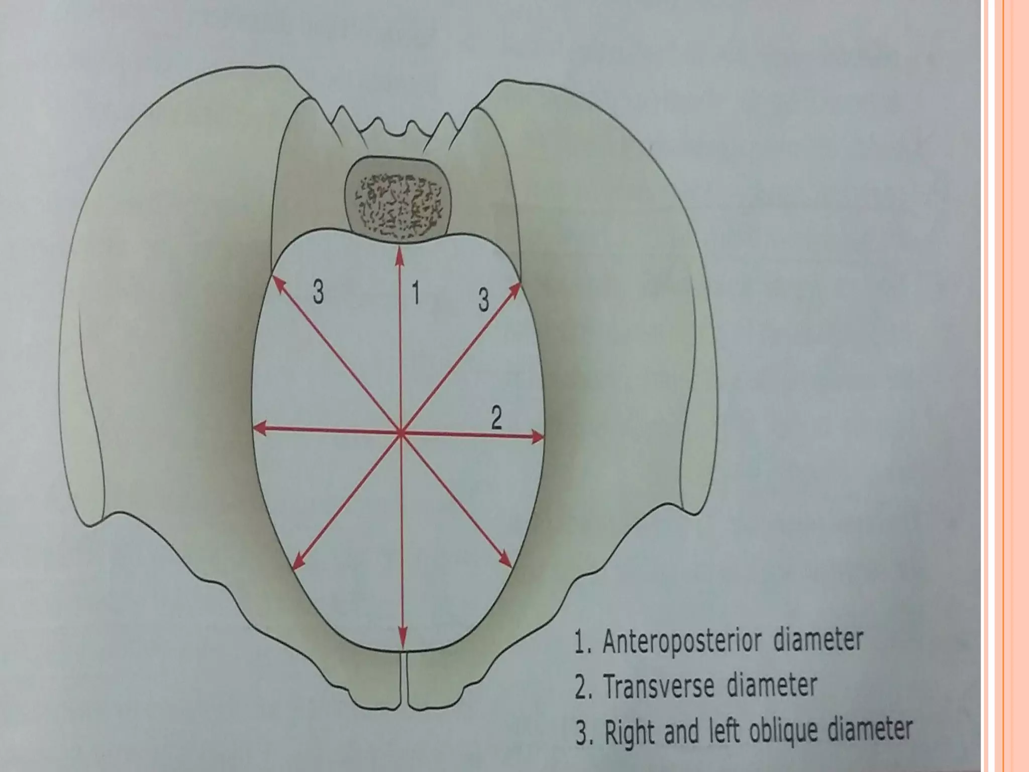

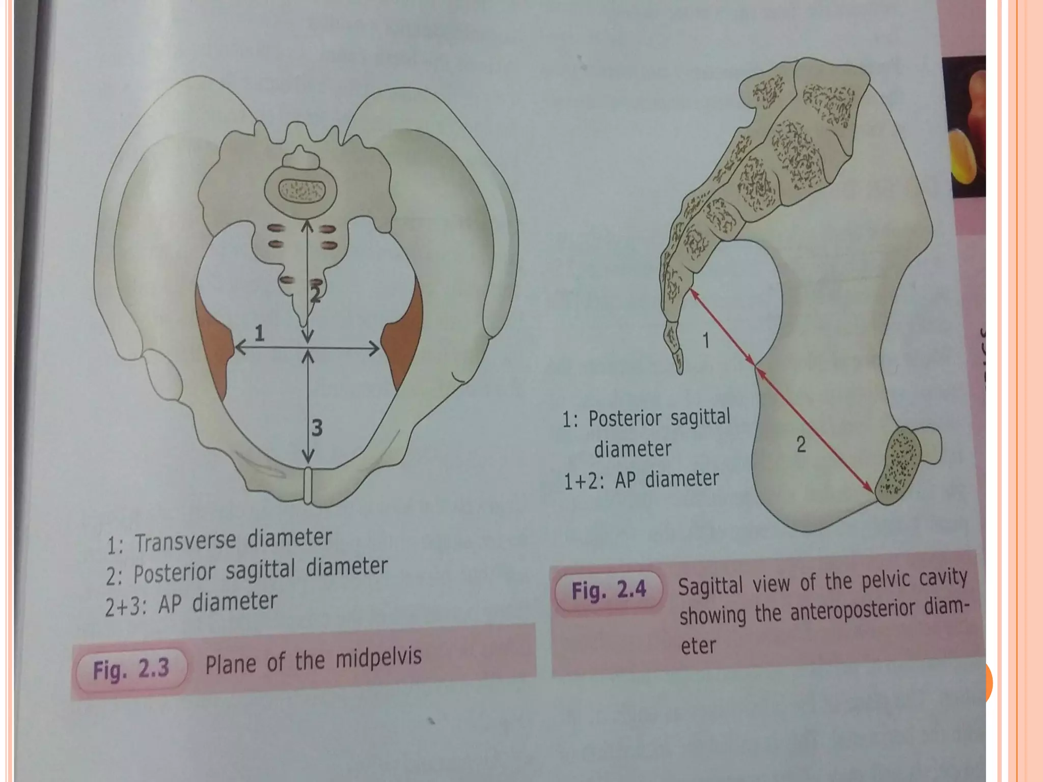
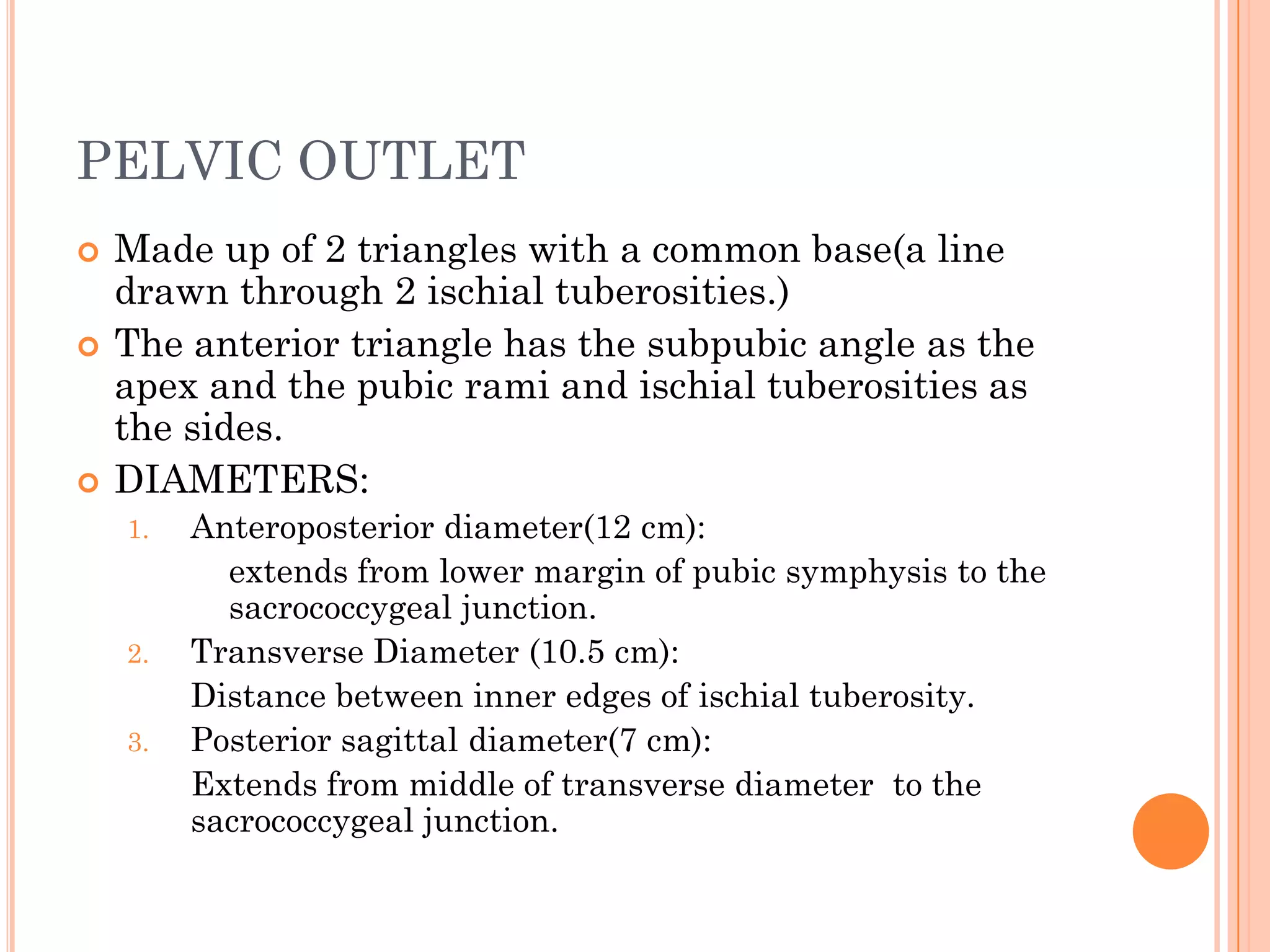
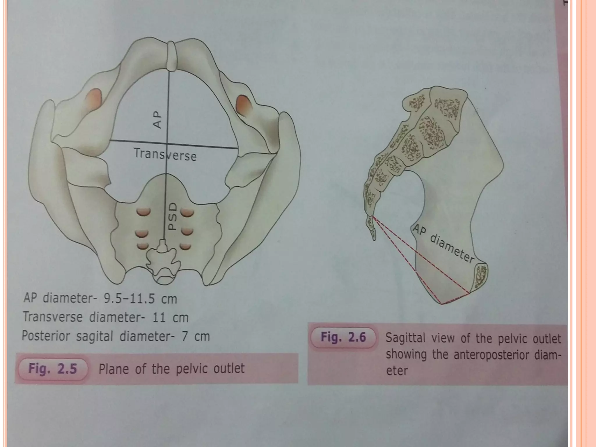
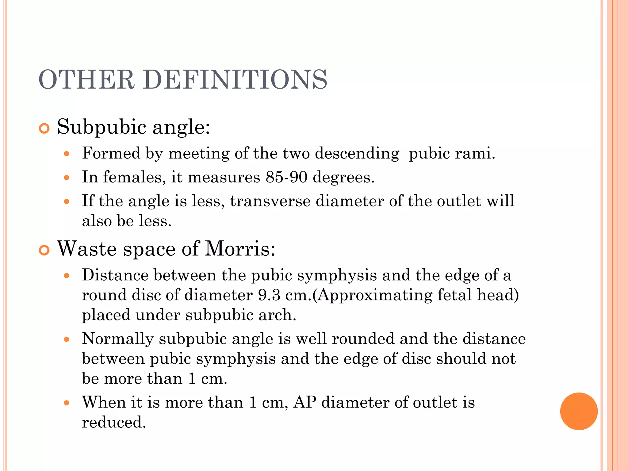


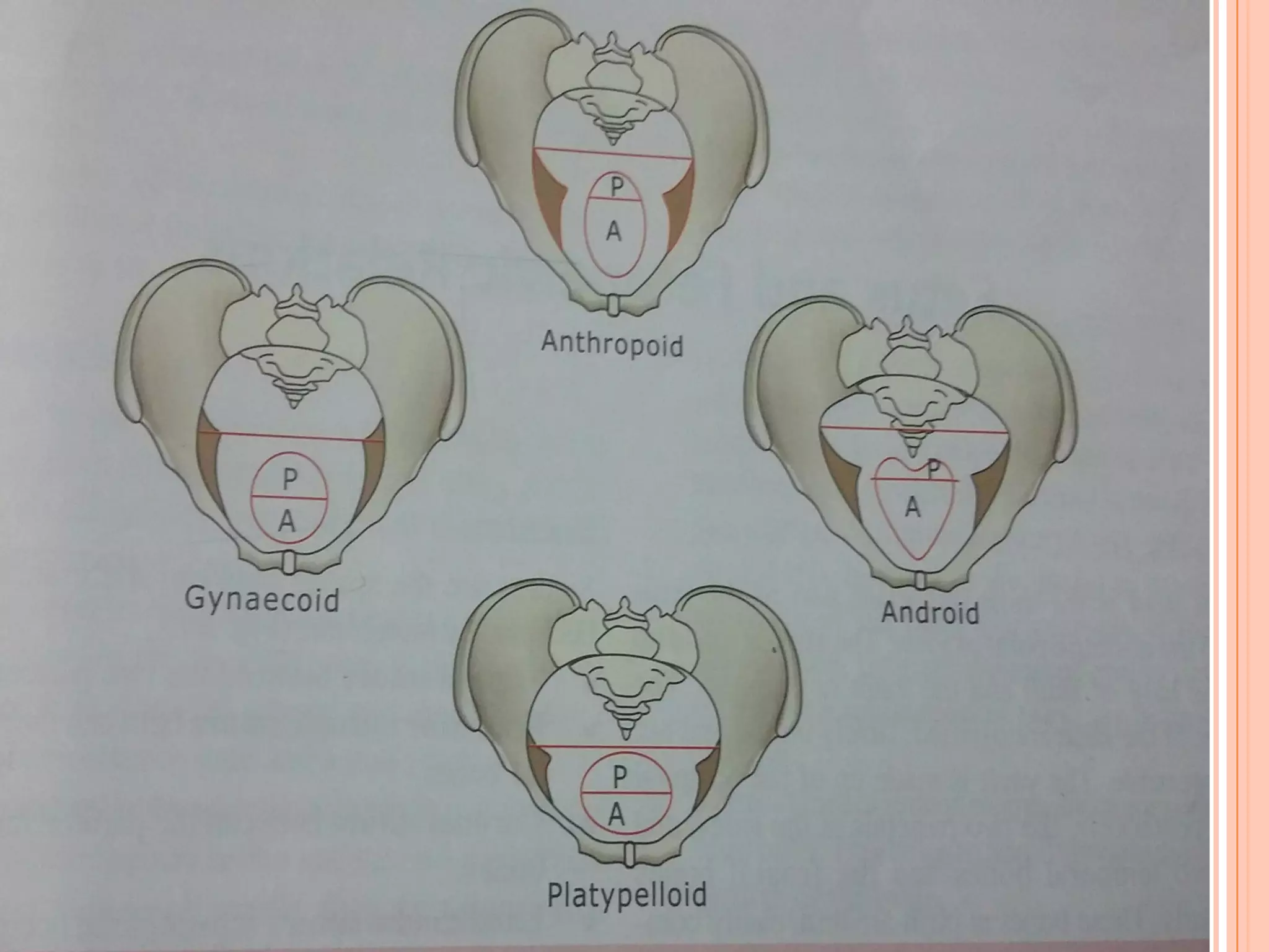


The pelvis is made of bones that form three areas - the inlet, cavity, and outlet. The inlet is oval shaped and its diameters include the obstetric conjugate and transverse diameter. The midpelvis is at the level of the ischial spines and its diameters are important for fetal engagement and rotation. The outlet has triangular shapes defined by the ischial tuberosities and subpubic arch. The pelvis can be classified based on the shape of these areas.


